. 2025 Mar;301(3):108216.
doi: 10.1016/j.jbc.2025.108216. Epub 2025 Jan 23.

The juxtamembrane sequence of small ankyrin 1 mediates the binding of its cytoplasmic domain to SERCA1 and is required for inhibitory activity

Affiliations

The juxtamembrane sequence of small ankyrin 1 mediates the binding of its cytoplasmic domain to SERCA1 and is required for inhibitory activity

Yi Li et al. J Biol Chem. 2025 Mar.

Abstract

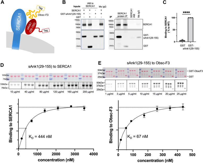
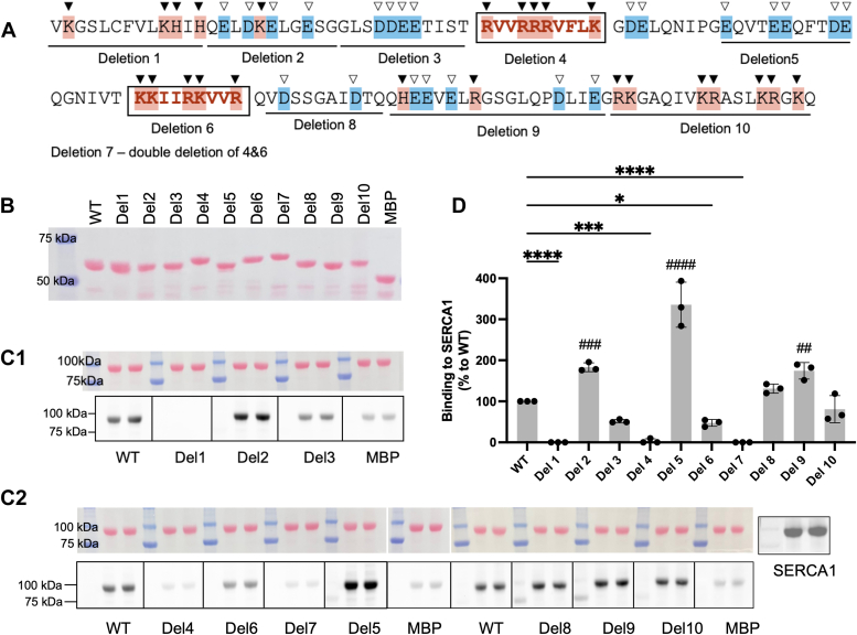
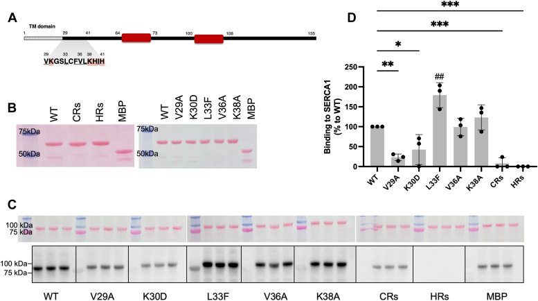
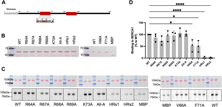
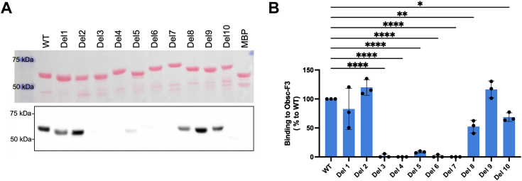
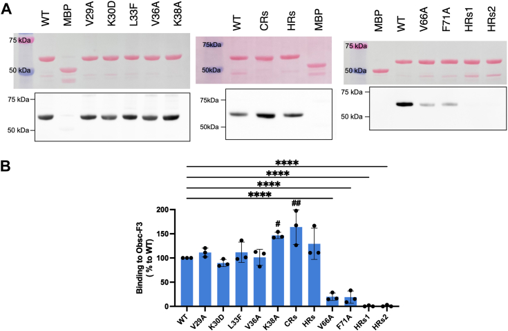
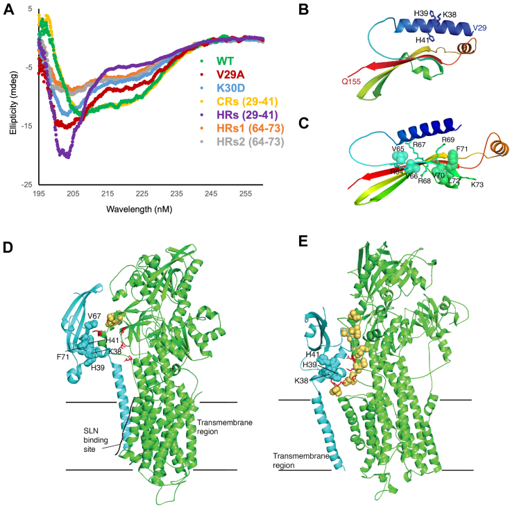
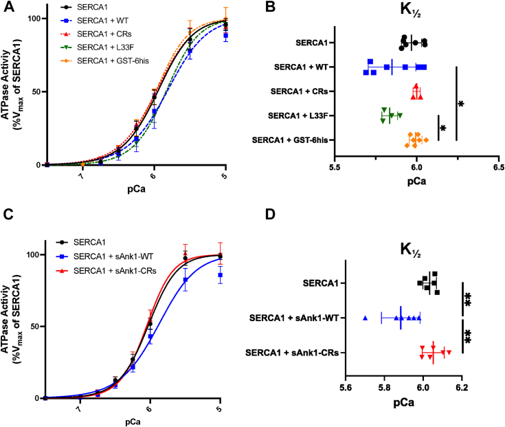
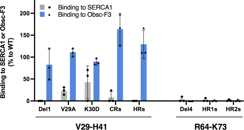
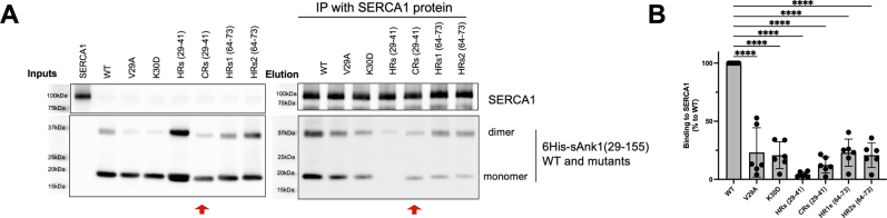
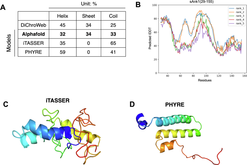
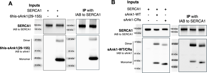
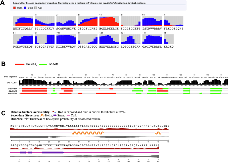
Sarcoplasmic/endoplasmic reticulum Ca2+-ATPase1 (SERCA1) is responsible for the clearance of cytosolic Ca2+ in skeletal muscle. Due to its vital importance in regulating Ca2+ homeostasis, the regulation of SERCA1 has been intensively studied. Small ankyrin 1 (sAnk1, Ank1.5), a 17 kDa muscle-specific isoform of ANK1, binds to SERCA1 directly via both its transmembrane and cytoplasmic domains and inhibits SERCA1's ATPase activity. Here, we characterize the interaction between the cytoplasmic domain of sAnk1 (sAnk1(29-155)) and SERCA1. The binding affinity for sAnk1 (29-155) to SERCA1 was 444 nM by blot overlay, about 7-fold weaker than the binding of sAnk1(29-155) to obscurin, a giant protein of the muscle cytoskeleton. Site-directed mutagenesis identified K38, H39, and H41, in the juxtamembrane region, as residues likely to mediate binding to SERCA1. These residues are not required for obscurin binding. Residues R64-K73, which do contribute to obscurin binding, are also required for binding to SERCA1, but only the hydrophobic residues in this sequence are required, not the positively charged residues necessary for obscurin binding. Circular dichroism analysis of sAnk1(29-155) indicates that most mutants show significant structural changes, with the exception of those containing alanines in place of K38, H39 and H41. Although the cytoplasmic domain of sAnk1 does not inhibit SERCA1's Ca2+-ATPase activity, with or without mutations in the juxtamembrane sequence, the inhibitory activity of full-length sAnk1 requires the WT juxtamembrane sequence. We used these data to model sAnk1 and the sAnk1-SERCA1 complex. Our results suggest that, in addition to its transmembrane domain, sAnk1 uses its juxtamembrane sequence and perhaps part of its obscurin binding site to bind to SERCA1, and that this binding contributes to their robust association in situ, as well as regulation of SERCA1's activity.

Keywords: AlphaFold; Ca-ATPase; ankyrin; circular dichroism (CD); complex; membrane transport; sarcoplasmic reticulum (SR); skeletal muscle; structural model.


Conflict of interest statement

Conflict of interest The authors declare that they have no conflicts of interest with the contents of this article.

Figures

Figure 1
Figure 1
sAnk1(29–155) binds directly to SERCA1 with a lower affinity than it binds to obscurin. A, schematic diagram showing the cytoplasmic domain of sAnk1 (sAnk1(29–155), red) bound to SERCA1 (blue) directly, as well as to obscurin (Obsc-F3, the F3 fragment of obscurin which contains the main binding sites for sAnk1, yellow), on the SR membrane (gray). In this work, sAnk1(29–155) WT and mutants were made as fusion proteins with MBP, GST, or 6His tags; Obsc-F3 was made as a fusion protein with a GST tag. B, Co-IP using beads coated with SERCA1 protein and affinity-purified GST-sAnk1(29–155). Inputs: beads coated with anti-SERCA1 or nonimmune mouse IgG, and incubated with SERCA1 were mixed with GST or GST-sAnk1(29–155). The mixtures were separated by SDS-PAGE and immunoblotted with the appropriate antibodies. Elution: Proteins bound to the beads were separated by SDS-PAGE and immunoblotted. C, Quantification: The amount of GST or GST-sAnk1(29–155) in the eluates was first normalized to the amount of SERCA1, then normalized to GST-sAnk1(29–155). Data are presented as % to WT, mean ± SD, n = 3; ∗∗∗∗, p < 0.0001. D and E, blot overlay assays and saturation binding curves of sAnk1(29–155) binding to SERCA1 (D) and GST-Obsc-F3. E, Top: results used to generate the binding curves. Blots were prepared with a constant amount of SERCA1, GST-Obsc-F3 or GST (shown by Ponceau S staining, samples in each membrane are separated by white bars) and overlaid with increasing concentrations of MBP-sAnk1(29–155) (separate samples are indicated by black lines). Bottom: binding curves and KD. D, for binding to SERCA1, KD = 444 ± 69.0 nM (mean ± SD, n = 3). E, for binding to Obsc-F3, KD = 66.6 ± 18.2 nM (mean ± SD, n = 3). The results showed that sAnk1(29–155) binds to SERCA1 directly, with an affinity 6.7-fold weaker than binding to GST-obsc-F3. Co-IP, co-immunoprecipitation; IgG, immunoglobulin G; MBP, maltose binding protein; sAnk1, small ankyrin 1 (Ank1.5); SERCA1, sarcoplasmic/endoplasmic reticulum Ca2+-ATPase1; SR, sarcoplasmic reticulum.
Figure 2
Figure 2
Binding of MBP-sAnk1(29–155) deletion mutants to SERCA1. A, full sequence of rat sAnk1(29–155). The positively charged residues are highlighted in red and indicated with ▼; the negatively charged residues are highlighted in blue and indicated with ▿. Two positively charged regions, which we previously showed mediate binding to obscurin, are bolded in red and highlighted with boxes. The sequences deleted in each deletion mutant are underlined and labeled. Deletion7 (Del7) is missing both positively charged regions. B, aliquots containing 1 μg affinity-purified MBP fusion proteins of the deletion mutants of sAnk1(29–155), Del1-10, and MBP alone, were separated by SDS-PAGE, transferred to nitrocellulose membrane and stained with Ponceau S. (C1 and C2) results of blot overlay assays of the MBP fusion proteins binding to SERCA1. Top, Ponceau S staining of blots of 1.5 μg SR vesicles per lane, immunoblotted for SERCA1 (shown below the Ponceau S staining in C1 and C2, each membrane is separated by white bars). Bottom, the blots were separated (indicated by solid black bars) and overlaid with 3 μg/ml MBP-sAnk1(29–155) (WT) and MBP fusion proteins of the deletion mutants, followed by extensive washing and then probed with the antibody to MBP. MBP protein alone was used as a control. All blots were imaged once. D, quantitation of blot overlay assays. All values are corrected for the value obtained from MBP alone, then normalized to WT (%), mean ± SD, n = 3. Statistical analysis and bar graphs were obtained using ordinary one-way ANOVA. ∗p < 0.05, ∗∗∗p < 0.0005, ∗∗∗∗p < 0.0001. For significantly increased groups, # was used instead of ∗ with the same scale for significance. The results show that V29-H41 (in Del1) and R64-K73 (in Del4) harbor potential binding sites for SERCA1. MBP, maltose binding protein; sAnk1, small ankyrin 1 (Ank1.5); SERCA1, sarcoplasmic/endoplasmic reticulum Ca2+-ATPase1; SR, sarcoplasmic reticulum.
Figure 3
Figure 3
Mutants of sAnk1(29–41) binding to SERCA1.A, cartoon depiction of the location and sequence of an N-terminal region of the cytoplasmic domain of sAnk1 that likely binds SERCA1, V29-H41. Hydrophobic and electropositive residues were underlined and highlighted in red, respectively. The two red boxes indicate the location of the clusters of positively charged residues that mediate binding to obscurin. B, aliquots containing 1 μg of affinity-purified MBP mutants of V29-H41 were stained for Ponceau S, to assess purity, as in Figure 2. C, blot overlays of the mutants in the V29 to 41 sequence for binding to SERCA1, as in Figure 2. D, quantitation of results in (C), mean ± SD, n = 3, as in Figure 2. The results showed the single amino acid substitutions, sAnk1(29-155)-V29A and -K30D to have minimal binding, and the multiple alanine mutants, sAnk1(29–155)-CRs and -HRs, to have little to no binding. MBP, maltose binding protein; sAnk1, small ankyrin 1 (Ank1.5); SERCA1, sarcoplasmic/endoplasmic reticulum Ca2+-ATPase1.
Figure 4
Figure 4
Mutants of sAnk1(64–73) binding to SERCA1.A, location and sequence of the C-central binding region, R64-K73, for SERCA1 in full-length sAnk1. Hydrophobic and electropositive residues were underlined and highlighted in red, respectively. Other features are as in Figures 2 and 3. B, Ponceau S staining of blots of 1 μg affinity-purified MBP fusion mutants of R64-K73. All-A has alanine mutations in all five charged residues (R64A/R67A/R68A/R69A/K73A). HRs1 (V65A/V66A) and HRs2 (F70A/F71A/L72A) have 2 and 3 alanine substitutions in hydrophobic residues, respectively. C, results of blot overlay assays testing mutants of R64-K73 binding to SERCA1. Top, alanine mutation in positively charged residues. Bottom, alanine mutations in hydrophobic residues. D, quantitation of blot overlay assays, mean ± SD, n = 3. The results showed that mutations in a single hydrophobic residue inhibited the binding significantly and that mutating two or three hydrophobic residues interrupted binding to SERCA1 completely. Mutation of the positively charged residues had no effect. MBP, maltose binding protein; sAnk1, small ankyrin 1 (Ank1.5); SERCA1, sarcoplasmic/endoplasmic reticulum Ca2+-ATPase1.
Figure 5
Figure 5
Deletion mutants of sAnk1(29–155) binding to Obsc-F3. Methods are as in Figures 2 and 3. A, blot overlay assay of mutants of MBP-sAnk1(29–155) binding to GST-Obsc-F3. Top, Ponceau S staining of membrane containing 1 μg MBP-sAnk1(29–155), deletion mutants, and MBP. Bottom, after overlaying the blot with 3 μg/ml GST-Obsc-F3, it was immunoblotted with the antibody to GST. B, quantitation of the assays, mean ± SD, n = 3. Readings were normalized to the loading of the sAnk1(29–155) mutants, obtained from Ponceau S staining, and presented as % of the binding to the WT. The results showed that Del1 bound to obscurin like the WT protein but did not bind to SERCA1, suggesting that V29-H41 binds uniquely to SERCA1, whereas Del4 failed to bind to both obscurin and SERCA1, suggesting it may harbor binding sites for both proteins. MBP, maltose binding protein; sAnk1, small ankyrin 1 (Ank1.5); SERCA1, sarcoplasmic/endoplasmic reticulum Ca2+-ATPase1.
Figure 6
Figure 6
Mutants in V29-H41 bind normally to GST-Obsc-F3, but mutants in R64-K73 do not. As in Figures 2 and 3. A, results of blot overlay assays of binding of mutants in MBP-sAnk1(29-155)-V29-H41 (left and middle panels) and -R64-K73 (right panel) to GST-Obsc-F3. Top, Ponceau S staining of 1 μg MBP-sAnk1 (29-155), mutants, and MBP protein. Bottom, after the blot was incubated with 3 μg/ml GST-Obsc-F3, it was immunoblotted with antibody to GST. B, quantitation of blot overlay assays, mean ± SD, n = 3. The significant changes in binding to GST-Obsc-F3 were labeled for significance as in Figure 2. The results showed that none of the mutations in V29-H41 affected the binding of sAnk1(29-155) to obscurin, whereas all four of the mutants in R64-K73 inhibited the binding of sAnk1(29-155) to obscurin and SERCA1 similarly. MBP, maltose binding protein; sAnk1, small ankyrin 1 (Ank1.5); SERCA1, sarcoplasmic/endoplasmic reticulum Ca2+-ATPase1.
Figure 7
Figure 7
Circular dichroism (CD) spectra of sAnk1(29–155) mutants, with models of sAnk1(29–155) and sAnk1 bound to SERCA1.A, CD spectra of 6His-sAnk1(29–155) and mutants that do not bind to SERCA1, analyzed at a concentration of 15 μM. Only the spectrum of CRs (K38A/H39A/H41A) overlapped with that of the WT protein (B and C) structural model of sAnk1(29–155) predicted by AlphaFold. The N-terminus is in blue, and the C-terminus is in red. The binding residues to SERCA1, K38, H39, and H41 are labeled in (B). The residues in R64-K73, both charged and hydrophobic, are labeled in (C). The structure shown in (C) suggests that R64-K73 could bind to SERCA1 and obscurin simultaneously. D and E, models of sAnk1 bound to SERCA. sAnk1 (blue) binds to SERCA (green) through interactions in the TM region, and through electrostatic interactions between sAnk1 K38, H39, and H41 (blue spheres) and negative charges on SERCA (red sticks), and through hydrophobic interactions (beige spheres on SERCA). D, Model1: sAnk1 occupied the same TM groove in SERCA as SLN does, and the cytoplasmic domain of sAnk1 binds to the A-domain of SERCA1. E, Model 2: sAnk1 binds to SERCA in TM sites different from SLN. sAnk1, small ankyrin 1 (Ank1.5); SERCA1, sarcoplasmic/endoplasmic reticulum Ca2+-ATPase1; SLN, sarcolipin; TM, transmembrane.
Figure 8
Figure 8
ATPase assay of SERCA1 in the presence of 6his-sAnk1(29–155) or mutant.A, microsomes of COS7 cells transfected to express SERCA1 (20 μg/ml) were mixed with 3.5 μM 6His-sAnk1(29–155) (WT), 6His-sAnk1(29–155)-K38A/H39A/H41A (CRs) or 6His-sAnk1(29–155)-L33F at different [Ca2+]free. Released Pi was normalized to the Vmax in SERCA1 alone, and fitted into the “Allosteric Sigmoidal” model to generate the curves and K½. B, pCa at which half-maximal activity (K½) for A, presented as mean ± SD. For SERCA1, SERCA1 + WT sAnk1(29–155) and SERCA1 + GST-6his, n = 7; for SERCA1 + sAnk1(29–155)-CRs, n = 3; for SERCA1 + sAnk1(29–155)-L33 F, n = 4. For comparisons, there are significant differences between SERCA1 + WT sAnk1(29–155) versus SERCA1 + GST-6his, p-value = 0.0322; SERCA1 + sAnk1(29–155)-L33F versus SERCA1 + GST-6his, p-value = 0.0493. There are no significant differences between other groups. C, microsomes (20 μg/ml) from COS7 cells transfected to express SERCA1 with or without full-length sAnk1-WT or sAnk1-CRs (K38A/H39A/H41A) were measured for Ca2+-ATPase activity. The released Pi, and reaction curves were measured and generated as above. D, pCa at which half-maximal activity (K½) for C, presented as mean ± SD, n = 6. For comparisons, SERCA1 versus SERCA1 + sAnk1-WT, p = 0.0054; SERCA1 + sAnk1-WT versus SERCA1 + sAnk1-CRs, p = 0.0021. There’s no significant difference of SERCA1 versus SERCA1 + sAnk1-CRs. sAnk1, small ankyrin 1 (Ank1.5); SERCA1, sarcoplasmic/endoplasmic reticulum Ca2+-ATPase1.
Fig. S1
Fig. S1
Fig. S2
Fig. S2
Fig. S3
Fig. S3
Fig. S4
Fig. S4
Fig. S5
Fig. S5

Similar articles

References

    1. Primeau J.O., Armanious G.P., Fisher M.E., Young H.S. The SarcoEndoplasmic reticulum calcium ATPase. Subcell. Biochem. 2018;87:229–258. - PubMed
    1. Lipskaia L., Hulot J.S., Lompre A.M. Role of sarco/endoplasmic reticulum calcium content and calcium ATPase activity in the control of cell growth and proliferation. Pflugers Arch. 2009;457:673–685. - PubMed
    1. Ait Ghezali L., Arbabian A., Roudot H., Brouland J.P., Baran-Marszak F., Salvaris E., et al. Induction of endoplasmic reticulum calcium pump expression during early leukemic B cell differentiation. J. Exp. Clin. Cancer Res. 2017;36:87. - PMC - PubMed
    1. Periasamy M., Maurya S.K., Sahoo S.K., Singh S., Sahoo S.K., Reis F.C.G., et al. Role of SERCA pump in muscle thermogenesis and metabolism. Compr. Physiol. 2017;7:879–890. - PubMed
    1. Gamu D., Juracic E.S., Hall K.J., Tupling A.R. The sarcoplasmic reticulum and SERCA: a nexus for muscular adaptive thermogenesis. Appl. Physiol. Nutr. Metab. 2020;45:1–10. - PubMed

MeSH terms

Substances

LinkOut - more resources