. 2025 Jan 25;15(1):3183.
doi: 10.1038/s41598-024-83624-9.

Poor fixation stability does not account for motion perception deficits in amblyopia

Affiliations

Poor fixation stability does not account for motion perception deficits in amblyopia

Kimberly Meier et al. Sci Rep. .

Abstract

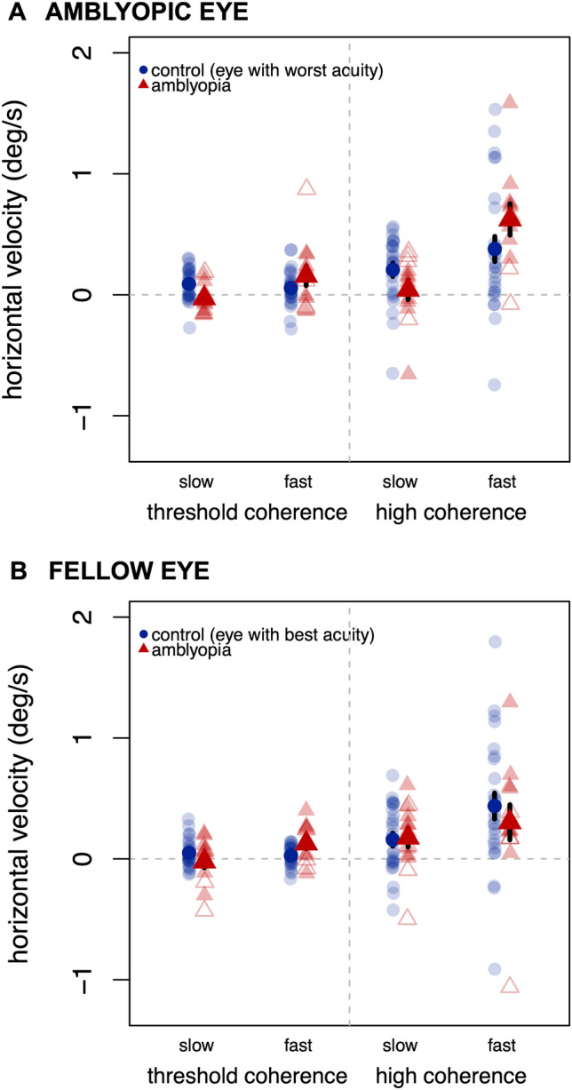
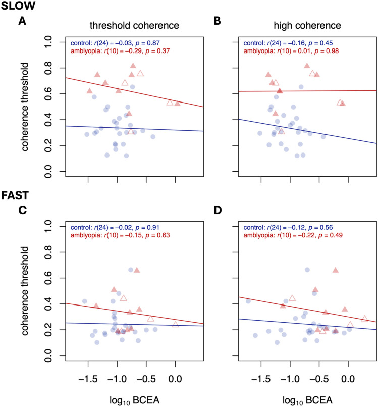
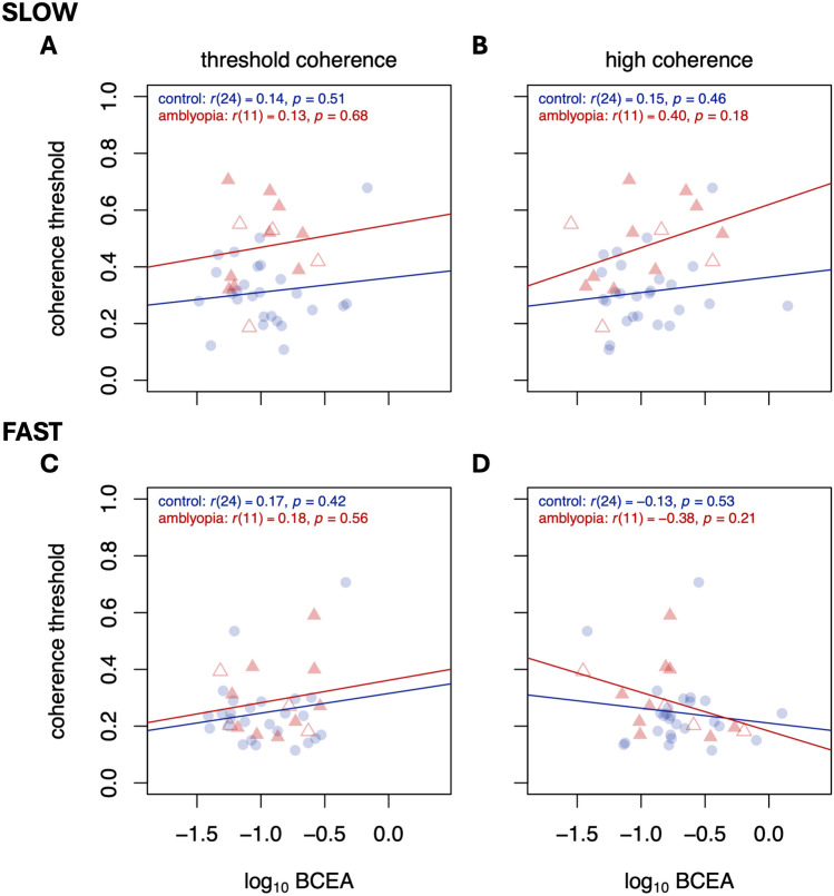
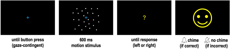
People with amblyopia show deficits in global motion perception, especially at slow speeds. These observers are also known to have unstable fixation when viewing stationary fixation targets, relative to healthy controls. It is possible that poor fixation stability during motion viewing interferes with the fidelity of the input to motion-sensitive neurons in visual cortex. To probe these mechanisms at a behavioral level, we assessed motion coherence thresholds in adults with amblyopia while measuring fixation stability. Consistent with prior work, participants with amblyopia had elevated coherence thresholds for the slow speed stimuli, but not the fast speed stimuli, using either the amblyopic or the fellow eye. Fixation stability was elevated in the amblyopic eye relative to controls across all motion stimuli, and not selective for conditions on which perceptual deficits were observed. Fixation stability was not related to visual acuity, nor did it predict coherence thresholds. These results suggest that motion perception deficits might not be a result of poor input to the motion processing system due to unstable fixation, but rather due to processing deficits in motion-sensitive visual areas.

Keywords: Amblyopia; Fixation stability; Global motion perception; Motion coherence; Speed.


Conflict of interest statement

Declarations. Competing interests: The authors declare no competing interests.

Figures

Fig. 1
Fig. 1
Mean direction discrimination thresholds for the slow (1 deg/s) and fast (30 deg/s) motion stimuli, separated by amblyopic eye (or eye with worst acuity) and fellow eye (or eye with best acuity). Lower values indicate better performance. Lighter markers indicate individual datapoints. Anisometropic amblyopia in filled triangles; strabismic or ansio-strabismic amblyopia in open triangles. Error bars reflect standard error.
Fig. 2
Fig. 2
Mean log10 bivariate contour ellipse area (BCEA) in (a) the amblyopic eye or (b) the fellow eye for slow (1 deg/s) and fast (30 deg/s) motion stimuli, separated for trials near each individual’s threshold coherence and trials at high coherence. Lower values indicate greater stability, as gaze points are dispersed over a smaller area. Lighter markers indicate individual datapoints. Anisometropic amblyopia in filled triangles; strabismic or ansio-strabismic amblyopia in open triangles. Error bars reflect standard error.
Fig. 3
Fig. 3
Mean horizontal eye velocity in (a) the amblyopic eye or (b) the fellow eye for slow (1 deg/s) and fast (30 deg/s) motion stimuli, separated for trials near each individual’s threshold coherence and trials at high coherence. Positive values indicate an eye movement in the same direction as the motion signal; negative values in the opposite direction. Lighter markers indicate individual datapoints. Anisometropic amblyopia in filled triangles; strabismic or ansio-strabismic amblyopia in open triangles. Error bars reflect standard error.
Fig. 4
Fig. 4
Motion coherence thresholds as a function of BCEA for slow (top row) and fast (bottom row) speeds, for trials near threshold (left column) and trials at high coherence (right column). Significance figures above are not adjusted for multiple comparisons. Data are from amblyopic eye/control best eye viewing. Colors and symbols are the same as Figs. 1, 2 and 3.
Fig. 5
Fig. 5
Same as Fig. 4, but for fellow eye/control worst eye viewing.
Fig. 6
Fig. 6
Trial sequence. Motion stimulus is schematic and not representative of dot size or density; see in-text for details.

Similar articles

References

    1. Birch, E. E. Amblyopia and binocular vision. Prog. Retin. Eye Res.33, 67–84. 10.1016/j.preteyeres.2012.11.001 (2013). - PMC - PubMed
    1. Levi, D. M. Crowding—an essential bottleneck for object recognition: A mini-review. Vis. Res.48, 635–654. 10.1016/j.visres.2007.12.009 (2008). - PMC - PubMed
    1. McKee, S. P., Levi, D. M. & Movshon, J. A. The pattern of visual deficits in amblyopia. J. Vis.3, 380–405. 10.1167/3.5.5 (2003). - PubMed
    1. Baker, D. H., Meese, T. S. & Hess, R. F. Contrast masking in strabismic amblyopia: Attenuation, noise, interocular suppression and binocular summation. Vis. Res.48, 1625–1640. 10.1016/j.visres.2008.04.017 (2008). - PubMed
    1. Hayward, J., Truong, G., Partanen, M. & Giaschi, D. Effects of speed, age, and amblyopia on the perception of motion-defined form. Vis. Res.51, 2216–2223. 10.1016/j.visres.2011.08.023 (2011). - PubMed

LinkOut - more resources