. 2025 Mar 5;33(3):1048-1072.
doi: 10.1016/j.ymthe.2025.01.038. Epub 2025 Jan 25.

Pharmacological blocking of microfibrillar-associated protein 4 reduces retinal neoangiogenesis and vascular leakage

Affiliations

Pharmacological blocking of microfibrillar-associated protein 4 reduces retinal neoangiogenesis and vascular leakage

Anders Schlosser et al. Mol Ther. .

Abstract

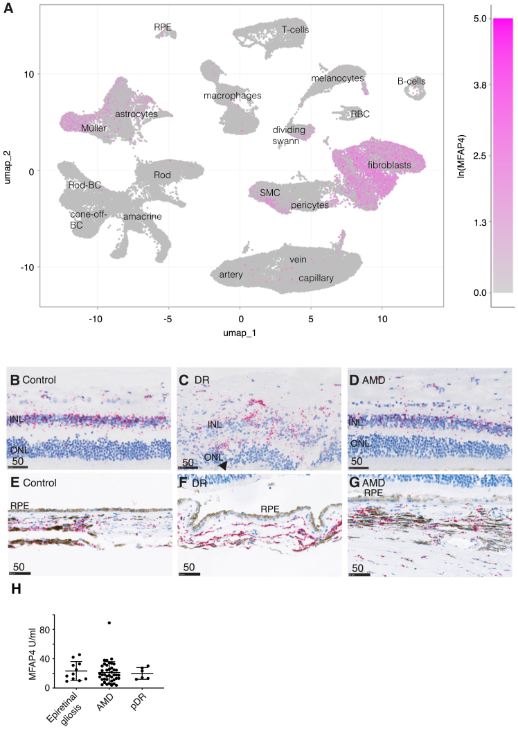
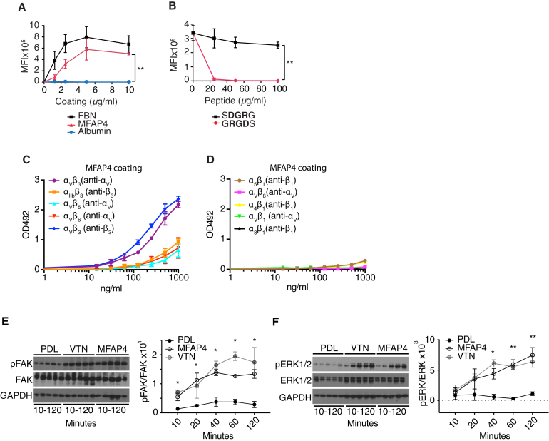
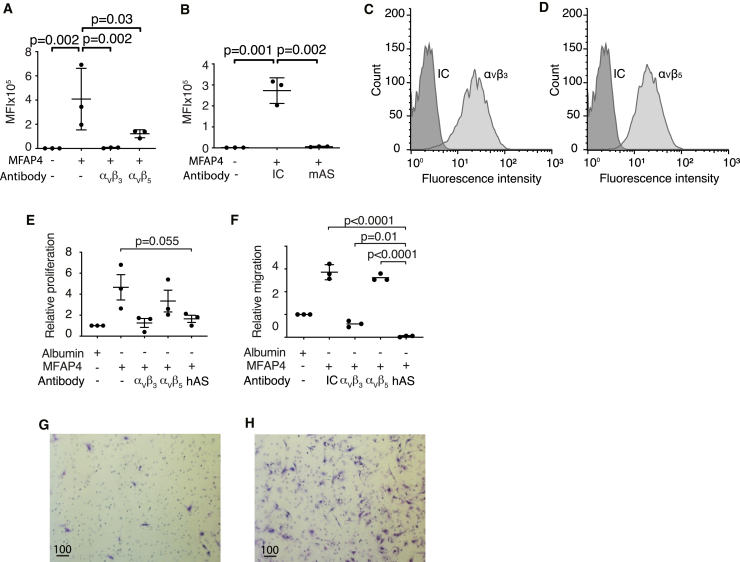
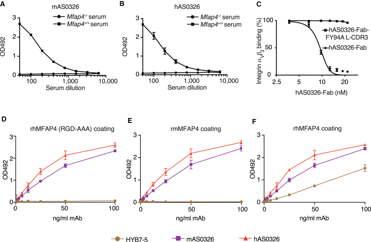
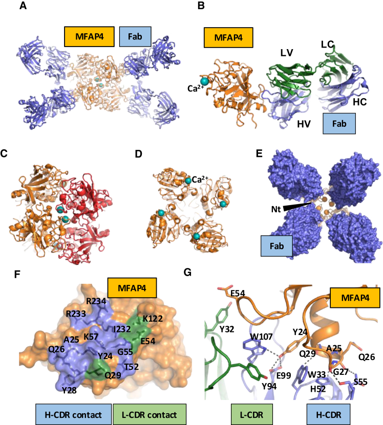
Neovascular age-related macular degeneration and diabetic macular edema are leading causes of vision loss evoked by retinal neovascularization and vascular leakage. The glycoprotein microfibrillar-associated protein 4 (MFAP4) is an integrin αVβ3/5/6 ligand present in the extracellular matrix. Single-cell transcriptomics reveal MFAP4 expression in cell types in close proximity to vascular endothelial cells, including choroidal vascular mural cells, retinal astrocytes, and Müller cells. Binding of the anti-MFAP4 antibody, hAS0326, makes MFAP4 inaccessible for integrin receptor interaction, and thereby hAS0326 blocked endothelial cell motility in vitro. Intravitreal hAS0326 inhibited retinal vascular lesion area and neovessel volume in a laser-induced choroidal neovascularization mouse model, vascular permeability in streptozotocin-induced retinopathy, and vascular leakage area in a chronic non-human primate model of DL-2-aminoadipic acid-induced retinopathy. One dose of hAS0326 showed duration of efficacy of at least 12 weeks in the latter model. Moreover, hAS0326 treatment significantly enriched Gene Ontology terms involving reduction of integrin binding. Our data suggest that hAS0326 constitutes a promising treatment of neovascularization and vascular leakage in retinal diseases.

Keywords: DME; MFAP4; diabetic macular edema; extracellular matrix; glycoprotein; integrin; microfibrillar-associated protein 4; nAMD; neovascular age-related macular degeneration; therapeutic antibody; vascular leakage; x-ray crystallography.


Conflict of interest statement

Declaration of interests A.S., U.H., and G.L.S are inventors on US Patent No. 9,988,442 and EP17199552.5 owned by University of Southern Denmark. D.O.B. is inventor on patents, shareholder, employee, and board member in Exonate Ltd. C.A. is an employee at Exonate Ltd. Exonate is a biopharmaceutical company focused on the discovery and development of small-molecule drugs targeting pathological blood vessel formation (angiogenesis) in ophthalmic conditions.

Figures

None
Graphical abstract
Figure 1
Figure 1
MFAP4 expression in the human eye (A) Expression of MFAP4 on Uniform Manifold Approximation and Projection (UMAP) from single-cell RNA sequencing analysis of ocular cells across human retina, RPE cells, and choroid. MFAP4 mRNA in situ hybridization (punctate pink staining) of human (B) control retina, (C) diabetic retinopathy (DR) retina (with distorted morphology), (D) AMD retina, (E) control choroid, (F) DR choroid, and (G) AMD choroid. INL, inner nuclear layer; ONL, outer nuclear layer; RPE, retinal pigment epithelium. Scale bars are in μm. (H) Relative vitreous MFAP4 levels in patients: nAMD (AMD), age-related macular degeneration; pDR, proliferative diabetic retinopathy. Individual measurements are shown with means (SD). Groups were compared using one-way ANOVA followed by Tukey’s multiple comparisons test.
Figure 2
Figure 2
MFAP4 binding and activation of RGD-dependent integrins (A) MFAP4 promotes adhesion of human pulmonary microvascular endothelial cells (HPMECs) in a dose-dependent manner. (B) MFAP4-mediated adhesion of HPMEC can be inhibited by RGD-containing peptide (GRGDS) but not control (SDGRG) peptide. Data are means (SD) of n = 3 independent experiments. Significance is calculated by two-way ANOVA. MFI, mean fluorescence intensity. Immobilized recombinant MFAP4 (2 μg/mL) was incubated with increasing concentrations of recombinant integrins. Integrin binding to recombinant MFAP4 was detected for (C) integrins αVβ3/5/6 and αIIbβ3 but not (D) integrins αVβ1/8 and α4/5/8β1. Used detection antibodies are shown in brackets. Data are shown as means (SD) of n = 3 independent experiments. Relative band density of (E) phosphorylated FAK (pFAK)/total FAK and (F) pERK/total ERK in HPMEC after cellular adhesion to poly-D-lysine (PDL, negative control), vitronectin (VTN, positive control), or MFAP4 are shown in representative western blots and quantitated mean (SD) of n = 3 independent experiments. Quantifications of western blotting was analyzed by two-way ANOVA for MFAP4 relative to negative control and the significance is provided for treatment factor only (independent of the significant time factor).
Figure 3
Figure 3
The AS0326 antibody blocks MFAP4s interaction with endothelial integrins (A) HPMECs were subjected to MFAP4-mediated adhesion, and inhibition of adhesion was tested with integrin αVβ3- or integrin αVβ5-blocking antibodies. (B) mAS0326 antibody (mAS) blocks HPMEC adhesion to MFAP4. MFI is of fluorescently labeled cells. IC, isotype control. Significance is calculated relative to the MFAP4-treated positive control in (A) and (B). Flow cytometry staining of (C) integrin αVβ3 and (D) integrin αVβ5 in HPMEC compared to IC. Representative histograms of n = 3 independent experiments are shown. Human primary retinal endothelial cells (RECs) were seeded on immobilized albumin or MFAP4. RECs were stimulated with VEGF and 24-h proliferation was assessed. (E) REC proliferation was co-treated with integrin αVβ3- or αVβ5-blocking antibodies or hAS0326 (hAS). REC migration for 3.5 h on MFAP4-coated surface was assessed by Transwell assay using VEGF as chemoattractant. (F) MFAP4-dependent migration was treated with integrin αVβ3- or αVβ5-blocking antibodies or hAS0326. Data are shown as individual datapoints with mean (SD) of n = 3 independent experiments. Data are normalized to albumin control. Significance is calculated relative to the hAS0326 treatment group for (E) and (F). Significance calculations are performed using one-way ANOVA followed by Dunnett’s multiple comparison test. Representative images of REC’s (purple) migrated through the pores of the Transwell assay inserts when migration on (G) albumin-coated insert or (H) MFAP4-coated insert. Bar, 100 μm. All antibodies were provided in 10 μg/mL doses for (A), (B), (E), and (F).
Figure 4
Figure 4
Specificity of the AS0326 antibody (A) mAS0326 and (B) hAS0326 efficiently detect MFAP4 in WT (Mfap4+/+) mouse serum, but not in MFAP4-deficient (Mfap4−/−) mouse serum. (C) Increasing concentrations of hAS0326-Fab or hAS0326Y94A L-CDR3 Fab variant were applied for inhibition of 1.5 nM MFAP4 binding to an excess of immobilized integrin αVβ3. (D–F) Brown symbols, HG-HYB7-5; purple symbols, mAS0326; red symbols; hAS0326. (D) Both mAS0326 and hAS0326 efficiently detect immobilized recombinant human MFAP4 (rhMFAP4 coating) variant carrying RGD-AAA mutation (RGD-AAA), while HG-HYB7-5 antibody show RGD-dependent binding. Both mAS0326 and hAS0326 efficiently detect immobilized (E) recombinant mouse (rm) MFAP4, and (F) rhMFAP4, while HG-HYB7-5 is rhMFAP4 specific. Data are shown as means (SD) of n = 3 independent experiments.
Figure 5
Figure 5
Structural basis for the hAS0326 Fab interaction with MFAP4 The hAS0326 Fab complex and the MFAP4 octamer. (A) Cartoon representation of the octameric MFAP4-Fab complex. (B) Each Fab contacts a single MFAP4 monomer at an epitope next to the MFAP4 N terminus but far from the binding site for the Ca2+ ion (cyan sphere). (C) The MFAP4 octamer is built from two FIBCD1-like tetramers (colored orange and red) with eight Ca2+ ions located at the tetramer-tetramer interface. (D) Inside view showing the concave face of the tetramer. (E) Top view of the convex face of the tetramer with the four bound Fab molecules. Orange spheres mark the first MFAP4 residue that can be located. Presumably, the disordered RGD integrin-binding motif at the N terminus of MFAP4 is trapped and inaccessible inside the funnel-shaped space formed by the Fab molecules. Nt, N terminus. (F) The epitope mapped on MFAP4. Residues primarily in contact with heavy-chain and light-chain CDRs are colored blue and green, respectively. (G) Details of the intermolecular interaction centered on MFAP4 residues 24–29. Putative polar interactions are indicated by dotted lines.
Figure 6
Figure 6
hAS0326 inhibits retinal vascular leakage in laser-induced mouse CNV and STZ-induced rat retinopathy (A) i.v.t. hAS0326 injection was performed on day 0 and 7 after laser burn in a 14-day mouse laser-induced CNV model. Representative (B) FFA and (C) IB4-stained images on laser-induced lesions from the different treatment groups. (D) Quantification of lesion size by FFA or (E) vascular volume (IB4-positive volume). Each data point represents a mean of 1–4 lesions per eye, n = 4–13 animals/group. (F) Type 1 diabetes was induced in Norway brown rats following a dose of STZ (50 mg/kg) and animals were maintained for 21 days. i.v.t. hAS0326 injection was performed on day 0 and 7. (G) FFA was used to calculate the vascular permeability coefficient in a 21-day time course after induction of diabetes. (H) The vascular permeability coefficient day 21. n = 7–8 animals/group (one eye per animal). Individual data points are shown with mean (SD) for all animal experiments. Significance is calculated relative to saline treatment. Significance calculations are performed using one-way ANOVA followed by Dunnett’s multiple comparison test.
Figure 7
Figure 7
i.v.t. hAS0326 reduces DL-AAA-induced retinal vascular leakage area in non-human primates (A) i.v.t. DL-AAA was provided in week 0 in each eye of African green monkeys. Eyes were randomized into treatment groups and 50 μL of vehicle, ranibizumab (positive control, 0.5 mg), or hAS0326 (2 mg) was administered i.v.t. by week 10. FFA imaging was obtained at week 8 post DL-AAA administration (pre-dose) and eight times following i.v.t. treatments (weeks 10, 12, 13, 14, 16, 18, 20, and 22). Fluorescein leakage was assessed by computer-based analysis of 1-min FFA intensity. (B) Vitreous MFAP4 levels are shown as means (SD) from week 0–22. Significance is calculated for the difference in MFAP4 levels at individual time points relative to week 0 in the two treatment groups. Significance was calculated by mixed-effects analysis followed by Dunnett’s multiple comparison test. Nd, not detected. (C) Representative FFA images obtained in week 10 (pre-dose), week 12 (maximal positive control efficacy), and week 22 (end of study) are shown for each treatment group. (D–F) Relative FFA-defined leakage areas in percentage of pre-dose area are shown as means (SD) from week 10–22 for vehicle, ranibizumab, and hAS0326 treatment, respectively. Data are normalized to the pre-dose leakage area. Significance is calculated for the difference in leakage from individual time points relative to week 10 in the respective treatment groups. Significance is estimated by mixed-effects analysis followed by Dunnett’s multiple comparison test. Relative leakage areas in percentage of pre-dose area (G) week 12 or (H) week 22. Individual data points are shown with mean (SD). Significance calculations are performed using one-way ANOVA followed by Tukey’s multiple comparison test. n = 6–8 eyes/group.
Figure 8
Figure 8
The macular proteome of the DL-AAA-induced model of chronic retinopathy supports integrin involvement Macular punches including choroid, RPE, and retina obtained at end-study (week 22) were analyzed by mass spectrometry and following analysis using clusterProfiler 4.0 package in R. The proteomes were generated from n = 3 eyes for non-diseased control, n = 5 eyes for DL-AAA treatment, and n = 5 eyes for hAS0326 treatment. (A) Over-representation analysis (ORA) showing the effect of DL-AAA treatment (relative to no DL-AAA treatment) and hAS0326 treatment of DL-AAA-treated eyes (relative to DL-AAA treatment), respectively. The plot was generated using compareCluster function with default settings. All ontologies with significant regulation post correction for multiple testing using the Benjamini-Hochberg procedure are shown. Dot sizes indicate the ratio (i.e., the coverage of a given term by proteins regulated for each comparison), and dot colors indicate the level of significance. (B–E) Volcano plots showing regulation of detected proteins underlying selected GO terms. For selected, significantly regulated gene ontologies, the ORA input proteins displaying significant regulation are highlighted in color. Protein IDs are shown for the top three proteins with lowest p belonging to a particular ontology. Moreover, protein IDs are shown for specific proteins of interest.

References

    1. Sedeh F.B., Scott D.A.R., Subhi Y., Sørensen T.L. Prevalence of neovascular age-related macular degeneration and geographic atrophy in Denmark. Dan. Med. J. 2017;64 - PubMed
    1. Ogurtsova K., da Rocha Fernandes J.D., Huang Y., Linnenkamp U., Guariguata L., Cho N.H., Cavan D., Shaw J.E., Makaroff L.E. IDF Diabetes Atlas: Global estimates for the prevalence of diabetes for 2015 and 2040. Diabetes Res. Clin. Pract. 2017;128:40–50. - PubMed
    1. Yau J.W.Y., Rogers S.L., Kawasaki R., Lamoureux E.L., Kowalski J.W., Bek T., Chen S.J., Dekker J.M., Fletcher A., Grauslund J., et al. Global prevalence and major risk factors of diabetic retinopathy. Diabetes Care. 2012;35:556–564. - PMC - PubMed
    1. Chandra S., Arpa C., Menon D., Khalid H., Hamilton R., Nicholson L., Pal B., Fasolo S., Hykin P., Keane P.A., Sivaprasad S. Ten-year outcomes of antivascular endothelial growth factor therapy in neovascular age-related macular degeneration. Eye (Lond) 2020;34:1888–1896. - PMC - PubMed
    1. Bhatwadekar A.D., Kansara V., Luo Q., Ciulla T. Anti-integrin therapy for retinovascular diseases. Expert Opin. Investig. Drugs. 2020;29:935–945. - PMC - PubMed

MeSH terms

Substances

LinkOut - more resources