

What is the impact of lateral nasal wall osteotomy depth on pterygomaxillary separation during a Le Fort I downfracture?
- PMID: 39864080
- PMCID: PMC11972640
- DOI: 10.4317/medoral.26939
What is the impact of lateral nasal wall osteotomy depth on pterygomaxillary separation during a Le Fort I downfracture?
Abstract
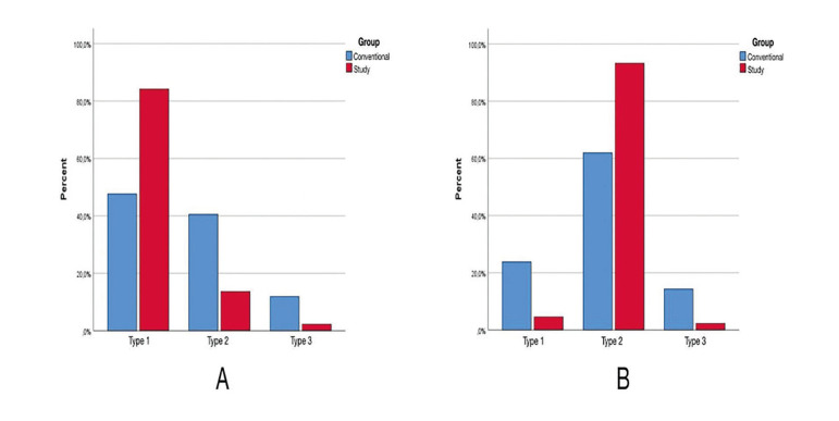
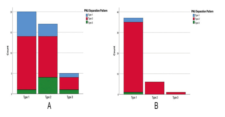
Background: The study aimed to investigate the effect of customized lateral nasal wall osteotomy (LNO) on the lateral nasal wall (LNW) and pterygomaxillary junction (PMJ) separation during Le Fort I. We hypothesized that customized LNO on the LNW affect the PMJ separation type.
Material and methods: This prospective, controlled, randomized study included forty-three patients were randomly assigned to either the conventional or customized (study) osteotomy groups. In the study group, LNW depth was measured before surgery in the axial section of the CT scan, and LNO was performed at a depth of 2 mm less than the measured distance. In the conventional osteotomy group, LNO was performed at 30 mm for females and 35 mm for males. Patients with cleft lip and palate, previous orthognathic surgery, or rhinoplasty were excluded. Separation types were classified as follows: LNW types; Type1-from the osteotomy line; Type2- 2-4 mm above the osteotomy line; Type3- 4 mm or more above the osteotomy line. PMJ types; Type1-including the tuber maxilla; Type2-from the pterygomaxillary junction; Type3-including the pterygoid plates. Chi-square tests were conducted to determine whether there was a significant correlation between groups and LNW separation types, groups and PMJ separation types and groups, and LNW separation type and PMJ separation type. A P value of < .05 was considered statistically significant.
Results: In both the conventional (P=0.052) and the study groups (p=0.828), there was no significant difference between LNW depth. Type 1 (P=0.0003) and Type 2 (P=0.0051) LNW separation types presented a significant difference between groups. A chi-square test showed a significant correlation between the surgical groups and PMJ separation patterns (P<0.05).
Conclusions: Customized LNO optimizes the LNW and PMJ separation. Facilitates the Le Fort I surgery and decrease unintentional fracture of the PMJ.
Conflict of interest statement
The authors declare no conflict of interest. All authors certify that they have no affiliations with or involvement in any organization or entity with any financial interest or non-financial interest in the subject matter or materials discussed in this manuscript.
Figures


Similar articles
-
What are the Pterygomaxillary Fracture Patterns in Cleft Orthognathic Surgery?J Oral Maxillofac Surg. 2025 Mar;83(3):307-316. doi: 10.1016/j.joms.2024.11.016. Epub 2024 Dec 9. J Oral Maxillofac Surg. 2025. PMID: 39710365
-
Pattern of pterygomaxillary disarticulation associated with Le Fort I maxillary osteotomy.Br J Oral Maxillofac Surg. 2022 Dec;60(10):1411-1416. doi: 10.1016/j.bjoms.2022.08.003. Epub 2022 Aug 22. Br J Oral Maxillofac Surg. 2022. PMID: 36175216
-
The Evaluation of the Pterygomaxillary Separation Pattern in Le Fort I Osteotomy Using Cone Beam Computed Tomography.J Craniofac Surg. 2025 Jun 1;36(4):1267-1270. doi: 10.1097/SCS.0000000000011088. Epub 2025 Jan 21. J Craniofac Surg. 2025. PMID: 39836008
-
Use of the 'shark-fin' osteotome in separation of the pterygomaxillary junction in Le Fort I osteotomy: a clinical and computerized tomography study.Int J Oral Maxillofac Surg. 2002 Feb;31(1):100-3. doi: 10.1054/ijom.2001.0179. Int J Oral Maxillofac Surg. 2002. PMID: 11936390 Review.
-
Blindness as a complication of Le Fort I osteotomy for maxillary distraction.Plast Reconstr Surg. 2002 Feb;109(2):688-98; discussion 699-700. doi: 10.1097/00006534-200202000-00041. Plast Reconstr Surg. 2002. PMID: 11818854 Review.
References
-
- Sakharia A, Muthusekar M. A comparative assessment of maxillary perfusion between two different Le Fort I osteotomy techniques. Int J Oral Maxillofac Surg. 2015;44:343–8. - PubMed
-
- Eshghpour M, Mianbandi V, Samieirad S. Intra- and Postoperative Complications of Le Fort I Maxillary Osteotomy. J Craniofac Surg. 2018;29:e797–803. - PubMed
-
- Li KK, Meara JG, Alexander Jr A. Location of the descending palatine artery in relation to the Le Fort I osteotomy. J Oral Maxillofac Surg. 1996;54:822–5. - PubMed
-
- van Otterloo JJdM, Tuinzing DB, Greebe RB, van der Kwast WA. Intra-and early postoperative complications of the Le Fort I osteotomy: a retrospective study on 410 cases. Journal of Cranio-Maxillofacial Surgery. 1991;19:217–22. - PubMed
-
- Tung T, Chen Y, Bendor-Samuel R. Surgical complications of the Le Fort I osteotomy--a retrospective review of 146 cases. Changgeng Yi Xue Za Zhi. 1995;18:102–7. - PubMed
Publication types
MeSH terms
LinkOut - more resources
Full Text Sources
