. 2025 Jan 26;16(1):47.
doi: 10.1038/s41419-025-07363-z.

Overexpression of miR-124 enhances the therapeutic benefit of TMZ treatment in the orthotopic GBM mice model by inhibition of DNA damage repair

Affiliations

Overexpression of miR-124 enhances the therapeutic benefit of TMZ treatment in the orthotopic GBM mice model by inhibition of DNA damage repair

Yuchen Wei et al. Cell Death Dis. .

Abstract

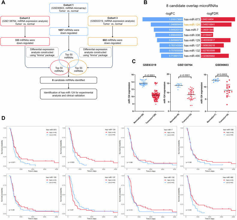
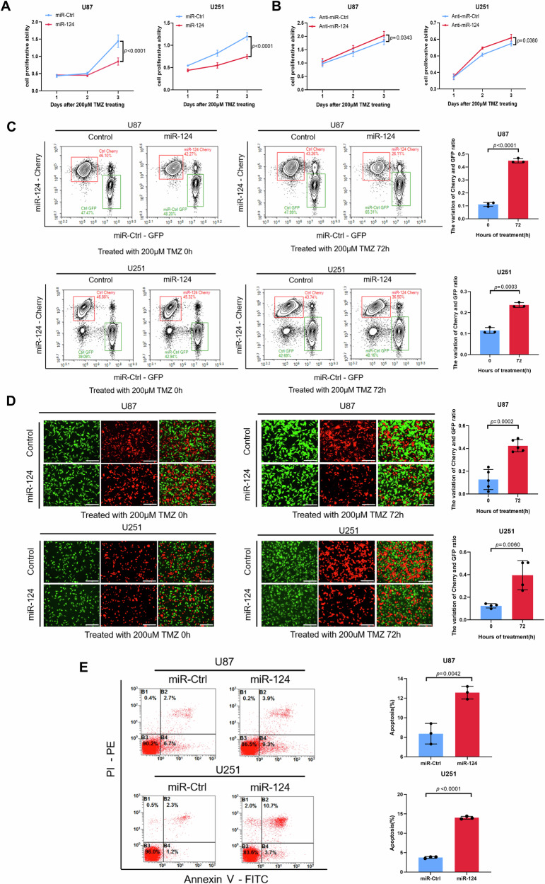
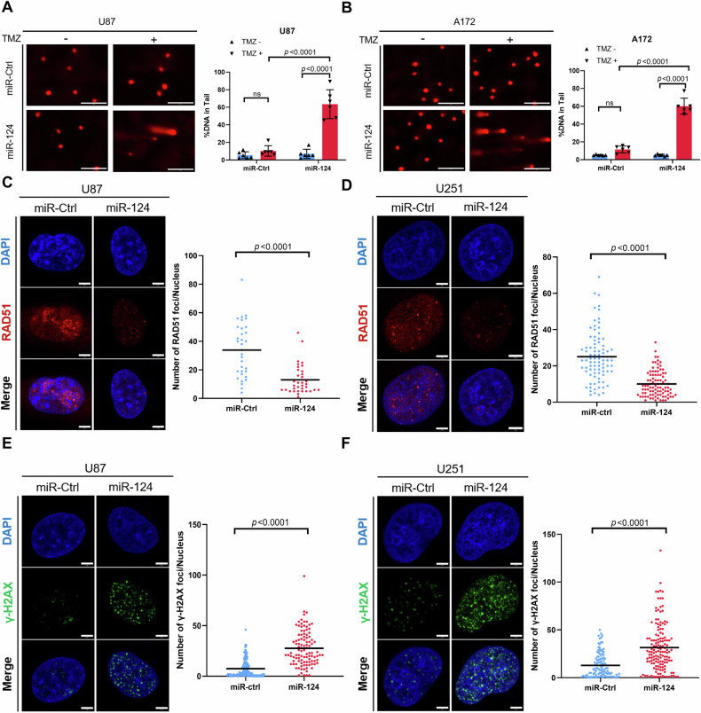
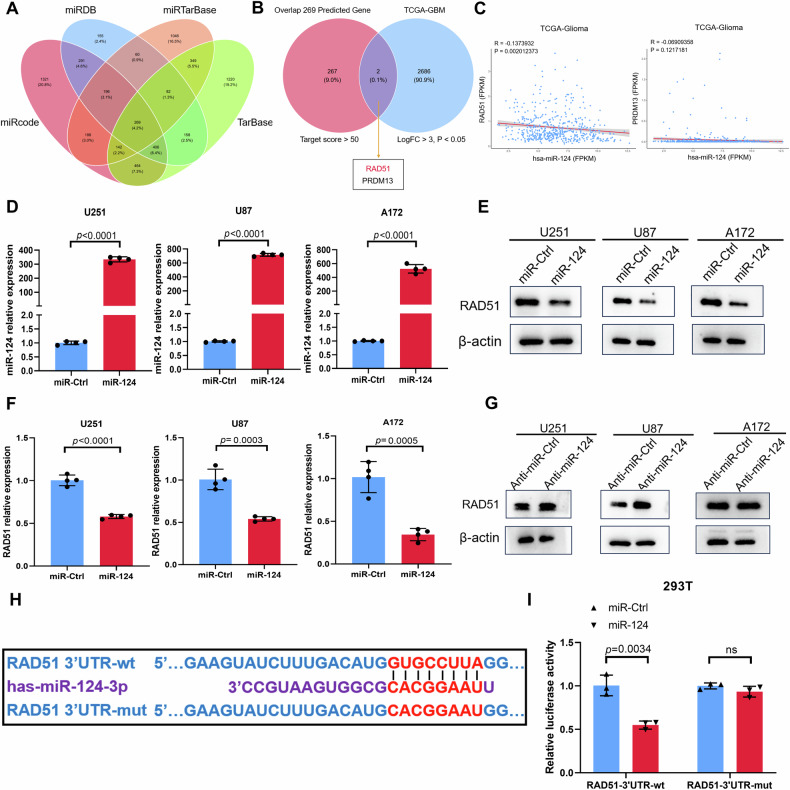
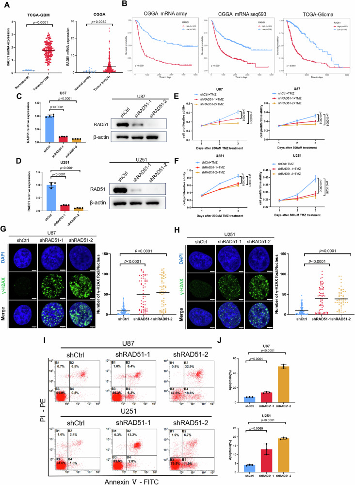
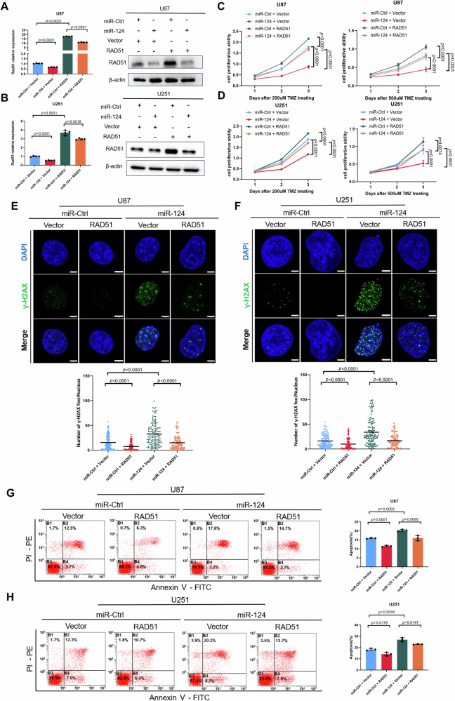
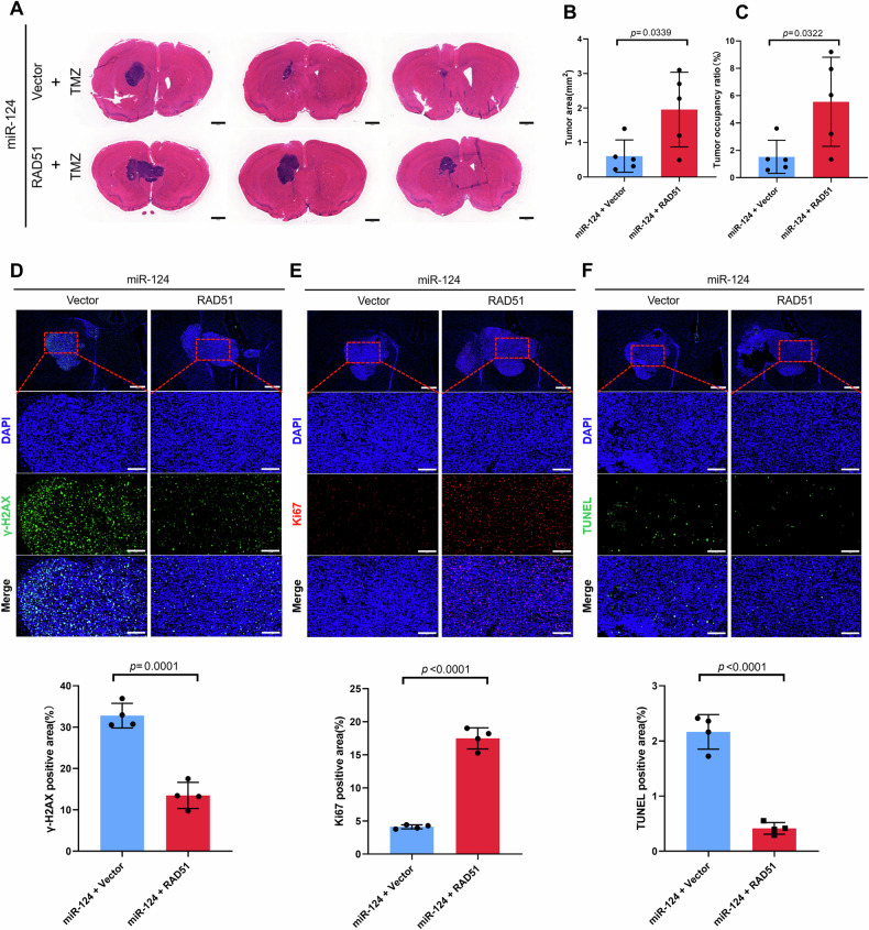
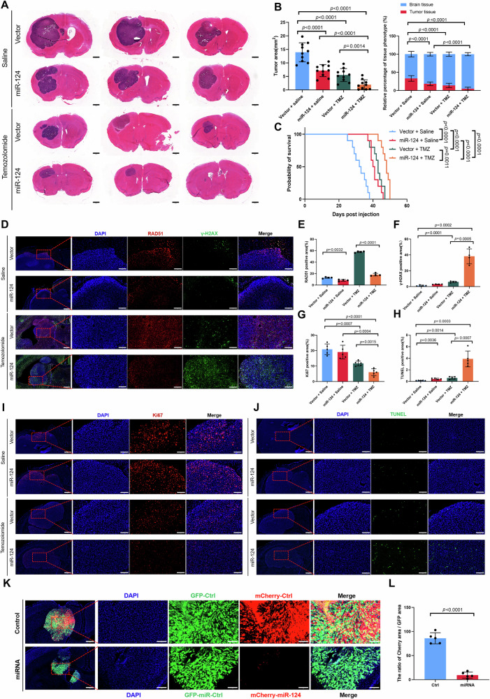
Glioblastoma (GBM) is the most common malignant primary brain cancer with poor prognosis due to the resistant to current treatments, including the first-line drug temozolomide (TMZ). Accordingly, it is urgent to clarify the mechanism of chemotherapeutic resistance to improve the survival rate of patients. In the present study, by integrating comprehensive non-coding RNA-seq data from multiple cohorts of GBM patients, we identified that a series of miRNAs are frequently downregulated in GBM patients compared with the control samples. Among them, a high level of miR-124 is closely associated with a favorable survival rate in the clinical patients. In the phenotype experiment, we demonstrated that miR-124 overexpression increases responsiveness of GBM cells to TMZ-induced cell death, and vice versa. In the mechanistic study, we for the first time identified that RAD51, a key functional molecule in DNA damage repair, is a novel and bona fide target of miR-124 in GBM cells. Given that other miR-124-regulated mechanisms on TMZ sensitivity have been reported, we performed recue experiment to demonstrate that RAD51 is essential for miR-124-mediated sensitivity to TMZ in GBM cells. More importantly, our in vivo functional experiment showed that combinational utilization of miR-124 overexpression and TMZ presents a synergetic therapeutic benefit in the orthotopic GBM mice model. Taken together, we rationally explained a novel and important mechanism of the miR-124-mediated high sensitivity to TMZ-induced cell death in GBM and provided evidence to support that miR-124-RAD51 regulatory axis could be a promising candidate in the comprehensive treatment with TMZ in GBM.


Conflict of interest statement

Competing interests: The authors declare no competing interests. Ethical approval: The protocol for the animal study was approved by the Animal Welfare and Ethics Committee of the Fourth Military Medical University (Xi’an, China).

Figures

Fig. 1
Fig. 1. MiR-124 is identified as a potential tumor suppressor in GBM.
A The workflow for the screening of downregulated microRNA in GBM. B Butterfly plot showing the median value of fold change (FC) and false discover rate (FDR) of screened 8 candidate microRNAs from intersection of three cohorts (GSE90603, GSE138764, GSE63319 datasets). C Scatter plot depicting the expression of miR-124 in normal versus tumor tissue samples from GEO database (GSE63319, GSE138764, GSE90603). D Kaplan–Meier survival analysis to investigate the associations of the expression levels of 8 candidate microRNAs with the overall survival of 90 grade IV patients from the CGGA database. The expression of 8 candidate microRNA was respectively divided into high and low groups according to their median values. P values were calculated using a log-rank test. The values of scatter plot were presented as means ± SD. P values were calculated using unpaired Student’s t-test.
Fig. 2
Fig. 2. MiR-124 enhances the sensitivity of GBM cell lines to TMZ treatment.
A, B CCK8 assay was used to assess the cell proliferative ability of different groups. U87 and U251 cells transfected with miR-Ctrl or miR-124 followed by TMZ treatment in (A), U87 and U251 cells transfected with anti-miR-Ctrl or anti-miR-124 followed by TMZ treatment in (B). Representative images of flow cytometry (C, left) and fluorescence imaging (D, left) and the variation ratio of Cherry/GFP cells (C and D, right) from co-culture cell competition assay in U87 and U251 cells exposed to 200 μM TMZ, with or without miR-124 overexpression. Scale bar, 300 μm. E PI/Annexin V-staining to detect the apoptosis of U87 and U251 cells exposed to TMZ (200 μM, 24 h) with or without miR-124 transfection (Representative images in left), Annexin V-stained (PI−/Annexin V+ and PI+/Annexin V+) cells were analyzed to measure the cell apoptosis rate (right). Above data are presented as means ± SD. All experiments were repeated at least 3 times. P values were calculated using unpaired Student’s t-test.
Fig. 3
Fig. 3. MiR-124 enhances the responses of TMZ in GBM cells by blocking DNA damage repair.
A, B Representative images (left) and DNA tailing rate (right) from comet assay in U87 and A172 cells exposed to TMZ (500 μM, 4 h), with or without miR-124 overexpression. Scale bar, 200 μm (% DNA in tail: Tail DNA Intensity/Cell DNA Intensity × 100, miR-ctrl plus TMZ vs miR-124 plus TMZ, P < 0.0001). C, D Representative images (left) and quantification of RAD51 fluorescence foci (right) of immunofluorescence analysis in U87 and U251 cells exposed to TMZ (200 μM, 24 h) with or without miR-124 transfection. Scale bar, 5 μm. E, F Representative images (left) and quantification of γ-H2AX fluorescence foci (right) of immunofluorescence analysis in U87 and U251 cells exposed to TMZ (200 μM, 24 h) with or without miR-124 transfection. Scale bar, 5 μm. All experiments were repeated at least 3 times. Above data are presented as means ± SD. P values were calculated using unpaired Student’s t-test.
Fig. 4
Fig. 4. RAD51 expression is regulated by miR-124 in GBM cells.
A Venn plot showing the overlap of four predicted miRNA-mRNA binding datasets (miRcode, miRTarBase, miRDB, TarBase). B Identification of potential target genes of miR-124. Only two genes (RAD51, PRDM13) were screened by overlapping the predicted genes (target score >50) in (A) with significant differential expression genes (LogFC >3, P < 0.05) in TCGA. C Correlation analysis between miR-124 and RAD51 or PRMD13 using TCGA database. A significant negative correlation was shown only for RAD51 and miR-124. P values were calculated using Pearson correlation test. D Quantitative real-time chain reaction (qRT-PCR) analysis of miR-124 expression in GBM cells transfected with miR-Ctrl or miR-124 mimics. E, F Western blot and qRT-PCR analysis of RAD51 expression in multiple GBM cell lines transfected with miR-Ctrl or miR-124 mimics. G Western blot analysis of RAD51 expression in GBM cells transfected with anti-miR-Ctrl or anti-miR-124. H Predication of one binding site of miR-124 on RAD51 3′-UTR by TargetScan. I Dual-luciferase reporter assay showing miR-124 directly targets the RAD51 3′-UTR. Firefly luciferase activity of the reporter was normalized to the internal Renilla luciferase activity. All experiments were repeated at least 3 times. All data are presented as means ± SD. P values were calculated using unpaired Student’s t-test.
Fig. 5
Fig. 5. Knockdown of RAD51 enhances TMZ chemosensitivity in GBM cells.
A Scatter plot showing the expression of RAD51 in normal versus tumor tissue samples from TCGA and CGGA databases. B Kaplan–Meier survival analysis investigate the associations of the expression levels of RAD51 with the overall survival from CGGA mRNA array (281 patients), CGGA mRNA-seq 693 (656 patients), TCGA (660 patients). The expression of RAD51 was divided into high and low groups according to its median value. P values were calculated using a log-rank test. C, D qRT-PCR (left) and western blot (right) validation of RAD51 expression in U87 and U251 cells with or without RAD51 knockdown. E, F CCK8 assay was used to assess cell viability of U87 and U251 cells with or without RAD51 knockdown treated with different dose of TMZ. G, H Representative images (left) and quantification of γ-H2AX fluorescence foci (right) of immunofluorescence analysis in U87 and U251 cells exposed to TMZ (200 μM, 24 h) with or without RAD51 knockdown. Scale bar, 5 μm. I, J Representative images (left) and apoptosis rate (right) of cell apoptosis assay in U87 and U251 cells exposed to TMZ (200 μM, 24 h) with or without RAD51 knockdown. All experiments were repeated at least 3 times. Above data are presented as means ± SD. P values were calculated using one-way ANOVA.
Fig. 6
Fig. 6. RAD51 is essential for miR-124 mediated sensitivity to TMZ treatment in GBM cells.
A, B qRT-PCR (left) and western blot (right) validation of RAD51 expression in U87 and U251 cells transfected with miR-Ctrl or miR-124 together with empty vector or RAD51. C, D CCK8 assay was used to assess cell proliferative ability of U87 and U251 cells with or without miR-124 transfection together with or without RAD51 overexpression and treated with different dose TMZ. E, F Representative images (top) and quantification of γ-H2AX fluorescence foci (bottom) of immunofluorescence analysis in U87 and U251 cells exposed to TMZ (200 μM, 24 h), transfected with miR-Ctrl or miR-124 together with empty vector or RAD51. Scale bar, 5 μm. G, H Representative images (left) and apoptosis rate (right) of cell apoptosis assay in U87 and U251 cells exposed to TMZ (200 μM, 24 h), transfected with miR-Ctrl or miR-124 together with empty vector or RAD51. All experiments were repeated at least 3 times. Above data are presented as means ± SD. P values were calculated using unpaired Student’s t-test.
Fig. 7
Fig. 7. RAD51 is required for GBM resistant to TMZ treatment in vivo.
A HE staining showing representative images (present 3 samples per group) of gross tumors (location near the lateral ventricle) from two rescue treatment groups in mouse orthotopic model. Scale bar, 1 mm. B, C Statistic of tumor size or tumor occupancy rate from two rescue treatment groups in (C) (n = 5 per group). DF Representative images (top) and quantification of γ-H2AX, Ki67 and TUNEL positive immunofluorescence area (bottom) in the two rescue treatment groups. Scale bar in low-power microscope 500 μm, in high-power microscope 150 μm. All experiments were repeated at least 3 times. Above data are presented as means ± SD. P values were calculated using unpaired Student’s t-test.
Fig. 8
Fig. 8. MiR-124 sensitizes TMZ therapy in GBM mouse model.
A Hematoxylin Eosin (HE) staining showing representative images (present 3 samples per group) of gross tumors (location near the lateral ventricle) from different treatment groups in mouse orthotopic model. Scale bar, 1 mm. B Statistic of tumor size or tissue phenotype percentage from different treatment group in (A) (n = 10 per group). C Kaplan–Meier survival analysis to assess survival of mice in the four-treatment groups. P values were calculated using a log-rank test. Representative images (D) and quantification of RAD51 (E) and γ-H2AX (F) positive immunofluorescence area in the four-treatment group. Scale bar in low-power microscope 500 μm, in high-power microscope 150 μm. Representative images (I, J) and quantification of Ki67 (G) and TUNEL (H) positive immunofluorescence area in the four-treatment group by continuous frozen section. Scale bar in low-power microscope 500 μm, in high-power microscope 150 μm. Representative images (K) and quantification of the Cherry/GFP area rate (L) of coculture fluorescence competition assay in vivo. Scale bar in low-power microscope 500 μm, in high-power microscope 150 μm. All experiments were repeated at least 3 times. Above data are presented as means ± SD. P values were calculated using unpaired Student’s t-test.

Similar articles

Cited by

References

    1. Schaff LR, Mellinghoff IK. Glioblastoma and other primary brain malignancies in adults: a review. JAMA. 2023;329:574–87. - PMC - PubMed
    1. Sarkaria JN, Hu LS, Parney IF, Pafundi DH, Brinkmann DH, Laack NN, et al. Is the blood-brain barrier really disrupted in all glioblastomas? A critical assessment of existing clinical data. Neuro Oncol. 2018;20:184–91. - PMC - PubMed
    1. Stupp R, Gander M, Leyvraz S, Newlands E. Current and future developments in the use of temozolomide for the treatment of brain tumours. Lancet Oncol. 2001;2:552–60. - PubMed
    1. Stupp R, Mason WP, van den Bent MJ, Weller M, Fisher B, Taphoorn MJ, et al. Radiotherapy plus concomitant and adjuvant temozolomide for glioblastoma. N Engl J Med. 2005;352:987–96. - PubMed
    1. Denny BJ, Wheelhouse RT, Stevens MF, Tsang LL, Slack JA. NMR and molecular modeling investigation of the mechanism of activation of the antitumor drug temozolomide and its interaction with DNA. Biochemistry. 1994;33:9045–51. - PubMed

MeSH terms