. 2025 Jan 26;16(1):26.
doi: 10.1186/s13287-025-04145-4.

Migrasomes derived from human umbilical cord mesenchymal stem cells: a new therapeutic agent for ovalbumin-induced asthma in mice

Affiliations

Migrasomes derived from human umbilical cord mesenchymal stem cells: a new therapeutic agent for ovalbumin-induced asthma in mice

Weifeng Gu et al. Stem Cell Res Ther. .

Abstract

Background: Asthma is a prevalent respiratory disease, and its management remains largely unsatisfactory. Mesenchymal stem cells (MSCs) have been demonstrated to be efficacious in reducing airway inflammation in experimental allergic diseases, representing a potential alternative treatment for asthma. Migrasomes are recently identified extracellular vesicles (EVs) generated in migrating cells and facilitate intercellular communication. The objective of this study was to investigate the therapeutic effects of migrasomes obtained from MSC in a model of asthma.

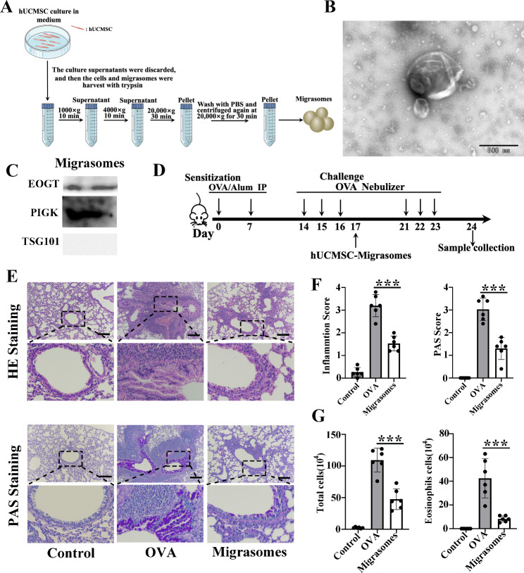
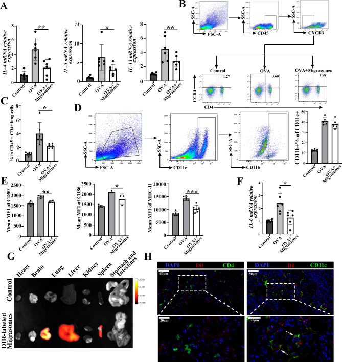
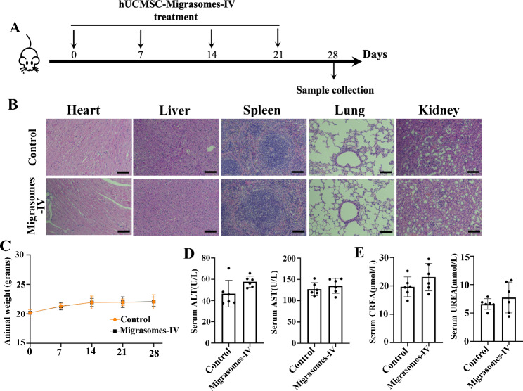
Methods: Migrasomes produced by human umbilical cord MSCs (hUCMSCs) were isolated by sequential centrifugation. Characterization of hUCMSC-derived migrasomes were carried out by transmission electron microscopy and western blot analysis. The therapeutic effects of migrasomes on airway inflammation in ovalbumin (OVA)-induced asthmatic mice were evaluated by hematoxylin-eosin (HE) and periodic-acid schiff (PAS) staining, and their mechanism were further testified by immunofluorescent staining, real-time PCR and flow cytometry.

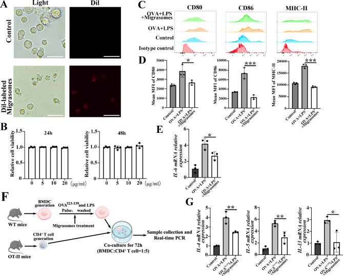
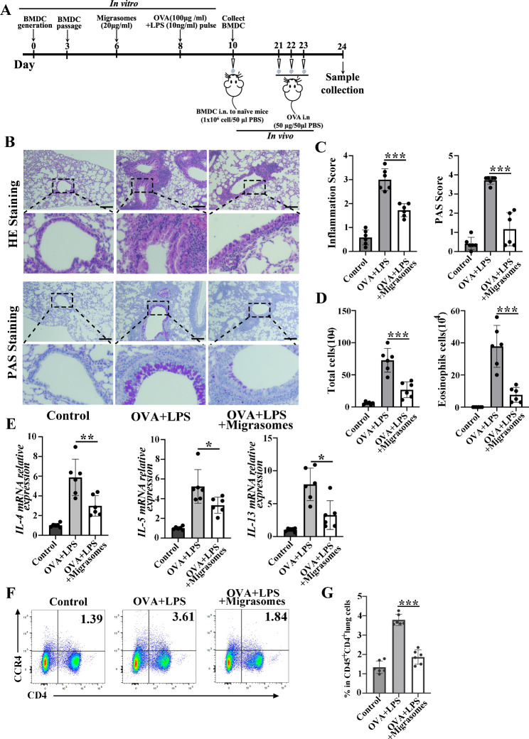
Results: Here, we showed that inhibition of migrasomes' production dramatically impaired the anti-inflammatory effects of hUCMSCs in OVA animals, as evidenced by a notable increase in both the infiltration of inflammatory cells and the number of epithelial goblet cells. We successfully isolated hUCMSC-migrasomes, which were morphologically intact and positive for the specific migrasomes markers. The administration of hUCMSC-migrasomes was observed to significantly ameliorate the symptoms of airway inflammation and mucus production in asthmatic mice. Additionally, the expression of Th2 cytokines (IL-4, IL-5 and IL-13) were found to be reduced, while the activation of dendritic cells (DCs) was inhibited. HUCMSC-migrasomes could possibly be delivered to lung region after injection, and were able to be taken in by DCs both in vivo and in vitro. Notably, in vitro, migraosmes decreased the capacity of BMDCs to stimulate OVA-specific Th2-cell responses. More importantly, we found that adoptive transfer of hUCMSC-migrasomes-treated BMDCs was sufficient to protect mice from allergic airway inflammation. In addition, we found that hUCMSC-migrasomes inhibited the receptor for advanced glycation end-products (RAGE) signal in OVA-treated BMDCs in vitro and in asthma mice lung in vivo.

Conclusion: Our results provided the first evidence that hUCMSC-migrasomes possess anti-inflammatory properties in OVA-induced allergic mice, which may provide a novel "MSC-cell free" therapeutic agent for the management of asthma.

Keywords: Asthma; Dendritic cells; Human umbilical cord mesenchymal stem cells; Migrasomes; Th2 cells.


Conflict of interest statement

Declarations. Ethics approval and consent to participate: Human umbilical cord samples were obtained from mothers who provided informed consent at the Affiliated Hospital of Jiangsu University, and the protocol was reviewed and approved by the Ethics Committee of the Affiliated Hospital of Jiangsu University (Project title: Extracellular vesicles released from hUCMSC inhibit IL-13 mediated HSC activation and alleviate schistosomiasis liver pathology; No: SWYXLL20200121-22; Date of approval: 2020-01-21). Animal experiments were approved by the Institutional Animal Care and Use Committee of Jiangsu University (Project title: Mechanism and clinical translation of hUCMSC-derived extracellular vesicles for asthma intervention; No. UJS-IACUC-AP-2023050902; Date of approval: 2023-05-09). Animal welfare was ensured in accordance with the Guide for the Care and Use of Laboratory Animals. Consent for publication: Not applicable. Competing interests: The authors declare that they have no competing interests.

Figures

Fig. 1
Fig. 1
HUCMSCs ameliorated OVA-induced airway inflammation in mice through migrasomes. A HUCMSCs of wild type and TSPAN4-KD were stained with 1 µg/ml WGA (Alexa 488) and observed under confocal microscopy. Scale bar = 10 μm. B Western blot analysis of TSPAN4 expression in hUCMSCs of wild type and TSPAN4-KD. C Experimental protocol for the development of murine allergic asthma model. D Representative images of lung sections stained with HE (up) and PAS (down) from each group are presented (black bar = 100 μm). E The inflammatory infiltration and goblet cell hyperplasia were quantified by HE (left) and PAS (right) scores (n = 6). F Statistical analysis was conducted on the total inflammatory cells (left) and eosinophils (right) in the BALF (n = 6). Data were presented as mean ± SD. A one-way analysis of variance (Tukey Kramer post hoc tests) was performed on the data. *P < 0.05, **P < 0.01, ***P < 0.001. Full-length blots are presented in supplemental Figure S6
Fig. 2
Fig. 2
HUCMSC-migrasome treatment significantly attenuated airway inflammation in OVA-induced asthma mice. A Experimental protocol for isolation of hUCMSC-migrasomes. B Micrographs of hUCMSC-migrasomes under transmission electron (scale bar = 500 nm). C Western blot analysis of PIGK, EOGT and TSG101 expression of hUCMSC-migrasomes. D Experimental protocol for the development of murine allergic asthma model. E Representative images of lung sections stained with HE (up) and PAS (down) from each group are presented (black bar = 100 μm). F The inflammatory infiltration and goblet cell hyperplasia were quantified by HE (left) and PAS (right) scores (n = 6). G Statistical analysis of the total inflammatory cells (left) and eosinophils (right) in the BALF (n = 6). Data were presented as mean ± SD. A one-way analysis of variance (Tukey Kramer post hoc tests) was performed on the data. ***P < 0.001. Full-length blots are presented in supplemental Figure S7
Fig. 3
Fig. 3
Administration of hUCMSC-migrasomes diminished Th2 response in allergic asthma by suppressing DC maturation. A The expression of IL-4, IL-5 and IL-13 in the lung of mice in each group were detected by real-time PCR (n = 6). B Gating strategy and representative flow cytometric images for the analysis of CD45+CD4+CCR4+CXCR3 Th2 cells in the lungs of mice from each group. C Graphs showing the % of CD45+CD4+CCR4+CXCR3Th2 cells in the lungs of mice from each group (n = 6). D Gating strategy and representative flow cytometric dot plots (left) for the analysis of CD11b+ DCs in the lungs of allergic mice and the numbers of CD11b+ DCs (right) in the lungs of mice from each group (n = 6). E The mean MFI of CD80, CD86 and MHC-II on CD11b+ DCs (n = 4–6). F The expression of IL-6 in the lung of mice were detected by real-time PCR (n = 6). G Distribution of Dir-labeled migrasomes in OVA-induced mice lung after tail vein administration (17-day injection and 18-day detected). H Representative immunofluorescence images of Dil-labeled migrasomes (red) taken up by CD11c+ cells or CD4+ cells. Data were presented as mean ± SD. A one-way analysis of variance (Tukey Kramer post hoc tests) was performed on the data. *P < 0.05, **P < 0.01, ***P < 0.001.
Fig. 4
Fig. 4
HUCMSC-migrasomes impaired DC maturation and function in vitro. A BMDCs incubated with Dil-labeled migrasomes under fluorescence microscope (white bar = 25 μm). B The cell viability of BMDCs treated by hUCMSC-migrasomes for 24 h (left) and 48 h (right) (n = 4). C BMDCs were isolated from wild-type BALB/c mice and cultured at a density of 2 × 10⁵ cells/mL. The cells were stimulated with OVA (100 µg/mL) and LPS (10 ng/mL) for 48 h, with or without hUCMSC-migrasomes (20 µg/mL) pre-treated. After that, cells were collected for the detection of DC maturation markers including CD80, CD86 and MHC-II (n = 3). The representative images for the detection of DC maturation markers are displayed. D Statistical analysis of the mean MFIs of CD80, CD86 and MHC-II of BMDCs (n = 3). E Statistical analysis of the expression of IL-6 in BMDCs by real-time PCR (n = 3). F BMDCs (2 × 104) from WT mice were pre-treated with or without migrasomes and then pulsed with OVA323–339 and LPS and incubated at a 1:5 ratio with splenic CD4+ T cells isolated from OT-II mice (1 × 105) for 72 h. G Statistical analysis of the expression of IL-4, IL-5, IL-13 in collected cell by real-time PCR (n = 3). Data were presented as mean ± SD. A one-way analysis of variance (Tukey Kramer post hoc tests) was performed on the data. *P < 0.05, **P < 0.01, ***P < 0.001.
Fig. 5
Fig. 5
Adoptive transfer of hUCMSC-migrasomes-treated DCs is sufficient to protect mice from allergic airway inflammation. A Experimental protocol for the development of murine allergic asthma model adoptive transfer of hUCMSC-migrasomes. BMDCs were pre-treated with or without hUCMSC-migrasomes and then pulsed with OVA and LPS, and then washed and transferred i.n. into naive mice. On days 21–23, mice were challenged i.n. with OVA (50 µg) and sacrifice at day 24. B Representative images of lung sections stained with HE (up) and PAS (down) from each group are presented (black bar = 100 μm). C The inflammatory infiltration and goblet cell hyperplasia were quantified by HE (left) and PAS (right) scores (n = 6). D Statistical analysis was conducted on the total inflammatory cells (left) and eosinophils (right) of the BALF (n = 6). E The expression of IL-4, IL-5, IL-6, IL-13 in the lung of mice were detected by real-time PCR (n = 6). F Representative flow cytometric images for the analysis of CD45+CD4+CCR4+CXCR3 Th2 cells in the lungs of mice from each group (n = 6). G Graph showing the % of CD45+CD4+CCR4+CXCR3 Th2 cells in the lungs of mice from each group (n = 6). Data were presented as mean ± SD. A one-way analysis of variance (Tukey Kramer post hoc tests) was performed on the data. *P < 0.05, **P < 0.01, ***P < 0.001.
Fig. 6
Fig. 6
The suppression effect of hUCMSC-migrasomes on BMDCs connected to the AGE-RAGE signaling. A Dot plot shows 651 significantly differentially expressed (P < 0.05 and|log2FC| > 1) genes with 227 downregulated and 424 upregulated in the migrasomes group. FC: fold change. B The graph shows changes of signature genes of BMDCs treated with migrasomes. C Real-time PCR verification of several of the list differentially expressed genes (n = 3). D Go enrichment analysis revealed differentially expressed genes (DEG) were significantly involved in the regulation of some biological processes, cellular components and molecular functions respectively. E KEGG enrichment shows top 20 downregulated pathway name in the migrasomes group. F, G Representative western blot of RAGE and P-P65 expression in BMDCs (F) and mice lung samples (G) from each group. H, I Statistical analysis of RAGE and P-P65 expression in BMDCs (H, n = 2-3) and mice lung samples (I, n = 4–6) from each group. Data were presented as mean ± SD. A one-way analysis of variance (Tukey Kramer post hoc tests) or the Student’s t test was performed on the data. *P < 0.05, **P < 0.01, ***P < 0.001. Full-length blots are presented in supplemental Figure S8. The mRNA sequencing data for A is available in the supplemental materials.
Fig. 7
Fig. 7
Biocompatibility evaluation of mice treated by hUCMSC-migrasomes. A Experimental protocol for hUCMSC-migrasomes-IV treatment. B Representative images of mice major organs (heart, brain, liver, spleen, lung, kidney, stomach, and intestines) by HE staining (scale bar = 100 μm). C Mouse body weights. D, E Indicators reflected the physiological function of the D liver (ALT and AST) and E kidney (CREA and UREA) were detected. ALT, glutamic pyruvic transaminase; AST, glutamic oxaloacetic transaminase; CREA, Creatinine. Each dot represents data from one animal and n = 6 per group. Data were presented as mean ± SD. The Student’s t test was performed on the data.

Similar articles

Cited by

References

    1. Hammad H, Lambrecht BN. The basic immunology of asthma. Cell. 2021;184(6):1469–85. 10.1016/j.cell.2021.02.016. - PubMed
    1. Komlosi ZI, van de Veen W, Kovacs N, Szucs G, Sokolowska M, O’Mahony L, et al. Cellular and molecular mechanisms of allergic asthma. Mol Aspects Med. 2022;85:100995. 10.1016/j.mam.2021.100995. - PubMed
    1. Wener RR, Bel EH. Severe refractory asthma: an update. Eur Respir Rev. 2013;22(129):227–35. 10.1183/09059180.00001913. - PMC - PubMed
    1. King-Biggs MB, Asthma. Ann Intern Med. 2019;171(7):ITC49–64. 10.7326/AITC201910010. - PubMed
    1. Fu X, Liu G, Halim A, Ju Y, Luo Q, Song AG. Mesenchymal stem cell Migration and tissue repair. Cells. 2019;8(8). 10.3390/cells8080784. - PMC - PubMed