

Obstructive sleep apnea and structural and functional brain alterations: a brain-wide investigation from clinical association to genetic causality
- PMID: 39865248
- PMCID: PMC11770961
- DOI: 10.1186/s12916-025-03876-8
Obstructive sleep apnea and structural and functional brain alterations: a brain-wide investigation from clinical association to genetic causality
Abstract
Background: Obstructive sleep apnea (OSA) is linked to brain alterations, but the specific regions affected and the causal associations between these changes remain unclear.
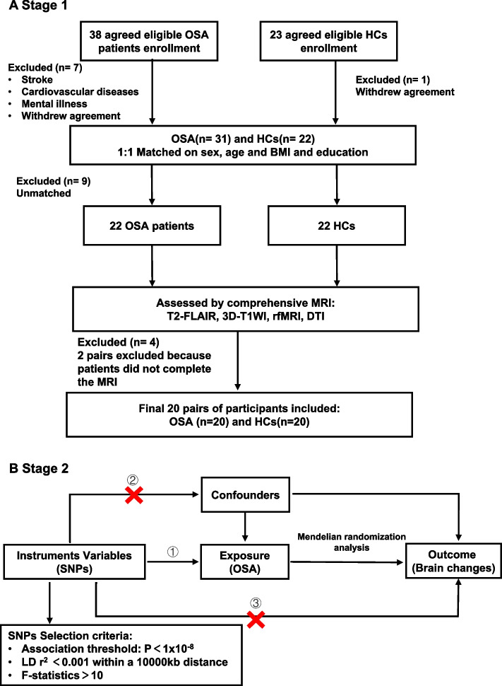
Methods: We studied 20 pairs of age-, sex-, BMI-, and education- matched OSA patients and healthy controls using multimodal magnetic resonance imaging (MRI) from August 2019 to February 2020. Additionally, large-scale Mendelian randomization analyses were performed using genome-wide association study (GWAS) data on OSA and 3935 brain imaging-derived phenotypes (IDPs), assessed in up to 33,224 individuals between December 2023 and March 2024, to explore potential genetic causality between OSA and alterations in whole brain structure and function.
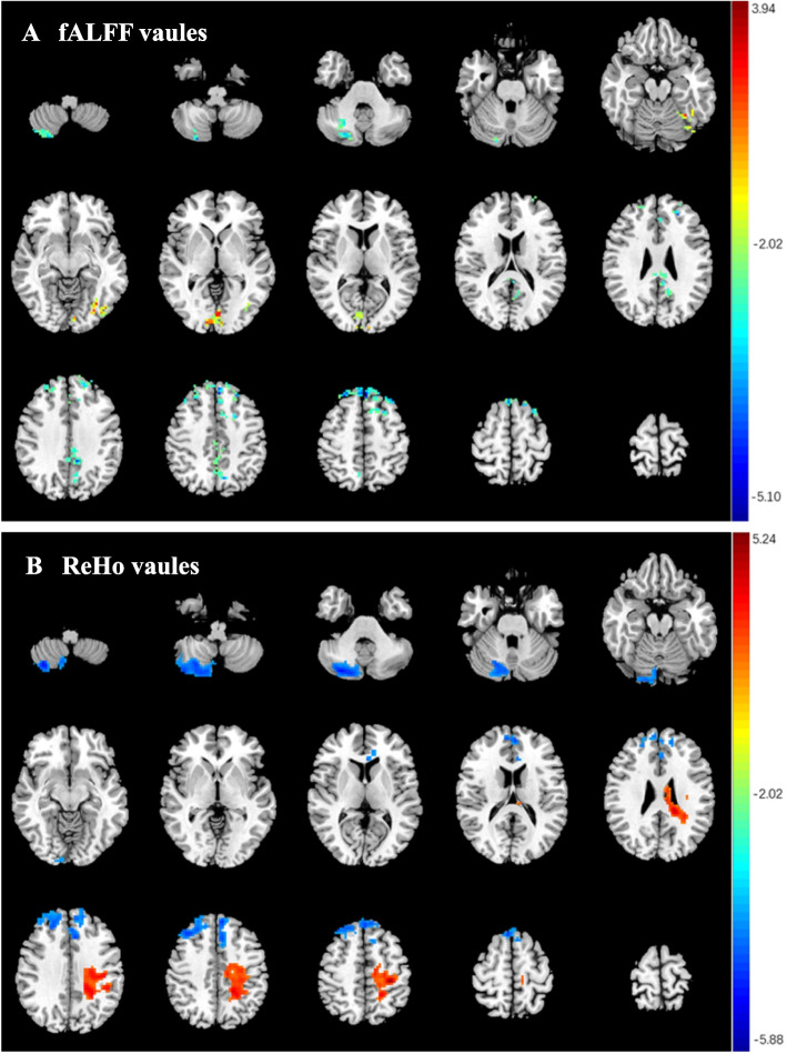
Results: In the cohort study, OSA patients exhibited significantly lower fractional amplitude of low-frequency fluctuation and regional homogeneity in the right posterior cerebellar lobe and bilateral superior and middle frontal gyrus, while showing higher levels in the left occipital lobe and left posterior central gyrus. Decreased fractional anisotropy (FA) but increased apparent diffusion coefficient (ADC) was shown in the bilateral superior longitudinal fasciculus. According to the results of Affiliation file 2: table s6, it is the ADC value of right superior longitudinal fasciculus was shown a positive correlation with the lowest oxygen saturation. In the Mendelian randomization analyses, the area of left inferior temporal sulcus (OR: 0.89; 95% CI: 0.82-0.96), rfMRI connectivity ICA100 edge 893 (OR: 0.88; 95% CI: 0.82-0.96), ICA100 edge 951 (OR: 0.89; 95% CI: 0.82-0.97), and ICA100 edge 1213 (OR: 0.89; 95% CI: 0.82-0.96) were significantly decreased in OSA. Conversely, mean thickness of G-front-inf-Triangul in right hemisphere (OR: 1.14; 95% CI: 1.05-1.23), mean orientation dispersion index in right tapetum (OR: 1.13; 95% CI: 1.04-1.23), and rfMRI connectivity ICA100 edge 258 (OR: 1.13; 95% CI: 1.04-1.22) showed opposite results.
Conclusions: Nerve fiber damage and imbalances in neuronal activity across multiple brain regions caused by hypoxia, particularly the frontal lobe, underlie the structural and the functional connectivity impairments in OSA.
Keywords: Brain structure and function; Mendelian randomization; Neuroimaging; Obstructive sleep apnea.
© 2025. The Author(s).
Conflict of interest statement
Declarations. Ethics approval and consent to participate: The ethics committee of the First Affiliated Hospital of Guangzhou Medical University strictly follows the Declaration of Helsinki and International Ethical Guidelines for Health-related Research Involving Humans, etc., to perform independent ethical review duties (Ethics number: 201705). All genome-wide association studies included in this study all originate from publicly published GWAS summary databases, which complies with the conditions for exemption from review as stated in the “Ethical Review Measures for Life Sciences and Medical Research Involving Humans.” Consent for publication: Not applicable. Competing interests: The authors declare no competing interests.
Figures


References
MeSH terms
LinkOut - more resources
Full Text Sources
