

Multiomic analyses direct hypotheses for Creutzfeldt-Jakob disease risk genes
- PMID: 39865733
- PMCID: PMC12404779
- DOI: 10.1093/brain/awaf032
Multiomic analyses direct hypotheses for Creutzfeldt-Jakob disease risk genes
Abstract
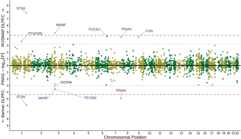
Prions are assemblies of misfolded prion protein that cause several fatal and transmissible neurodegenerative diseases, with the most common phenotype in humans being sporadic Creutzfeldt-Jakob disease (sCJD). Aside from variation of the prion protein itself, molecular risk factors are not well understood. Prion and prion-like mechanisms are thought to underpin common neurodegenerative disorders meaning that the elucidation of mechanisms could have broad relevance. Herein we sought to further develop our understanding of the factors that confer risk of sCJD using a systematic gene prioritization and functional interpretation pipeline based on multiomic integrative analyses. We integrated the published sCJD genome-wide association study summary statistics with publicly available bulk brain and brain cell type gene and protein expression datasets. We performed multiple transcriptome and proteome-wide association studies and Bayesian genetic colocalization analyses between sCJD risk association signals and multiple brain molecular quantitative trait loci signals. We then applied our systematic gene prioritization pipeline to the obtained results and nominated prioritized sCJD risk genes with risk-associated molecular mechanisms in a transcriptome and proteome-wide manner. Genetic upregulation of both gene and protein expression of syntaxin-6 (STX6) in the brain was associated with sCJD risk in multiple datasets, with a risk-associated gene expression regulation specific to oligodendrocytes. Similarly, increased gene and protein expression of protein disulfide isomerase family A member 4 (PDIA4), involved in the unfolded protein response, was linked to increased disease risk, particularly in excitatory neurons. Protein expression of mesencephalic astrocyte derived neurotrophic factor (MANF), involved in protection against endoplasmic reticulum stress and sulfatide binding (linking to the enzyme in the final step of sulfatide synthesis, encoded by sCJD risk gene GAL3ST1), was identified as protective against sCJD. In total 32 genes were prioritized into two tiers based on the level of evidence and confidence for further studies. This study provides insights into the genetically-associated molecular mechanisms underlying sCJD susceptibility and prioritizes several specific hypotheses for exploration beyond the prion protein itself, as well as beyond the previously highlighted sCJD risk loci, through the newly prioritized sCJD risk genes and mechanisms. These findings highlight the importance of glial cells, sulfatides and the excitatory neuron unfolded protein response in sCJD pathogenesis.
Keywords: multiomics; neurodegeneration; proteome-wide association studies (PWAS); sporadic Creutzfeldt-Jakob disease (sCJD); transcriptome-wide association studies (TWAS).
© The Author(s) 2025. Published by Oxford University Press on behalf of the Guarantors of Brain.
Conflict of interest statement
The authors report no competing interests.
Figures


References
-
- Collinge J. Mammalian prions and their wider relevance in neurodegenerative diseases. Nature. 2016;539:217–226. - PubMed
-
- Ladogana A, Puopolo M, Croes EA, et al. Mortality from Creutzfeldt-Jakob disease and related disorders in Europe, Australia, and Canada. Neurology. 2005;64:1586–1591. - PubMed
-
- NCJDRSU . Annual Report. 2022: https://www.cjd.ed.ac.uk/sites/default/files/report29.pdf
-
- Collinge J. Prion diseases of humans and animals: Their causes and molecular basis. Annu Rev Neurosci. 2001;24:519–550. - PubMed
MeSH terms
Supplementary concepts
Grants and funding
- P30 AG072975/AG/NIA NIH HHS/United States
- University of Antwerp
- RF1 AG057440/AG/NIA NIH HHS/United States
- P50 AG016574/AG/NIA NIH HHS/United States
- R01 AG017917/AG/NIA NIH HHS/United States
- P50 AG005136/AG/NIA NIH HHS/United States
- BOF 49758/National Institute for Health and Care Research
- P30 AG010161/AG/NIA NIH HHS/United States
- R01 NS080820/NS/NINDS NIH HHS/United States
- P30 AG066509/AG/NIA NIH HHS/United States
- U01 AG006781/AG/NIA NIH HHS/United States
- R01 AG018023/AG/NIA NIH HHS/United States
- MRC_/Medical Research Council/United Kingdom
- U01 AG046152/AG/NIA NIH HHS/United States
- Brein Instituut
- U01 AG061356/AG/NIA NIH HHS/United States
- U51 CK000100/CK/NCEZID CDC HHS/United States
- U01 AG046170/AG/NIA NIH HHS/United States
- U24 AG061340/AG/NIA NIH HHS/United States
- R01 AG032990/AG/NIA NIH HHS/United States
- U19 AG060909/AG/NIA NIH HHS/United States
- R01 NS074317/NS/NINDS NIH HHS/United States
- U01 AG046139/AG/NIA NIH HHS/United States
- P01 AG003949/AG/NIA NIH HHS/United States
- U24 NS072026/NS/NINDS NIH HHS/United States
- P30 AG019610/AG/NIA NIH HHS/United States
- P50 AG025711/AG/NIA NIH HHS/United States
- U19 AG066567/AG/NIA NIH HHS/United States
- P01 AG017216/AG/NIA NIH HHS/United States
- U01 AG006786/AG/NIA NIH HHS/United States
- R01 NS103848/NS/NINDS NIH HHS/United States
- R01 AG036836/AG/NIA NIH HHS/United States
- R01 AG015819/AG/NIA NIH HHS/United States
LinkOut - more resources
Full Text Sources
Medical
Miscellaneous
