. 2025 Mar;12(11):e2409641.
doi: 10.1002/advs.202409641. Epub 2025 Jan 27.

Endothelial TRIM35-Regulated MMP10 Release Exacerbates Calcification of Vascular Grafts

Affiliations

Endothelial TRIM35-Regulated MMP10 Release Exacerbates Calcification of Vascular Grafts

Yiming Leng et al. Adv Sci (Weinh). 2025 Mar.

Abstract

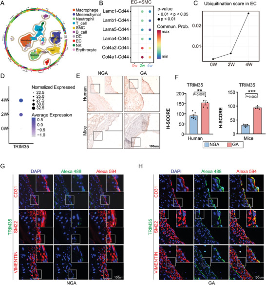
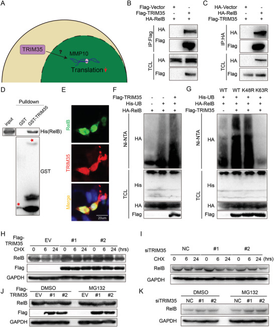
Vascular calcification is a highly regulated process in cardiovascular disease (CVD) and is strongly correlated with morbidity and mortality, especially in the adverse stage of vascular remodeling after coronary artery bypass graft surgery (CABG). However, the pathogenesis of vascular graft calcification, particularly the role of endothelial-smooth muscle cell interaction, is still unclear. To test how ECs interact with SMCs in artery grafts, single-cell analysis of wild-type mice is first performed using an arterial isograft mouse model and found robust cytokine-mediated signaling pathway activation and SMC proliferation, together with upregulated endothelial tripartite motif 35 (TRIM35) expression. Unexpectedly, severe SMC calcification in artery grafts is found in TRIM35 conditional endothelial knockout (cKO) mice. Calcified medium (comprising calcium chloride and beta-glycerophosphate)-induced calcium deposition in vitro is also found in SMCs cocultured with TRIM35 knockout endothelium. This extraordinary phenomenon is further confirmed to be induced by increased MMP10 secretion. Mechanistically, endothelial TRIM35 inhibits MMP10 expression and secretion by promoting K63-linked ubiquitination of RelB and maintaining its nuclear localization, consequently inhibiting nuclear transcription of MMP10 through the noncanonical NF-κB signaling pathway. Targeting MMP10 in situ in arterial isografts can effectively alleviate vascular calcification caused by conditional endothelial TRIM35 knockout. These findings demonstrated that TRIM35 inhibited vascular calcification during arterial isograft remodeling, a process that is driven by the aberrant secretion of endothelial MMP10. Targeting MMP10 pathway may be a potential therapeutic strategy for vascular calcification in vessel grafts.

Keywords: MMP10; TRIM35; calcification; endothelial cells; smooth muscle cells.


Conflict of interest statement

The authors declare no conflict of interest.

Figures

Figure 1
Figure 1
Endothelial TRIM35 is upregulated after CABG. A) UMAP plot for 10 major cell clusters in the aortic graft, n = 39,787 cells. B) Dotplot for ligand‐receptor interaction pairs between EC ligand‐expressing and SMC receptor‐expressing cluster. C) Line chart showing changes of ubiquitination score in EC cluster after surgery. D) Dotplot displaying changes of TRIM35 expression level in EC cluster after surgery. E,F) Immunohistochemistry (IHC) staining for TRIM35 in arteries from patients with graft atherosclerosis (GA) post‐CABG and non‐graft atherosclerosis (NGA) post‐CABG, as well as on arteries from mice with GA and NGA. (F) H‐Score for TRIM35 in human and mice intima, n = 5 in human group and n = 3 in mice group. G,H) Immunofluorescence (IF) staining for TRIM35 (green) and CD31 (red), SM22 (red) or VIMENTIN (red) in arteries from mice with graft atherosclerosis (GA) post‐CABG and non‐graft atherosclerosis (NGA) post‐CABG. Yellow indicates the co‐localization region. Data are means and SEM; *p < 0.05, **p < 0.01, ***p < 0.001.
Figure 2
Figure 2
Endothelial TRIM35 Knockout Exacerbates Vascular Calcification. A) Schematic procedure for transplanting TRIM35flox or TRIM35cKO aortic segments to TRIM35flox mice for 4 or 8 weeks. B) Immunofluorescence (IF) staining for TRIM35 (green) and CD31 (red) in arteries from TRIM35flox or TRIM35cKO mice. C) Evan’ s Blue staining in arterial grafts for 3 days or 2 weeks, n = 3. D,E) Hematoxylin and Eosin (HE) staining of isograft arteries from 4W or 8W TRIM35flox and TRIM35cKO mice (D). The above images were all taken under a 10X objective lens. Quantification of neointimal and luminal areas were shown in E, n = 6 in each group. Data are means and SEM. F) Masson staining of isograft arteries from 4W or 8W TRIM35flox and TRIM35cKO mice. The above images were all taken under a 10X objective lens. G) Von Kossa and Nuclear Fast Red staining of isograft arteries from 4W or 8W TRIM35flox and TRIM35cKO mice. The above images were all taken under a 20X objective lens. Data are means and SEM; *p < 0.05, **p < 0.01, ***p < 0.001.
Figure 3
Figure 3
TRIM35 Knockout Induces EC‐driven VSMC Calcification During Phenotype Switching. A) Schematic model for strategy to explore the effect of EC on VSMC osteogenic switching. B) IF staining for TRIM35 (green) and CD31 (red) of pEC isolated from TRIM35flox and TRIM35cKO mice. C–E) Alizarin Red staining for calcium nodules in pVSMC indirectly co‐cultured with TRIM35flox or TRIM35cKO pEC (C). Perspective under the microscope (UTM) was shown in D. Quantification of Alizarin Red staining in the pVSMCs was shown in E, n = 3 in each group. F) Quantification of alkaline phosphatase (ALP) activity of pVSMC co‐cultured with conditioned medium extracted from TRIM35flox or TRIM35cKO pEC and DMEM or calcification medium, n = 3 in each group. G,H) QPCR (G) and Western‐blotting (H) illustrating the RNA and protein levels of BMP2 and RUNX2 in pVSMCs co‐cultured with conditioned medium extracted from TRIM35flox or TRIM35cKO pECs and treated with either DMEM or calcification medium. Each group consisted of n = 3 samples. Data are means and SEM, *p < 0.05, **p < 0.01, ***p < 0.001, ****p < 0.0001.
Figure 4
Figure 4
MMP10 Upregulation in TRIM35‐KO ECs Stimulated VSMC Calcification. A) Schematic model illustrating strategies for screening and validating potential effector proteins involved in EC‐mediated VSMC osteogenic switching. B) Heatmap displaying the top six cytokine related genes with the highest levels of upregulation and downregulation between TRIM35flox and TRIM35cKO pEC. C) Volcano plot showing gene features TRIM35flox and TRIM35cKO pEC. Selected DEGs (with P adj value < 0.05; log10 (fold change) >2 or log2 (fold change) ←2) were highlighted in blue or red. D,E) Western‐blotting (D) and qPCR (E) illustrating MMP10 protein and RNA level of TRIM35flox and TRIM35cKO pEC, n = 3 in each group. F) Enzyme‐linked immunosorbent assay (ELISA) was performed to measure secretion level of MMP10 in conditioned medium collected from TRIM35flox and TRIM35cKO pEC. G,H) IF staining (G) for MMP10 (green) and CD31 (red) in isografts from TRIM35flox or TRIM35cKO mice. Quantification of positive cell counting in intima layer of each section (H), n = 6 in each rgroup. I,J) MMP10 in vitro stimulation experiment. Alizarin Red staining for calcium nodules in pVSMC stimulated by MMP10 was shown in I. Quantification of Alizarin Red staining in the pVSMCs was shown in J, n = 3. Data are means and SEM, *p < 0.05, **p < 0.01, ***p < 0.001, ****p < 0.0001.
Figure 5
Figure 5
TRIM35 Induces the Ubiquitination of RelB K63 without Affecting Protein Stability or Degradation. A) Schematic for exploring potential binding protein regulating TRIM35‐mediated MMP10 expression. B,C) HUVEC were transfected with indicated plasmids, followed by inmunoprecipitation (IP) and Western‐blotting. D) Purified GST‐TRIM35 fusion protein was incubated with cell lysate containing His‐RelB and GST‐beads, followed by Western‐blotting. E) IF staining for TRIM35 (red), RelB (green) and DAPI (blue) in HUVEC. F) HUVEC were transfected with Flag‐TRIM35, HA‐RelB and His‐UB plasmids, followed by in vivo ubiquitination assay and Western‐blotting. G) HUVEC were transfected with Flag‐TRIM35, HA‐RelB (wild‐type, K48R or K63R) and His‐UB plasmids as indicated, followed by in vivo ubiquitination assay and Western‐blotting. H,I) TRIM35‐overexpressing HUVEC cell lines (#1, #2) in H and TRIM35 knockdown HUVEC cell lines with siRNA in I treated with CHX (100 ug mL−1) for 0h, 6h, 24h, followed by Western‐blotting. J,K) TRIM35‐overexpressing HUVEC cell lines (#1, #2) in J and TRIM35 knockdown HUVEC cell lines with siRNA in K treated with DMSO or MG132 for 5 h respectively, followed by Western‐blotting.
Figure 6
Figure 6
TRIM35 Promotes the Noncanonical NF‐κB Signaling Pathway through K63‐ubiquitinated RelB at K242/K327 and Regulates MMP10 Expression. A) Potential ubiquitination sites of RelB predicted by phosphositePlus. B) HUVEC were transfected with Flag‐TRIM35, HA‐RelB (wild‐type, K242R, K327R and K242/327R) and His‐UB plasmids as indicated, followed by in vivo ubiquitination assay and Western‐blotting. C,D) HUVEC were transfected with plasmids as indicated and treated with BAFF (50ng mL−1) (C) or CD40L (0.1µg mL−1) (D) for 0h, 6h, 12h, followed by cytoplasmic‐nuclear fractionation assay. E) HUVEC were transfected with plasmids as indicated and followed by ICC for TRIM35 (red), RelB (green) and DAPI (blue). F) HUVEC were transfected with plasmids as indicated and followed by cytoplasmic‐nuclear fractionation assay. G) IF staining for p52 (green) and CD31 (red) in isografts from TRIM35flox or TRIM35cKO mice. H) HUVEC were transfected with negative control (siNC) or siNIK (#1, #2, #3). I)EC isolated from TRIM35cKO were transfected with HA‐empty vector (EV), HA‐RelB (wild‐type), and HA‐RelB‐2KR, followed by detection with indicated antibodies. J) Schematic for endothelial TRIM35 protect grafts against calcification via ubiquitinating RelB to regulate MMP10 synthesis and secretion.
Figure 7
Figure 7
Inhibiting the Physiological Function of MMP10 Effectively Alleviates Vascular Calcification. A‐C) Alizarin Red (A) staining for calcium nodules in pVSMC treated with DMSO or GM6001 (10uM) indirectly co‐cultured with TRIM35flox or TRIM35cKO pEC. UTM was shown in B. Quantification of Alizarin Red staining was shown in C, n = 3. D‐G) Von Kossa and Nuclear Fast Red staining (D), Masson staining (E) and HE staining (F and G) of isograft arteries from 8W TRIM35cKO mice locally administered GM6001 or DMSO, n = 6 in each group. H,I) IF staining for p52 (green, in H), MMP10 (green, in I) and CD31 (red) in isograft arteries from 8W TRIM35cKO mice locally administered GM6001 or DMSO. Data are means and SEM, *p < 0.05, **p < 0.01, ***p < 0.001, ****p < 0.0001.
Figure 8
Figure 8
Plasma MMP10 Predicts Unfavorable Prognosis in CABG Patients. A) Schematic for analysis flowchart. B) Heatmaps showing MMP10 was highly associated with risk factors of adverse outcomes in CABG patients. C) Association between risk of CVD mortality, incident MACE and MMP10 in crude model or adjusted model. D,E) ROC curve predicted the 10‐year mortality risk and incidence MACE risk prediction of MMP10.

Similar articles

References

    1. Mohr F., Morice M., Kappetein A., Feldman T., Ståhle E., Colombo A., Mack M., Holmes D., Morel M., Van Dyck N., Houle V., Dawkins K., Serruys P., Lancet. 2013, 381, 629. - PubMed
    1. Aldea G., Bakaeen F., Pal J., Fremes S., Head S., Sabik J., Rosengart T., Kappetein A., Thourani V., Firestone S., Mitchell J., Ann. Thoracic Surge. 2016, 101, 801. - PubMed
    1. Gaudino M., Benedetto U., Fremes S., Ballman K., Biondi‐Zoccai G., Sedrakyan A., Nasso G., Raman J., Buxton B., Hayward P., Moat N., Collins P., Webb C., Peric M., Petrovic I., Yoo K., Hameed I., Di Franco A., Moscarelli M., Speziale G., Puskas J., Girardi L., Hare D., Taggart D., JAMA, J. Am. Med. Assoc. 2020, 324, 179. - PMC - PubMed
    1. Pober J., Sessa W., Nat. Rev. Immunol. 2007, 7, 803. - PubMed
    1. Mitchell R., Libby P., Circ. Res. 2007, 100, 967. - PubMed

Substances

LinkOut - more resources