. 2025 Jan 24;7(6):1524-1542.
doi: 10.1039/d4na00595c. eCollection 2025 Mar 11.

Fabrication of water-dispersible dye/polymer matrix-stabilized β-FeOOH (Rh-B/F127@β-FeOOH) nanoparticles: synthesis, characterization and therapeutic applications

Affiliations

Fabrication of water-dispersible dye/polymer matrix-stabilized β-FeOOH (Rh-B/F127@β-FeOOH) nanoparticles: synthesis, characterization and therapeutic applications

Neela Mohan Chidambaram et al. Nanoscale Adv. .

Abstract

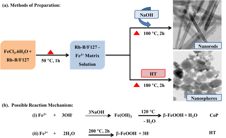
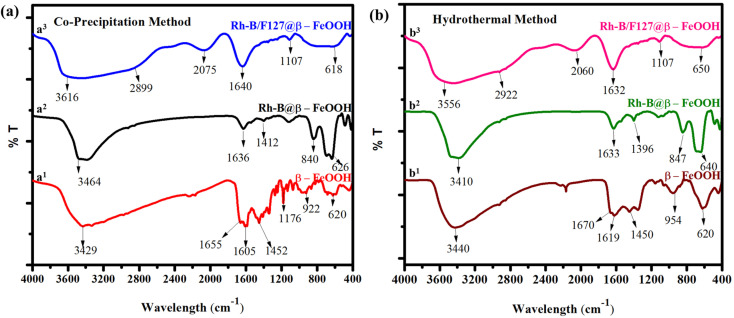
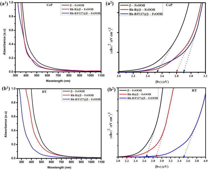
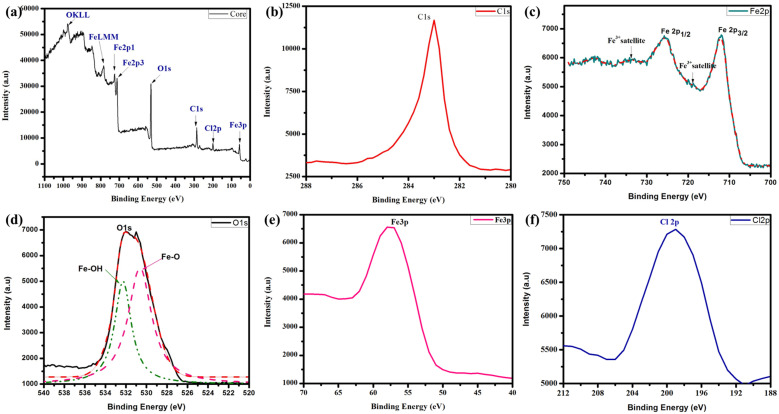
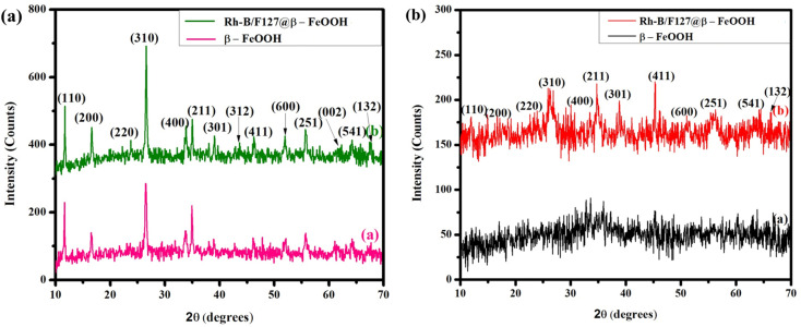
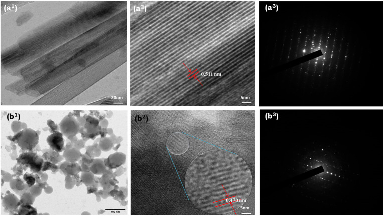
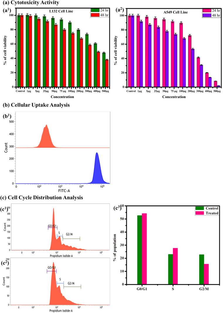
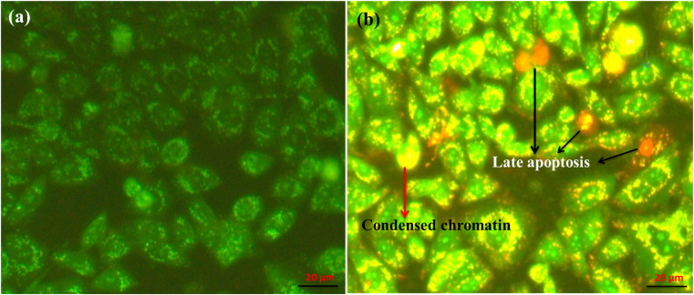
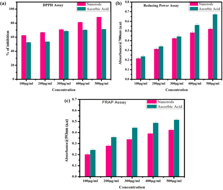
In this study, dye/polymer matrix-stabilized β-FeOOH nanomaterials were fabricated for therapeutic applications. Rh-B/F127@β-FeOOH nanomaterials were synthesized using two different methods: co-precipitation (CoP) and hydrothermal (HT) methods. The as-synthesized nanoparticles were characterized using various spectroscopic techniques, including FT-IR, UV-Vis, PL, XRD, HR-TEM, and XPS analysis. The functional groups and optical properties were confirmed by FT-IR spectroscopy, UV-Vis and fluorescence spectroscopy. The Rh-B/F127@β-FeOOH nanomaterials exhibited both rod-like and sphere-like morphology, as confirmed by HR-TEM analysis. Unlike the nanorods, the nanospheres produced multi-colored emissions at 407, 446, 482 and 520 nm. The oxidative states and elements were confirmed by XPS spectroscopy. MTT assays were used to analyze the cytotoxicity of the nanospheres against A549 cells. The reactive oxygen species (ROS) generation and apoptotic cell death caused by the β-FeOOH nanospheres were evaluated by flow cytometry. Cell cycle analysis indicated that the treatment of nanospheres-induced S-phase cell cycle arrest in A549 cells. The synthesized nanospheres induced late-stage apoptosis in the A549 cell line, with a cell death rate of up to 30.37% at the IC50 concentration. Additionally, the antioxidant activities of the synthesized nanorods showed a high scavenging activity against free radicals, as examined by different assays such as such as DPPH, RP, and FRAP. The above results suggest that the synthesized nanorods and nanospheres are promising and efficient material for therapeutic applications.


Conflict of interest statement

We declare that we have no known competing financial interests or personal relationships that could have appeared to influence the work reported in this paper.

Figures

Scheme 1
Scheme 1. Schematic diagram of the synthesis routes for β-FeOOH nanoparticles. (a) Synthesis of β-FeOOH nanomaterials by co-precipitation (CoP) and hydrothermal (HT) methods; the corresponding TEM images confirm the rod-like and spherical-like morphologies. (b) Proposed mechanisms for the formation of β-FeOOH nanorods and nanospheres.
Fig. 1
Fig. 1. FT-IR spectra of β-FeOOH NPs prepared by (a) co-precipitation and (b) hydrothermal methods; (a1 and b1) urea-capped β-FeOOH NPs, (a2 and b2) Rh-B@β-FeOOH, and (a3 and b3) Rh-B/F127@β-FeOOH NPs.
Fig. 2
Fig. 2. (a1 and b1) UV-Vis spectra and (a2 and b2) bandgaps of the Rh-B/F127@β-FeOOH nanorods and nanospheres. The bandgap of the nanoparticles was calculated by the Tauc equation. The optical bandgaps were obtained by extrapolating the linear portion of (αhν)2versus hν and were found to be between 2.52 and 3.50 eV.
Fig. 3
Fig. 3. (a) PL spectra of β-FeOOH nanorods using an excitation wavelength of 380 nm, (b1) PL spectra of β-FeOOH nanospheres using an excitation wavelength of 320 nm; and (b2) Gaussian band deconvolution of the PL spectra of the Rh-B/F127@β-FeOOH nanospheres.
Fig. 4
Fig. 4. (a–f) XPS spectra of the Rh-B/F127-matrix-stabilized β-FeOOH nanospheres; (a) XPS survey spectrum; (b) XPS spectrum of C 1s; (c) XPS spectrum of Fe 2p; (d) XPS spectrum of O 1s. (e) XPS spectrum of Fe 3p and (f) XPS spectrum of Cl 2p.
Fig. 5
Fig. 5. XRD patterns of β-FeOOH and Rh-B/F127@β-FeOOH nanoparticles synthesized by (a) CoP and (b) HT methods. The observed peaks well matched with the tetragonal structure of the β-FeOOH phase and JCPDS card no. 34-1266.
Fig. 6
Fig. 6. HR-TEM micrographs of the Rh-B/F127@β-FeOOH nanomaterials prepared by (a) CoP and (b) HT methods. (a1 and b1) TEM images showing the nanorods- and nanospheres-like morphologies of Rh-B/F127@β-FeOOH nanoparticles; (a2 and b2) interplanar spacings, and (a3 and b3) SAED patterns of the Rh-B/F127@β-FeOOH nanorods and nanospheres.
Fig. 7
Fig. 7. Cytotoxicity analysis. (a) Cytotoxicity activities: (a1) L132 cell line and (a2) A549 cell lines at 24 and 48 h treatment with Rh-B/F127@β-FeOOH nanospheres as revealed by MTT assay. (b). Cellular uptake analysis: (b1) flow cytometry quantification of Rh-B/F127@β-FeOOH nanospheres uptake by A549 cells. (c). Cell cycle distribution analysis: representative histograms showing the cell population according to the DNA content as determined by propidium iodide staining. (c1) Control and (c2) treated cells with Rh-B/F127@β-FeOOH nanospheres; (c3) bar diagram of cell distribution in the G0/G1, S, and G2/M phases. The results indicated that the nanoparticles-induced cell cycle arrest in the S phase.
Fig. 8
Fig. 8. Apoptosis study. (a) Rh-B/F127@β-FeOOH nanospheres-induced ROS generation in A549 cells: (a1) control, (a2) ROS induced by nanoparticles, (a3) FCM analysis of intracellular ROS using a DFCH-DA probe. (b) Evaluation of apoptosis in A549 cells using the annexin-V/dead cell assay: A549 cells were treated with Rh-B/F127@β-FeOOH nanospheres for 24 h and apoptosis was assayed via flow cytometry using the annexin-V/dead cell assay; (b1) control, (b2) treated, and (b3) histogram showing % cell death. (c) Morphology of A549 cell nuclei observed using DAPI staining: (c1) control, and (c2) treated cells. DAPI images were recorded using laser light excitation at 360 nm. The scale bars represent 100 μm. (d) Proposed mechanism for ROS generation and apoptosis cell death by the overproduction of ROS generation (generation of OH and ˙OH radicals through Fenton and/or Haber–Weiss reactions).
Fig. 9
Fig. 9. Fluorescence images of A549 cells stained with acridine orange/ethidium bromide (AO/EB). (a) Untreated cells (control); (b) cells incubated with Rh-B/F127@β-FeOOH nanoparticles. The cells were observed at an excitation wavelength of 480 nm. The bright-green emissions originated from the live cells (untreated cells), while yellow-orange indicated early-stage apoptotic cells. Red emissions originated from late apoptosis. The scale bars represent 20 μm.
Fig. 10
Fig. 10. Antioxidant activity of Rh-B/F127@β-FeOOH nanorods. (a) DPPH assay, (b) reducing power assay, and (c) FRAP assay. The antioxidant activity of β-FeOOH nanorods was found to increase with the increase in concentration from 100 to 500 mg mL−1.

References

    1. Bae C. Kim H. Kook Y.-M. Lee C. Kim C. Yang C. Park M. H. Piao Y. Koh W.-G. Lee K. Induction of ferroptosis using functionalized iron-based nanoparticles for anti-cancer therapy. Mater. Today Bio. 2022;17:100457. doi: 10.1016/j.mtbio.2022.100457. - DOI - PMC - PubMed
    1. Zhao H. Dong Y. Jiang P. Wang G. Zhang J. Highly dispersed CeO2 on TiO2 nanotube: a synergistic nanocomposite with superior peroxidase-like activity. ACS Appl. Mater. Interfaces. 2015;7:6451–6461. doi: 10.1021/acsami.5b00023. - DOI - PubMed
    1. Zhang Z. Hao J. Yang W. Lu B. Ke X. Zhang B. Tang J. Porous Co3O4 nanorods–reduced graphene oxide with intrinsic peroxidase-like activity and catalysis in the degradation of methylene blue. ACS Appl. Mater. Interfaces. 2013;5:3809–3815. doi: 10.1021/am4004655. - DOI - PubMed
    1. Hu M. Korschelt K. Daniel P. Landfester K. Tremel W. Bannwarth M. B. Fibrous nanozyme dressings with catalase-like activity for H2O2 reduction to promote wound healing. ACS Appl. Mater. Interfaces. 2017;9:38024–38031. doi: 10.1021/acsami.7b12212. - DOI - PubMed
    1. Champion J. A. Katare Y. K. Mitragotri S. Particle shape: a new design parameter for micro- and nanoscale drug delivery carriers. J. Controlled Release. 2007;121:3–9. doi: 10.1016/j.jconrel.2007.03.022. - DOI - PMC - PubMed

LinkOut - more resources