. 2025 Jan 16;33(2):357-367.
doi: 10.32604/or.2024.050878. eCollection 2025.

Melanoma cell line-derived exosomal miR-424-5p: a key promoter of angiogenesis through LATS2 interaction

Affiliations

Melanoma cell line-derived exosomal miR-424-5p: a key promoter of angiogenesis through LATS2 interaction

Junwei DU et al. Oncol Res. .

Abstract

Objectives: Melanoma is a highly aggressive and metastatic form of cancer, and the role of exosomal microRNAs (miRNAs) in its progression remains largely unexplored. This study aimed to investigate the effects of melanoma cell-derived exosomal miR-424-5p on angiogenesis and its underlying mechanisms.

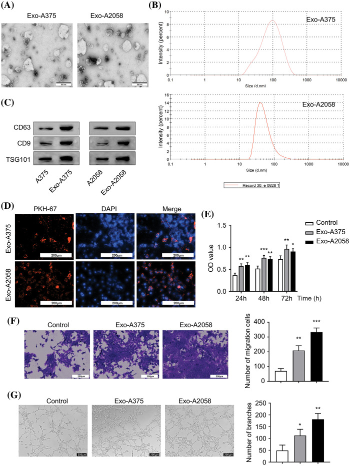
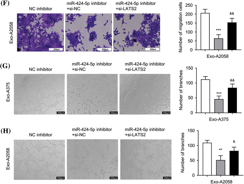
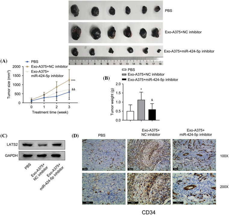
Methods: Exosomes were isolated from melanoma cell lines A375 and A2058, and their effects on the proliferation, migration, and angiogenesis of human umbilical vein endothelial cells (HUVECs) were examined. The interaction between miR-424-5p and its target gene, large tumor suppressor kinase 2 (LATS2), was analyzed using luciferase reporter assays and functional experiments. In vivo, tumor growth and angiogenesis were studied in a xenograft model using nude mice.

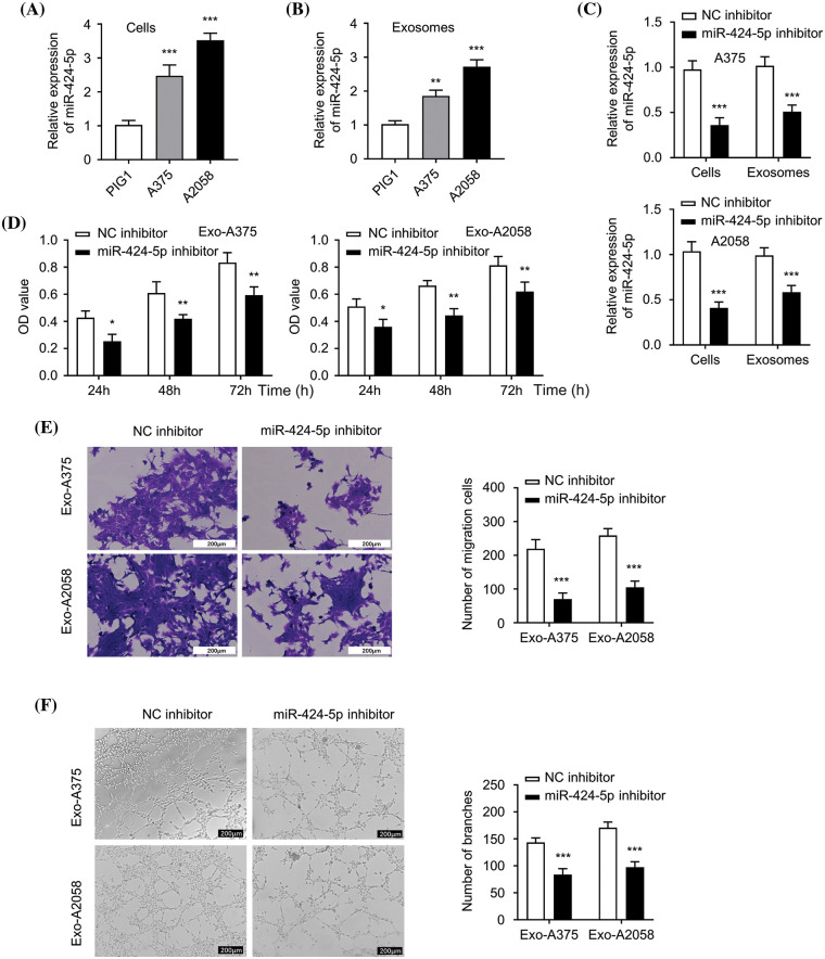
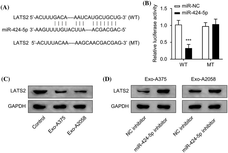
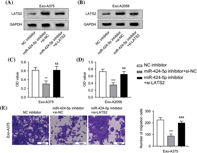
Results: Melanoma cell-derived exosomes could be internalized by HUVECs, which promoted proliferation, migration, and angiogenesis. miR-424-5p was highly expressed in melanoma cells and their exosomes, and its inhibition in exosomes suppressed HUVEC proliferation, migration, and angiogenesis. LATS2 was identified as a direct target of miR-424-5p, and its silencing reversed the inhibitory effects of miR-424-5p inhibition on HUVEC functions. In vivo, exosomes derived from miR-424-5p-inhibited melanoma cells suppressed tumor growth and angiogenesis in xenograft models.

Conclusions: Melanoma cell-derived exosomal miR-424-5p promotes angiogenesis by targeting LATS2, contributing to melanoma progression. Targeting the exosomal miR-424-5p/LATS2 axis could be a potential therapeutic strategy for melanoma.

Keywords: Cancer progression; Cell proliferation; Exosomal miR-424-5p; Large tumor suppressor kinase 2 (LATS2); Therapeutic targets.


Conflict of interest statement

The authors declare no conflicts of interest to report regarding the present study.

Figures

Figure 1
Figure 1. Melanoma cell-derived exosomes promote proliferation, migration and angiogenesis of HUVEC cells. (A): Exosomes were visualized using electron microscopy. (B): Nanoparticle Tracking Analysis (NTA) was conducted for determining exosome size originating from A375 and A2058 cells. (C): Exosomal markers (CD63, CD9, and TSG101) levels in A375 and Exo-A375 exosomes, as well as in A2058 cells and Exo-A2058 exosomes, was assessed using Western blot analysis. (D): The internalization of PKH67-labeled Exo-A375 and Exo-A2058 exosomes in HUVEC cells was detected through immunofluorescence analysis. (E): CCK-8 assay was utilized for evaluating proliferation levels of HUVEC cells co-cultured with different groups (control, Exo-A375, and Exo-A2058) throughout 24, 48, and 72 h. (F): A Transwell assay was conducted for measuring HUVEC migration after co-culture with different groups (control, Exo-A375, and Exo-A2058) after 24 h. (G): Tube formation assay in different groups of HUVECs (control, Exo-A375, and Exo-A2058). Every assay was carried out thrice. *p < 0.05, **p < 0.01, ***p < 0.001, vs. control group.
Figure 2
Figure 2. Exosome derived from melanoma cells with miR-424-5p knockdown suppresses HUVEC proliferation, migration and angiogenesis. (A): miR-424-5p expression levels within healthy human epidermal melanocytes and melanoma cells (A375 and A2058) were quantified using qRT-PCR. (B): miR-424-5p expression within exosomes (Exo-miR-424-5p) derived from normal human epidermal melanocytes and in exosomes (Exo-A375 and Exo-A2058) derived from melanoma cell lines A375 and A2058 were measured using qRT-PCR. (C): A375 and A2058 cells were transfected into NC inhibitor and miR-424-5p inhibitor, then miR-424-5p level within these cells and exosomes derived from these cells was assessed using qRT-PCR. (D): HUVEC cell proliferation after co-culture with different groups of exosomes as indicated for 24/48/72 h was determined using the CCK-8 assay. (E): Migration level of each group of cells was evaluated by a Transwell assay. (F): The tube formation level of each group of cells was assessed by a tube formation assay. Every assay was completed thrice. *p < 0.05, **p < 0.01, ***p < 0.001, vs. NC inhibitor group.
Figure 3
Figure 3. miR-424-5p targets LATS2. (A): Starbase prediction indicates that miR-424-5p targets LATS2 sites. (B): The luciferase reporter gene assay was conducted for confirming miR-424-5p binding to LATS2 3′-UTR. (C): Western-blotting analysis was performed for assessing LATS2 protein expression in HUVEC cells co-cultured with different groups (control, Exo-A375, and Exo-A2058) for 24 h. (D): Western-blotting assay was also conducted for measuring LATS2 protein level in HUVEC cells co-cultured with Exo-A375 and Exo-A2058 for 24 h. Every assay was carried out thrice. ***p < 0.001, vs. miR-NC group.
Figure 4
Figure 4. Exosome-derived miR-424-5p regulates HUVEC proliferation, migration and angiogenesis of HUVEC cells via LATS2. A: Western-blotting analysis was conducted for measuring LATS2 protein levels in different groups of HUVECs cultivated with Exo-A375 (NC inhibitor, miR-424-5p inhibitor+si-NC, and miR-424-5p inhibitor+si-LATS2) after 24 h. B: Western-blotting assay was also conducted for assessing LATS2 protein expression within different groups of HUVECs cultivated with Exo-A2058 (NC inhibitor, miR-424-5p inhibitor+si-NC, and miR-424-5p inhibitor+si-LATS2) after 24 h. C: CCK-8 assay was carried out for evaluating HUVEC proliferation in different groups of HUVECs cultivated with Exo-A375 (NC inhibitor, miR-424-5p inhibitor+si-NC, and miR-424-5p inhibitor+si-LATS2). D: CCK-8 assay was also carried out for measuring HUVEC proliferation in different groups cultivated with Exo-A2058 (NC inhibitor, miR-424-5p inhibitor+si-NC, and miR-424-5p inhibitor+si-LATS2). E: The Transwell assay was conducted for assessing HUVECs cultivated with Exo-A375 (NC inhibitor, miR-424-5p inhibitor+si-NC, and miR-424-5p inhibitor+si-LATS2). F: The Transwell assay was conducted for assessing HUVECs cultivated with Exo-A2058 (NC inhibitor, miR-424-5p inhibitor+si-NC, and miR-424-5p inhibitor+si-LATS2). G: The tube formation assay was performed for evaluating tube-forming ability in HUVECs cultivated with Exo-A375 (NC inhibitor, miR-424-5p inhibitor+si-NC, and miR-424-5p inhibitor+si-LATS2). H: The tube formation assay was performed for evaluating tube-forming ability in HUVECs cultivated with Exo-A2058 (NC inhibitor, miR-424-5p inhibitor+si-NC, and miR-424-5p inhibitor+si-LATS2). Every assay was carried out thrice. **p < 0.05, ***p < 0.001, vs. NC inhibitor group; &p < 0.05, &&p < 0.01, &&&p < 0.001, vs. miR-424-5p inhibitor+si-NC group.
Figure 5
Figure 5. Melanoma cell-derived exosomes promote melanoma cell proliferation and angiogenesis in vivo. (A and B): A subcutaneous tumor model was established with A375 cells, then tumor volume and mass were measured. (C): Western-blotting analysis was conducted for detecting LATS2 protein expression in subcutaneous tumors of each group. (D): Immunohistochemistry (IHC) was carried out for detecting CD34 protein level within subcutaneous tumors in different groups. N = 6 in each group. *p < 0.05, ***p < 0.001, vs. PBS group; &p < 0.05, &&p < 0.01, vs. Exo-A375-NC inhibitor group.

Similar articles

References

    1. Centeno PP, Pavet V, Marais R. The journey from melanocytes to melanoma. Nat Rev Cancer. 2023;23(6):372–90; - PubMed
    1. Guo J, Qin S, Liang J, Lin T, Si L, Chen X, et al. . Chinese guidelines on the diagnosis and treatment of melanoma (2015 Edition). Ann Transl Med. 2015;3(21):322; - PMC - PubMed
    1. Sung H, Ferlay J, Siegel RL, Laversanne M, Soerjomataram I, Jemal A, et al. . Global cancer statistics 2020: GLOBOCAN estimates of incidence and mortality worldwide for 36 cancers in 185 countries. CA Cancer J Clin. 2021;71(3):209–49; - PubMed
    1. Marzagalli M, Ebelt ND, Manuel ER. Unraveling the crosstalk between melanoma and immune cells in the tumor microenvironment. Semin Cancer Biol. 2019;59(2):236–50. doi:10.1016/j.semcancer.2019.08.002; - DOI - PubMed
    1. Luke JJ, Flaherty KT, Ribas A, Long GV. Targeted agents and immunotherapies: optimizing outcomes in melanoma. Nat Rev Clin Oncol. 2017;14(8):463–82; - PubMed

MeSH terms

LinkOut - more resources