Review
. 2024 Dec 31:30:101438.
doi: 10.1016/j.mtbio.2024.101438. eCollection 2025 Feb.

Biomaterial-assisted organoid technology for disease modeling and drug screening

Affiliations
Review

Biomaterial-assisted organoid technology for disease modeling and drug screening

Yunyuan Shao et al. Mater Today Bio. .

Abstract

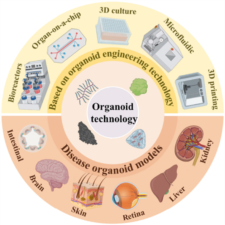
Developing disease models and screening for effective drugs are key areas of modern medical research. Traditional methodologies frequently fall short in precisely replicating the intricate architecture of bodily tissues and organs. Nevertheless, recent advancements in biomaterial-assisted organoid technology have ushered in a paradigm shift in biomedical research. This innovative approach enables the cultivation of three-dimensional cellular structures in vitro that closely emulate the structural and functional attributes of organs, offering physiologically superior models compared to conventional techniques. The evolution of biomaterials plays a pivotal role in supporting the culture and development of organ tissues, thereby facilitating more accurate disease state modeling and the rigorous evaluation of drug efficacy and safety profiles. In this review, we will explore the roles that various biomaterials play in organoid development, examine the fundamental principles and advantages of utilizing these technologies in constructing disease models, and highlight recent advances and practical applications in drug screening using disease-specific organoid models. Additionally, the challenges and future directions of organoid technology are discussed. Through continued research and innovation, we aim to make remarkable strides in disease treatment and drug development, ultimately enhancing patient quality of life.

Keywords: Biomaterial; Disease modeling; Drug screening; Organoid technology.


Conflict of interest statement

The authors declare that they have no known competing financial interests or personal relationships that could have appeared to influence the work reported in this paper.

Figures

Image 1
Graphical abstract
Fig. 1
Fig. 1
Organoid Generation and Hair Follicle Histology (A) Schematic diagram of an organoid generation procedure that can be generated by TDC or iPSC. This diagram is reproduced with minor modifications from Ref. [65], Copyright © 2022 Nat Rev Methods Primers. published by Springer Nature. (B) Hair follicles were subjected to histological examination, which involved sectioning and staining with antibodies that were fluorescently labelled. Specifically, the antibodies targeted K14, AE13, and versican to visualize these proteins within the follicloids. This diagram is reproduced with minor modifications from Ref. [64], Copyright © 2023 Sci Rep. published by Nature Portfolio.
Fig. 2
Fig. 2
Histological Staining and 3D Cell Culture Techniques (A) Optical appearance and microstructure of HA, HAD, HAR, and HADR hydrogels, respectively (scale bar: 100 μm and 50 μm). This figure is reproduced with minor modifications from Ref. [80], Copyright © 2024 Int J Biol Macromol. Published by ELSEVIER (B) Brightfield images of gastric organoids grown in decellularized stomach derived ECM (SEM) hydrogels and Matrigel (MAT) at day 5 [104], Copyright ©2024 J Biomed Sci. published by BioMed Central.
Fig. 3
Fig. 3
Bioprinting and Organ-on-a-Chip Technologies (A) Three-dimensional bioprinting [129],Copyright © 2018 Journal of Biomedical Science, published by Elsevier. (B) Conceptual diagram of organoid manipulation using the proposed microfluidic device. The figure is reproduced with minor modifications from Ref. [130], copyright © Micromachines (Basel). Published by MDPI (C) The organ-on-a-chip platform facilitates controlled cell cultivation within a microstructured, organotypic environment based on 3D culture techniques. This figure is reproduced with minor modifications from Ref. [131], Copyright © 2021 Trends Pharmacol Sci. Published by Elsevier Ltd.
Fig. 4
Fig. 4
Advancements in Gut Organoid Technology and Intestinal Ecology (A) Time course of crypt-like organ growth. Differential interference contrast images show granule-containing Pan cells (red arrows) at the ectodermal site of neocrypt formation. Lgr5-GFP (green) stem cells expand at the base of the crypt close to the Pan cells. Zoom bars: 50 μm. this figure is reproduced with minor modifications from Ref. [153], Copyright © 2011 Nature. published by Nature Publishing Group. (B) An in vivo intestinal microarray in which human villous epithelium and vascular endothelium are arranged on opposite sides of a pliable porous surface in response to fluid flow and peristaltic-like stimuli. This figure is reproduced with minor changes from Ref. [140], copyright ©2018 Cell Mol Gastroenterol Hepatol. By Elsevier.
Fig. 5
Fig. 5
Brain Organoid Culture System and Zika Virus Impact on Neural Stem Cells (A) Brain-like organ culture system with example images of each stage. This figure has been slightly altered and is reproduced from Ref. [165]. Copyright © 2013 Nature. Published by Nature Publishing Group. (B) Zika virus infects neural stem cells and causes microcephaly. Figure slightly adapted from Ref. [172], Copyright © 2016 Cell. by Elsevier Publishing.
Fig. 6
Fig. 6
PDX Production, PDO Applications, and Tumor Tissue Analysis (A) PDX Production method steps and PDO applications. Tumour-like tissues containing cancer cells, fibroblasts, myofibroblasts or stem cells are mechanically and chemically dissociated into very small fragments, cell membranes or single cells and cultured under appropriate three-dimensional conditions in a gel system containing ECM components. This figure is reproduced with minor modifications from Ref. [180], copyright Cell. Published by Cell. (B) PDX in the new era of cancer treatment. This figure shows the current conundrums of cancer treatment including restricted beneficiaries, tumor heterogeneity, drug resistance as well as tumor metastasis and recurrence, and shows the versatile functions of PDX in developing therapeutics against cancer [181]. This figure is reproduced with minor modifications from reference Copyright ©2023 Signal Transduct Target Ther. by Springer Nature.
Fig. 7
Fig. 7
Methods for skin organoid preparation and culturing (A) The figure discloses a method and application for preparing an organoid. Specifically, the method comprises culturing at least one organoid derived from a patient during a period from day 3 to day 8 to form clusters of organoids comprising a plurality of organoids. These organoid clusters are then further cultured during a period from day 9 to day 13 to form organoids having a diameter greater than 1 mm. The Figure is reproduced with minor adaptations from Ref. [209] with permission, Copyright ©2023 Nat Protoc. Published by Nature Publishing Group. (B) The present figure provides a method of preparing a class organ, characterised in that it comprises: culturing at least one class organ derived from a patient in a skin class organ medium of W25, W35 and/or W45 (W25, day 25; W35, day 35; W45, day 45); and culturing said class organ in a mixture of an inducer of differentiation (IDC), said IDC being selected from W20, W30, W40, W50, W60 and W70 of skin organoid medium (W20, day 20; W30, day 30; W40, day 40; W50, day 50; W60, day 60; W70, day 70). The Figure is reproduced with minor adaptations from Ref. [209] with permission, Copyright ©2023 Nat Protoc. Published by Springer Nature.
Fig. 8
Fig. 8
Retinal Organoid Culture and Layer Simulation (A) This figure provides a retinal organoid culture mould inserted for retinal development in vivo. The upper row shows some features of retinal development and some key signal transduction interactions. Photographs capturing both fluorescence (API) and field intensity in the same row illustrate the morphology of retinal organoid tissue at each respective developmental stage. The final column presents the timing of each developmental phase in both mouse (m) and human (h) retinal organoid cultures. This figure is reproduced with minor modifications from Ref. [216]. Copyright © 2021 Front Cell Neurosci. (B) Simulation of retinal layers in vitro. This figure is reproduced with minor modifications from Ref. [217] with permission, Copyright ©20 Development. published by The Biologist, Inc.
Fig. 9
Fig. 9
Liver Anatomy and Hybrid Live Organoid Model (A) Diagram of the anatomy of the liver. Localised hepatic endothelial cells (LSEC) surrounding the hepatic Sinusoid. The Disse space, which harbors stellate cells, separates hepatocytes from the inner cortical layer. The hepatic macrophages, known as Kupffer cells, maintain close proximity to liver sinusoidal endothelial cells (LSECs), facing the bloodstream. Miliary cells coat the internal spaces within the biliary tree. The Figure is reproduced with minor adaptations from Ref. [9] with permission, Copyright ©2020 Int J Mol Sci. Published by MDPI. (B) Scheme of the different steps involved in obtaining a hybrid multicellular liver organoid model using our agarose micromoulding technique. Image of immunofluorescence of the progression of differentiated multicellular liver organoids. Differentiated human induced pluripotent stem cells (hiPSC) labelled with nuclear staining DAPI; human stellate cells (HSC) labelled with GFP; and hepatic sinusoidal endothelial cells (LSEC) labelled with RFP. scale bar 50 μm [220]. with permission, Copyright ©2021 Nature Communications. Published by Nature.
Fig. 10
Fig. 10
Organoid Formation and Viral Infection Mitigation (A) The organism formation protocol depicted in this figure, which has undergone slight modifications, is adapted from Ref. [238], copyright Micromachines (Basel), published by MDPI (Multidisciplinary Digital Publishing Institute). (B) A clinical-grade version of recombinant human ACE2 has been shown to diminish SARS-CoV-2 infection in cellular and various human organoid models. This figure is reproduced with minor modifications from Ref. [224], Copyright ©20 Cell. published by Cell Press. (C) Developing kidne organs cultured in vitro at high fluid flow rates exhibit enhanced angiogenesis during nephrogenesis. Developing renal organoid tissues were placed on engineered extracellular matrix (ECM) in a perfusable millifluidic chip with controlled fluidic shear stress (FSS) applied. This figure is reproduced with minor changes from Ref. [239], Copyright © 2018 Cell. by Cell Press.
Fig. 11
Fig. 11
MSC Isolation and Hydrogel Strategies for Cartilage Repair (A) MSCs are easily detached from bone marrow and adipose tissue, however, as part of the microvascular system, all tissues contain MSC-like cells. This figure is reproduced with minor changes from Ref. [250], Copyright © 2019 NPJ Regen Med. by Nature Publishing Group. (B) Hydrogel strategies for targeting articular cartilage. The biodistribution patterns of diverse targeted (either passive or active) hydrogel particles are examined in both healthy and osteoarthritic joint tissues. This figure is reproduced with minor modifications from Ref. [251], Copyright ©20 Acta Biomater. published by Elsevier BV.

Similar articles

Cited by

References

    1. Saorin G., Caligiuri I., Rizzolio F. Microfluidic organoids-on-a-chip: the future of human models. Semin. Cell Dev. Biol. 2023;144:41–54. doi: 10.1016/j.semcdb.2022.10.001. - DOI - PubMed
    1. Hong Z.X., Zhu S.T., Li H., Luo J.Z., Yang Y., An Y., Wang X., Wang K. Bioengineered skin organoids: from development to applications. Mil Med Res. 2023;10(1):40. doi: 10.1186/s40779-023-00475-7. - DOI - PMC - PubMed
    1. Takahashi T., Fujishima K., Kengaku M. Modeling intestinal stem cell function with organoids. Int. J. Mol. Sci. 2021;22(20) doi: 10.3390/ijms222010912. - DOI - PMC - PubMed
    1. Simian M., Bissell M.J. Organoids: a historical perspective of thinking in three dimensions. J. Cell Biol. 2017;216(1):31–40. doi: 10.1083/jcb.201610056. - DOI - PMC - PubMed
    1. Tang X.Y., Wu S., Wang D., Chu C., Hong Y., Tao M., Hu H., Xu M., Guo X., Liu Y. Human organoids in basic research and clinical applications. Signal Transduct. Targeted Ther. 2022;7(1):168. doi: 10.1038/s41392-022-01024-9. - DOI - PMC - PubMed

LinkOut - more resources