Review
. 2025 Jan 24:16:20417314241310541.
doi: 10.1177/20417314241310541. eCollection 2025 Jan-Dec.

Bioengineering strategies targeting angiogenesis: Innovative solutions for osteonecrosis of the femoral head

Affiliations
Review

Bioengineering strategies targeting angiogenesis: Innovative solutions for osteonecrosis of the femoral head

Weihong Zhu et al. J Tissue Eng. .

Abstract

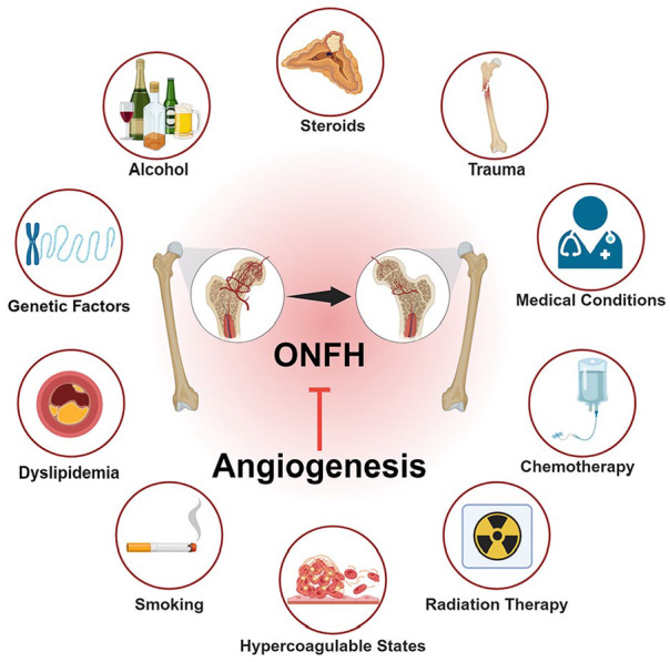
Osteonecrosis of the femoral head (ONFH) is a prevalent orthopedic disorder characterized primarily by compromised blood supply. This vascular deficit results in cell apoptosis, trabecular bone loss, and structural collapse of the femoral head at late stage, significantly impairing joint function. While MRI is a highly effective tool for diagnosing ONFH in its early stages, challenges remain due to the limited availability and high cost of MRI, as well as the absence of routine MRI screening in asymptomatic patients. . In addition, current therapeutic strategies predominantly only relieve symptoms while disease-modifying ONFH drugs are still under investigation/development. Considering that blood supply of the femoral head plays a key role in the pathology of ONFH, angiogenic therapies have been put forward as promising treatment options. Emerging bioengineering interventions targeting angiogenesis hold promising potential for ONFH treatment. In this review, we introduce the advances in research into the pathology of ONFH and summarize novel bioengineering interventions targeting angiogenesis. This review sheds light upon new directions for future research into ONFH.

Keywords: Osteonecrosis; angiogenesis; biomaterial scaffold; delivery system; femoral head.


Conflict of interest statement

The author declared no potential conflicts of interest with respect to the research, authorship, and/or publication of this article.

Figures

Graphical abstract
Graphical abstract
Figure 1.
Figure 1.
Angiogenic process and key molecular regulators involved in the formation and maturation of new blood vessels. Angiogenesis triggered by internal and external stresses such as low oxygen levels (hypoxia) lead to the release of angiogenic factors. Tip cells sense the stimuli and transmit stimuli signals to neighboring endothelial cells (ECs) through cell-to-cell communication. ECs respond to angiogenic stimuli, resulting in vasodilation and degradation of the basement membrane facilitated by matrix metalloproteinases (MMPs). ECs then migrate and proliferate to form new capillary tubes. The newly formed vessels undergo maturation and stabilization, aided by pericytes and smooth muscle cells. Several factors influence the angiogenesis process via targeting different stages. Created with biorender.com.
Figure 2.
Figure 2.
Illustration of delivery systems for angiogenesis. There are diverse strategies employed to deliver angiogenic factors, each with unique advantages and applications. These advanced delivery systems are pivotal in the development of therapeutic approaches for enhancing angiogenesis, with significant implications for regenerative medicine and the treatment of various vascular diseases. Created with biorender.com.
Figure 3.
Figure 3.
Scaffold-based bioengineering approaches to ONFH management. (A) The CaO2/gelatin-based, oxygen-releasing microspheres and the 3D printed PCL/nHA tubular scaffold were molded into rod-shaped complexes. Micro-CT confirmed bone regeneration and immunohistochemical staining of CD 31 indicated angiogenesis in the femoral heads in rabbit ONFH model 4 weeks after implantation. Sc, PCL/nHA Scaffold; Hy, hydrogel; Cc, calcium carbonate; CPO, calcium peroxide. Each value is the mean ± standard error of mean (n = 3); Figures reproduced with permission from Wang et al., Copyright 2021, RSC. (B) Images of toluidine blue-stained cobalt bioactive glass/collagen-glycosaminoglycan scaffolds. These two scaffolds, with different structural integrity and porosity, are prepared at a controlled freezing rate of 1 and 4℃/min, respectively. Scanning electron microscope images revealed that these scaffolds, loaded with small- and large-diameter bioactive glass particles, elute cobalt effectively. Figures reproduced witi perrmission from Quinlan et al., Copyright 2015, Elsevier Ltd. (C) Schematic illustration of the fabrication of a versatile MCFS nanosheet-functionalized 3D-printed BGS for periprosthetic infection preven tion/treatment and vascularized osteogenesis confirmed by CLSM and digital images. Figures reproduced witi permission from Bian et al. (D) Scanning electron micrographs illustrating the structural changes in MBG-PCL-zol scaffolds before and after 21 and 30 days of drug release tests, respectively. Confocal images of the morphology of osteoclast-like cells 1 and 6 days after immersing the MBG-PCL-zol scaffolds in the medium of osteoclast cultures. Actin was stained with rhodamine-phalloidin (red) and cell nuclei with DAPI (blue). Figures reproduced with permission from Gómez-Cerezo et al., Copyright 2019, Elsevier Ltd.
Figure 4.
Figure 4.
4D bioprinted self-folding vascular structures. (A) 4D bioprinting of cell-laden tubular structures: (a) Schematic of the 4D bioprinting process for cell-laden AlgMA or HA-MA structures. Cells are mixed with AlgMA or HA-MA and printed on a substrate. Figures reproduced with permission from Kirillova et al., Copyright 2017, Wiley-VCH. (b) The printed structures are crosslinked using green light, followed by drying. (c) The dried constructs are folded into tubular shapes upon immersion in water, PBS, or cell culture media. (d) Microscopic images of the cell-laden tubular structures after folding, demonstrating structural integrity and cell viability (scale bar: 400 µm). (B) Self-folding PEGDA bilayer structures: (d–f) The first and second layers are photocrosslinked sequentially, leading to a self-folding construct when immersed in water. (g and h) Microscopic images showing various self-folded micropatterns in the bilayer structures. (i) Fluorescent imaging of cell-laden scaffolds indicates successful cell seeding and cell distribution (scale bar: 200 µm). Figures reproduced with permission from Jamal et al., Copyright 2013, Wiley-VCH. (C) 4D printing and transformation of vascular structures: 3D bioprinting or PDMS molding to create a grid-like scaffold, followed by transformation into complex vascular shapes in aqueous solutions. Magnetically driven shape transformation showing the 2D-to-3D transition to form branching vascular structures. Confocal microscopy images of the constructed vascular channels showing network formation by NHLFs and HUVECs within the matrix (scale bar: 100 µm). Figures reproduced with permission from Xie et al.
Figure 5.
Figure 5.
Cell and cellular component-based therapy on angiogenesis and osteogenesis. (A) Schematic diagram illustrating the sources and types of stem cells and cell-derived component used as advanced therapeutics. Stem cells can be derived from bone marrow, adipose tissue, muscle, neonatal tissues, dental pulp, and skin. Different types of stem cells have been employed in relevant previous studies, including iPSCs, bone marrow stem cells, embryonic stem cells, and adipose-derived stem cells. The processes may involve cell culture, identification, intervention, and extraction of required components. (B) Effects of PRP-Exos on angiogenesis of cells or tissue treated with glucocorticoids. Osteogenesis and angiogenesis of the femoral head in each group were analyzed by immunohistochemical staining. The red arrows indicate vessels. Scale bars = 100 μm. 3D micro-CT images showing the subchondral region of the femoral head in each group. Figures reproduced with permission from Xu et al., Copyright 2021, Wiley Periodicals LLC. (C) The angiogenesis of HMEC-1 cells treated with DEX alone or with Dex + PRP-Exos was evaluated using a tube formation assay. Angiographic images show the blood supply in the different groups. Scale bar: 100 μm. Figures reproduced with permission from Tao et al., Copyright 2017, Ivyspring International Publisher.(D) Angiographic images showing the blood supply in a rat model of ONFH treated with different interventions. The density and structure of vessels in each group are visualized in red. Figures reproduced with permission from Zuo et al.

Similar articles

References

    1. Malizos KN, Karantanas AH, Varitimidis SE, et al.. Osteonecrosis of the femoral head: etiology, imaging and treatment. Eur J Radiol 2007; 63(1): 16–28. - PubMed
    1. Mont MA, Cherian JJ, Sierra RJ, et al.. Nontraumatic osteonecrosis of the femoral head: where do we stand today? A ten-year update. J Bone Joint Surg Am 2015; 97(19): 1604–1627. - PubMed
    1. Lespasio MJ, Sodhi N, Mont MA. Osteonecrosis of the hip: a primer. Perm J 2019; 23(8): 1–7. DOI: 10.7812/TPP/18-100. - DOI - PMC - PubMed
    1. Mont MA, Salem HS, Piuzzi NS, et al.. Nontraumatic osteonecrosis of the femoral head: where do we stand today?: a 5-year update. J Bone Joint Surg Am 2020; 102(12): 1084–1099. - PMC - PubMed
    1. Zhao D, Zhang F, Wang B, et al.. Guidelines for clinical diagnosis and treatment of osteonecrosis of the femoral head in adults (2019 version). J Orthop Translat 2020; 21: 100–110. - PMC - PubMed

LinkOut - more resources