Use of Indocyanine Green (ICG) to Assess Myocardial Perfusion and Territorial Distribution of Vein Grafts Implanted on Coronary Arteries in an Ex-vivo Porcine Model. A Potential Adjunct to Assist Revascularization Strategies and Training in Coronary Artery Bypass Grafting
- PMID: 39867206
- PMCID: PMC11759963
- DOI: 10.31083/RCM25778
Use of Indocyanine Green (ICG) to Assess Myocardial Perfusion and Territorial Distribution of Vein Grafts Implanted on Coronary Arteries in an Ex-vivo Porcine Model. A Potential Adjunct to Assist Revascularization Strategies and Training in Coronary Artery Bypass Grafting
Abstract
Background: The fluorescent dye indocyanine green (ICG) has been used to identify anatomical structures intraoperatively in coronary artery bypass grafting (CABG). This study aimed to evaluate the feasibility of using ICG to assess graft patency and territorial distribution of myocardial reperfusion during CABG.
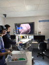
Methods: Porcine arrested hearts (n = 18) were used to evaluate territorial distribution of native coronary arteries and of a coronary bypass constructed with porcine saphenous vein graft (SVG) using ICG. Coronary ostia were dissected and selectively cannulated for ICG injection. Sequential fluorescence was assessed in the epicardial coronary arteries, myocardium and coronary veins using an infrared-sensitive charge-coupled device (CCD) camera system. In a separate set of experiments, SVG was used for anastomosis in end-to-side fashion to a terminal obtuse marginal (OM) branch. This approach was used to avoid bias in the assessment of territorial distribution. The anastomosis was injected with ICG; graft patency and territorial distribution was assessed using an infrared-sensitive CCD camera system from 30 cm above the field, as previously described. Native circulation and SVG grafts were assessed using real-time video recording and fluorescence intensity mapping that was averaged into a graded scoring system. The heart was divided into functional regions: anterior wall, lateral wall, inferior wall and right ventricle. All experiments were performed in triplicates.
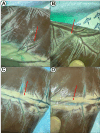
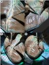
Results: After ICG injection into the individual coronary ostia, perfusion of the native coronary artery was visible. Portions of the vessels embedded into the epicardial fat could be easily visualized on the surface of the heart and the dissection facilitated via fluorescence guidance. The territorial distribution reflected the expected regional perfusion. The SVG graft was anastomosed to an OM branch. ICG visualization allowed for assessment of graft patency excluding potential technical anastomosis problems or graft twisting or dissection. The myocardial perfusion observed in real-time confirmed regional distribution to the entire lateral wall and minimally to the inferior wall. These findings were confirmed in all the specimens used in the study.
Conclusions: Besides assisting the identification of intramyocardial vessels, ICG can provide information on the native coronary circulation status and the territorial distribution of the perfusion before and after grafting. It enables visualization of collaterals and the territory of distribution subtended by a graft offering real-time assessment and guidance on the grafting strategy.
Keywords: arteriogenesis; coronary artery bypass grafting; fluorescent imaging; fractional flow reserve; graft patency; incomplete revascularization; indocyanine green; neoangiogenesis.
Copyright: © 2025 The Author(s). Published by IMR Press.
Conflict of interest statement
The authors declare no conflict of interest. Cristiano Spadaccio is serving as Guest Editor of this journal. We declare that Cristiano Spadaccio had no involvement in the peer review of this article and has no access to information regarding its peer review. Full responsibility for the editorial process for this article was delegated to Francesco Pelliccia.
Figures


References
-
- Desai ND, Miwa S, Kodama D, Koyama T, Cohen G, Pelletier MP, et al. A randomized comparison of intraoperative indocyanine green angiography and transit-time flow measurement to detect technical errors in coronary bypass grafts. The Journal of Thoracic and Cardiovascular Surgery . 2006;132:585–594. doi: 10.1016/j.jtcvs.2005.09.061. - DOI - PubMed
LinkOut - more resources
Full Text Sources

