. 2024 Aug 21;5(1):47-61.
doi: 10.1021/acsphyschemau.4c00069. eCollection 2025 Jan 22.

An Unexpected Water Channel in the Light-Harvesting Complex of a Diatom: Implications for the Switch between Light Harvesting and Photoprotection

Affiliations

An Unexpected Water Channel in the Light-Harvesting Complex of a Diatom: Implications for the Switch between Light Harvesting and Photoprotection

Vangelis Daskalakis et al. ACS Phys Chem Au. .

Abstract

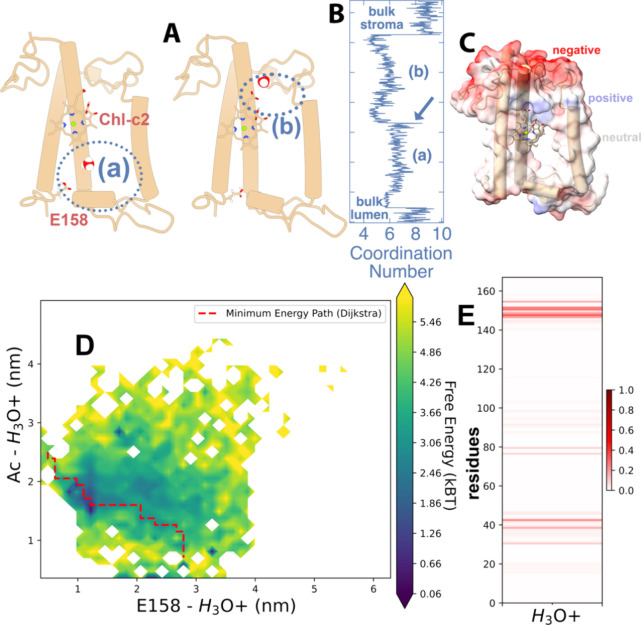
Many important processes in cells depend on the transfer of protons through water wires embedded in transmembrane proteins. Herein, we have performed more than 55 μs all-atom simulations of the light-harvesting complex of a diatom, i.e., the fucoxanthin and chlorophyll a/c binding protein (FCP) from the marine diatom Phaeodactylum tricornutum. Diatoms are unique models to study natural photosynthesis as they exert an efficient light-harvesting machinery with a robust pH-dependent photoprotective mechanism. The present study reports on the dynamics of an FCP monomer, a dimer, and a tetramer at varying pH values. Surprisingly, we have identified at low pH a water channel across FCP that selectively hydrates and protonates the acrylate of a Chl-c2 pigment located in the middle of the membrane. These results are further supported by QM/MM calculations and steered MD simulations on the proton dynamics. It is shown that proton hopping events between the lumenal and stromal sides of the membrane through the observed water channel are highly disfavored. This hindrance is due to the presence of residues Arg31 and Lys82 close to the acrylate, along with an hydronium desolvation penalty that shows close similarities to the water conductance in aquaporins. Furthermore, we provide strong evidence that this identified water channel is governing the transition between light-harvesting and photoprotective states of the major FCP complex in the diatom P. tricornutum.


Conflict of interest statement

The authors declare no competing financial interest.

Figures

Figure 1
Figure 1
(A) Crystal structure of a monomer FCP from the diatom Phaeodactylum tricornutum (pdb: 6a2w). The polypeptide chain is depicted in green cartoon representations. Chlorophyll a (Chl-a) molecules are shown in faded green stick representations, selected fucoxanthins are labeled and shown in red ball and stick, and the two chlorophyll-c pigments (Chl-c1 and c2) are shown in gray. On the right, we show only the Chl-c1 and c2 chromophores of the whole pigment network for reference. Also shown are selected residues in a hydrogen bond distance to the acrylate of Chl-c1, i.e., Lys-136 (K136), as well as to the acrylate of Chl-c2, i.e., Arg31 (R31). Moreover, the helices a1 (residues 25–56) and a3 (residues 129–161) are marked for reference. (B) Side view of the FCP monomer embedded in a thylakoid membrane patch. Lipids are drawn as white licorice moieties and the water phases using dense blue-white sticks. (C, D) Top views of the FCP dimer and tetramer models embedded in thylakoid membrane patches. The lipids are depicted as white licorice moieties, while the water molecules have been omitted for clarity.
Figure 2
Figure 2
(A) Free energy of the protein scaffold spanned over the tICA components IC1 and IC2. The macrostates S1 to S4 are highlighted with labels denoting the qualitative contribution of each different state (monomer, dimer, tetramer, neutral-low pH) to the FES. The energy values are given in units of kBT with kB being the Boltzmann constant and T the temperature. The configurational changes from S1 (green cartoons) to S4 (blue cartoons) are provided in the inset with arrows indicating the expansion of the protein scaffold. (B) FCP monomer in the different S1–S4 conformations color coded as in panel A. Selected pigments are also shown as superimposed from the crystal structure; Chls-c pigments are colored gray, Chl-a 409 green and the fucoxanthin carotenoid Fx-301 red. The FCP helixes are marked for reference (helix a1: residues 25–56, helix a1′: 75–83, helix a2:84–103, and helix a3:129–161).
Figure 3
Figure 3
(A) Distributions of the distances between the acrylate of Chl-c2 and residue Arg31 (R31) for the different FCP states. (B) Radial distribution functions (RDFs) of the water molecules around the Chl-c2 acrylate averaged over 500 ns windows and over the monomers in each dimer or tetramer trajectory. The colored shaded areas represent the standard deviations of the averaging.
Figure 4
Figure 4
(A) The FCP monomer is shown in cartoon representations along with the Chl-c2 pigment. The path of the identified water channel is indicated by a large blue/mesh surface in a qualitative manner. Selected residues are shown as sticks along the channel. The “STROMA” and “LUMEN” labels indicate the positions of the respective aquatic phases. (B) Radius of the water channels along the z-coordinate across the thylakoid membrane (membrane normal) for the MSM-predicted FCP states. The z-axis has been replaced by the FCP protein for reference. (C) Order parameter of the water molecules along the membrane normal for the S1, S2, and S4 states. As in (B), the protein structure in panel A is provided as reference for the position along the membrane normal. (D) The residue Arg31 (R31) is shown to be in close contact with the acrylate of Chl-c2 along with an adjacent Lys28 (K28) on helix a2 of FCP. A wire of water molecules that change their orientations is also depicted out of the MD simulation. The associated region in the (A) FCP crystal structure is highlighted by a red dotted circle.
Figure 5
Figure 5
The FCP dimer from the X-ray crystal structure is shown in green cartoon along with the Chl-c2 pigments in solid ball and stick representations. The aquatic phases are shown in pale blue and transparent ball-and-stick representation from a snapshot of the pH 7.0 runs. The colored wired spheres represent waters with large scattering densities that indicate immobile waters at pH 5.5 (blue), pH 5.5* (red), and pH 7.0 (green).
Figure 6
Figure 6
(A) FCP monomer (pdb: 6a2w, green) and selected helices of spinach aquaporin (pdb: 4ia4, grayish) superimposed in different orientations (A, B) to each other.
Figure 7
Figure 7
Steered MD simulation for an H3O+ along the identified water channel. (A) Two snapshots of the hydronium trajectory through FCP highlighting (a) a region before and (b) a region after the binding of H3O+ to the acrylate of the Chl-c2 molecule. The hydronium ion is shown with red oxygen and white hydrogen spheres. Glu-158 (E158) and Chl-c2 are also shown for reference. (B) Coordination number (solvation) for H3O+ along the trajectory including bulk and channel with regions (a) and (b) highlighted. A blue arrow indicates the binding of H3O+ to the Chl-c2 acrylate. The y-axis roughly indicates the position along the membrane normal in the scale of the structures in (A). (C) The electrostatic potential is superimposed on the FCP structure, indicating the negative (red), neutral (white), and blue (positive) regions. (D) Free energy of the H3O+ on two coordinates; the H3O+ distance to (i) the Chl-c2 acrylate (Ac, carboxyl group) and (ii) to the Glu-158 (E158) side chain carboxyl group. The energy values are given in units of kBT with kB being the Boltzmann constant and T the temperature. A minimum energy pathway is shown as calculated by the Dijkstra algorithm. (E) Protein residues (horizontal axis) that interact with H3O+ associated with the energy surface of D. The color legend spans from increased strength and large residence times (dark red) to no interaction (white).
Figure 8
Figure 8
| Positions of protons (H+) along with Arg31 (R31) and Chl-c2 observed at different pH values, i.e., at (A) pH 7.0, (B) pH 5.5* in the absence of H3O+, and (C) in the presence of H3O+ in the QM region and along the QM/MM MD trajectory at three time points. The protein environment and the lipids have been omitted for clarity. Only Chl-c2, Arg31, and water molecules in a radius of 0.8 nm around the acrylate of Chl-c2 and Arg31 are shown. (De)protonation events are highlighted by circles. Solid red arrows indicate the site of the Chl-c2 acrylate. (D) Distribution of Chl-c2 (acrylate)–proton and Chl-c2 (acrylate)–R31 distance for the different cases in (A), (B), and (C) after the 25 ps time frame.
Figure 9
Figure 9
| Quenching mechanism. The distributions of Qy excitation energies (A) and transition dipole moments (B) for the protonated (pH 5.5) and deprotonated (pH 7.0) Chl-c2 within the FCP complex along the 1 ns QM(DFTB)/MM MD trajectory. Protonated and deprotonated structures of Chl-c2 are also shown for reference in the inset of (A, B). The excitonic couplings for the Chl-a 409/Fx-301 pigment pair (C) and the Chl-c2/Fx-306 pigment pair (D) along the steered MD trajectory as projected onto two coordinates: the distance of the H3O+ (oxygen) to (i) the Chl-c2 acrylate (carboxyl group) and (ii) to the Glu-158 (E158) side chain carboxyl group. The sizes of the circles indicate the magnitude of the coupling values, as also indicated by the color code. (E) Representative interactions between the Chl-c2 acrylate, Arg31 (R31), and the ring of Fx-306 for two different states (S1, S2). For reference, the locations of these states are shown in (D) as well.

Similar articles

Cited by

References

    1. Kratochvil H. T.; Watkins L. C.; Mravic M.; Thomaston J. L.; Nicoludis J. M.; Somberg N. H.; Liu L.; Hong M.; Voth G. A.; DeGrado W. F. Transient Water Wires Mediate Selective Proton Transport in Designed Channel Proteins. Nat. Chem. 2023, 15 (7), 1012–1021. 10.1038/s41557-023-01210-4. - DOI - PMC - PubMed
    1. Swanson J. M. J.; Maupin C. M.; Chen H.; Petersen M. K.; Xu J.; Wu Y.; Voth G. A. Proton Solvation and Transport in Aqueous and Biomolecular Systems: Insights from Computer Simulations. J. Phys. Chem. B 2007, 111 (17), 4300–4314. 10.1021/jp070104x. - DOI - PMC - PubMed
    1. Supekar S.; Kaila V. R. I. Dewetting Transitions Coupled to K-Channel Activation in Cytochrome c Oxidase. Chem. Sci. 2018, 9 (32), 6703–6710. 10.1039/C8SC01587B. - DOI - PMC - PubMed
    1. Supekar S.; Gamiz-Hernandez A. P.; Kaila V. R. I. A Protonated Water Cluster as a Transient Proton-Loading Site in Cytochrome c Oxidase. Angewandte Chemie - International Edition 2016, 55 (39), 11940–11944. 10.1002/anie.201603606. - DOI - PubMed
    1. de Groot B. L.; Grubmüller H. Water Permeation Across Biological Membranes: Mechanism and Dynamics of Aquaporin-1 and GlpF. Science (1979) 2001, 294 (5550), 2353–2357. 10.1126/science.1066115. - DOI - PubMed

LinkOut - more resources