. 2025 Jan 19:2025:4955849.
doi: 10.1155/mi/4955849. eCollection 2025.

METTL3/miR-192-5p/SCD1 Axis Regulates Lipid Metabolism to Affect T Cell Differentiation in Asthma

Affiliations

METTL3/miR-192-5p/SCD1 Axis Regulates Lipid Metabolism to Affect T Cell Differentiation in Asthma

Zhengrong Chen et al. Mediators Inflamm. .

Abstract

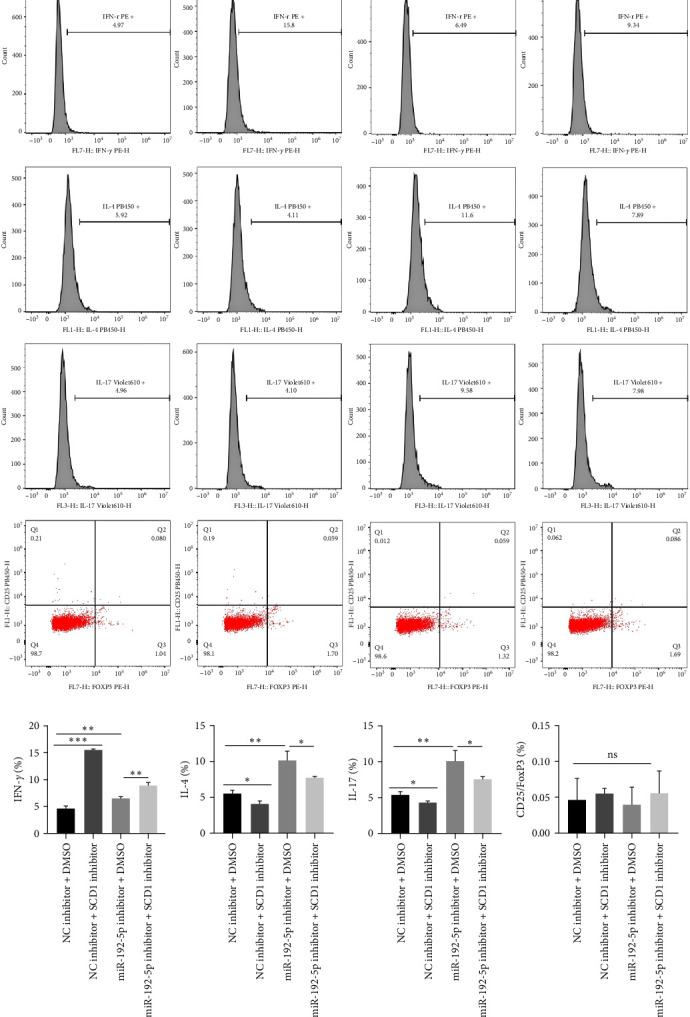
Background: This study aimed to explore the mechanisms underlying T-cell differentiation in asthma. Methods and Results: Flow cytometry was performed to detect Th cells. LC-MS/MS was performed to assess lipid metabolism. HE staining was performed to assess the pathological changes of the lung tissues. ELISA was performed to detect cytokine levels. The results of quantitative real-time polymerase chain reaction (qRT-PCR) and western blot showed that miR-192-5p expression was decreased, while SCD1 expression was increased in CD4+T cells isolated from the peripheral blood of children with asthma. The dual luciferase reporter assay determined the direct interaction between miR-192-5p and SCD1. MiR-192-5p inhibitor reduced ASCL3 and PPARα, increased FASN and SREBP1c mRNA expression and protein levels in mouse spleen CD4+T cells, and elevated Th2 and Th17 cells, but these effects were reversed by the SCD1 inhibitor. Oleic acid (OA) reduced Th1 cells and increased Th2 and Th17 cells in mouse spleen CD4+T cells treated with an SCD1 inhibitor. Additionally, pri-miR-192-5p expression was increased in CD4+T cells isolated from the peripheral blood of asthmatic children, and the deletion of METTL3 upregulated pri-miR-192-5p expression in an m6A-dependent manner. MiR-192-5p mimic and inhibitor both reversed miR-192-5p and SCD1 expression affected by overexpression or deletion of METTL3, both in vivo and in vitro. Furthermore, METTL3 overexpression attenuated lung inflammation, elevated Th1 cells, and reduced Th2 and Th17 cells in CD4+T cells isolated from the peripheral blood of asthmatic mice. These effects were reversed by the miR-192-5p inhibitor. Conclusion: These results suggest that METTL3/miR-192-5p/SCD1 axis regulates lipid metabolism and affects T cell differentiation, thus affecting asthma progression. This study may provide novel insights into the pathogenesis of asthma and a new treatment strategy.

Keywords: METTL3; SCD1; T cell differentiation; asthma; lipid metabolism; miR-192-5p.


Conflict of interest statement

The authors declare no conflicts of interest.

Figures

Figure 1
Figure 1
MiR-192-5p-reduced SCD1 expression via targeting SCD1. CD4+T cells were isolated from the peripheral blood of children with asthma and healthy children (n = 12), and qRT-PCR was performed to examine miR-192-5p (A) and SCD1 (B) expression in CD4+T cells; (C) the predicted binding sites between miR-192-5p and SCD1; (D) the dual luciferase reporter assay was employed to assess the interaction between miR-192-5p and SCD1 in HEK-293 T cells. Mouse spleen CD4+T cells were isolated and transfected with miR-192-5p inhibitor, qRT-PCR, and western blotting were performed to examine miR-192-5p expression (E), SCD1 mRNA expression (F), and protein level (G). ∗∗p  < 0.01, ∗∗∗p  < 0.001. All experiments were repeated for three times, and all values were exhibited as mean ± SD. The differences between groups were analyzed using unpaired Student's t-test and followed by Tukey's post hoc test.
Figure 2
Figure 2
The heat map of lipid metabolism. Mouse spleen CD4+T cells were isolated and transfected with an miR-192-5p inhibitor, and LC-MS/MS was performed to assess lipid metabolism.
Figure 3
Figure 3
MiR-192-5p regulated lipid metabolism via targeting SCD1. Mouse spleen CD4+T cells were isolated and transfected with miR-192-5p or SCD1 inhibitors. qRT-PCR (A–E) and western blotting (F–J) were performed to examine miR-192-5p, ASCL3, PPARα, FASN, and SREBP1c expression levels. All experiments were repeated for three times, and all values were exhibited as mean ± SD. The differences between groups were analyzed using ANOVA and followed by Tukey's post hoc test. p  < 0.05, ∗∗p  < 0.01, ∗∗∗p  < 0.001.
Figure 4
Figure 4
MiR-192-5p/SCD1 axis regulated T cell differentiation. Mouse spleen CD4+T cells were isolated and transfected with miR-192-5p inhibitor or SCD1 inhibitor, and flow cytometry was employed to detect Th1, Th2, Th17, and Treg cells (A–E). All experiments were repeated for three times, and all values were exhibited as mean ± SD. The differences between groups were analyzed using ANOVA and followed by Tukey's post hoc test. p  < 0.05, ∗∗p  < 0.01, ∗∗∗p  < 0.001.
Figure 5
Figure 5
Lipid metabolism affected T cell differentiation. Mouse spleen CD4+T cells were transfected with SCD1 inhibitor and oleic acid (OA) was added. Flow cytometry was used to detect Th1, Th2, and Th17 cells (A–D). All experiments were repeated for three times, and all values were exhibited as mean ± SD. The differences between groups were analyzed using the unpaired Student's t-test, and followed by Tukey's post hoc test. ∗∗p  < 0.01.
Figure 6
Figure 6
Sh-METTL3 inhibited the processing of pri-miR-192-5p in an m6A-dependent manner to reduce miR-192-5p expression in vitro. CD4+T cells were isolated from the peripheral blood of children with asthma and healthy children. qRT-PCR (A) was performed to examine pri-miR-192-5p expression in CD4+T cells. The mouse spleen CD4+T cells were transfected with sh-METTL3, qRT-PCR (B), and RIP (C) to examine pri-miR-192-5p expression. Mouse spleen CD4+T cells transfected with sh-METTL3 were treated with miR-192-5p mimic, qRT-PCR (D–F), and western blotting (G–I) were performed to examine METTL3, miR-192-5p, and SCD1 expression. All experiments were repeated for three times, and all values were exhibited as mean ± SD. The differences between groups were analyzed using ANOVA and followed by Tukey's post hoc test. p  < 0.05, ∗∗p  < 0.01, ∗∗∗p  < 0.001.
Figure 7
Figure 7
METTL3/miR-192-5p regulated SCD1 expression in vivo. The asthma model was established (n = 6), and the model mice were administered with miR-192-5p inhibitor or oe-METTL3 via the tail vein. CD4+T cells were isolated from the peripheral blood of the mice, qRT-PCR (A–C) and western blotting (D–F) were performed to examine METTL3, miR-192-5p, and SCD1 expression, and (G) ELISA was performed to detect serum OA levels. All experiments were repeated for three times, and all values were exhibited as mean ± SD. The differences between groups were analyzed using ANOVA and followed by Tukey's post hoc test. p  < 0.05, ∗∗p  < 0.01, ∗∗∗p  < 0.001.
Figure 8
Figure 8
METTL3/miR-192-5p/SCD1 axis affected lung inflammation. The model mice were administered with miR-192-5p inhibitor or oe-METTL3 via the tail vein, and BALF and lung tissues were collected (n = 6). (A) HE staining was performed to assess the pathological morphological changes in lung tissues. ELISA was performed to detect IFN-γ (B) and IL-4 (C) levels in the BALF. All experiments were repeated for three times, and all values were exhibited as mean ± SD. The differences between groups were analyzed using ANOVA and followed by Tukey's post hoc test. ∗∗∗p  < 0.001.
Figure 9
Figure 9
METTL3/miR-192-5p/SCD1 axis regulated T cell differentiation in vivo. The model mice were administered the miR-192-5p inhibitor or oe-METTL3 via the tail vein, and CD4+T cells were isolated from the peripheral blood of the mice (n = 6). Flow cytometry was used to detect Th1, Th2, and Th17 cells (A–D). All experiments were repeated for three times, and all values were exhibited as mean ± SD. The differences between groups were analyzed using ANOVA and followed by Tukey's post hoc test. ∗∗p  < 0.01, ∗∗∗p  < 0.001.

Similar articles

References

    1. Rehman A., Amin F., Sadeeqa S. Prevalence of Asthma and Its Management: A Review. JPMA. The Journal of the Pakistan Medical Association . 2018;68(12):1823–1827. - PubMed
    1. Romagnani S. Immunologic Influences on Allergy and the TH1/TH2 Balance. Journal of Allergy and Clinical Immunology . 2004;113(3):395–400. doi: 10.1016/j.jaci.2003.11.025. - DOI - PubMed
    1. Borish L. The Immunology of Asthma: Asthma Phenotypes and Their Implications for Personalized Treatment. Annals of Allergy, Asthma & Immunology . 2016;117(2):108–114. doi: 10.1016/j.anai.2016.04.022. - DOI - PMC - PubMed
    1. Chen L., Gao D., Shao Z., Zheng Q., Yu Q. miR-155 Indicates the Fate of CD4+T Cells. Immunology Letters . 2020;224:40–49. doi: 10.1016/j.imlet.2020.05.003. - DOI - PubMed
    1. Ahmadian-Elmi M., Bidmeshki Pour A., Naghavian R., et al. miR-27a and miR-214 Exert Opposite Regulatory Roles in Th17 Differentiation via Mediating Different Signaling Pathways in Peripheral Blood CD4+T Lymphocytes of Patients With Relapsing-Remitting Multiple Sclerosis. Immunogenetics . 2016;68(1):43–54. doi: 10.1007/s00251-015-0881-y. - DOI - PubMed