. 2020 May 28;2(6):300-317.
doi: 10.1038/s42254-020-0184-6.

Measuring mechanical stress in living tissues

Affiliations

Measuring mechanical stress in living tissues

Manuel Gómez-González et al. Nat Rev Phys. .

Abstract

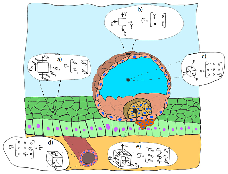
Living tissues are active multifunctional materials capable of generating, sensing, withstanding and responding to mechanical stress. These capabilities enable tissues to adopt complex shapes during development, to sustain those shapes during homeostasis, and to restore them during healing and regeneration. Abnormal stress is associated with a broad range of pathologies, including developmental defects, inflammatory diseases, tumor growth and metastasis. Here we review techniques that measure mechanical stress in living tissues with cellular and subcellular resolution. We begin with 2D techniques to map stress in cultured cell monolayers, which provide the highest resolution and accessibility. These techniques include 2D traction microscopy, micro-pillar arrays, monolayer stress microscopy, and monolayer stretching between flexible cantilevers. We next focus on 3D traction microscopy and the micro-bulge test, which enable mapping forces in tissues cultured in 3D. Finally, we review techniques to measure stress in vivo, including servo-null methods for measuring luminal pressure, deformable inclusions, FRET sensors, laser ablation and computational methods for force inference. Whereas these techniques remain far from becoming everyday tools in biomedical laboratories, their rapid development is fostering key advances in the way we understand the role of mechanics in morphogenesis, homeostasis and disease.


Figures

Box 1
Box 1. Geometric representation of the traction vector, T (red), acting at point A of a body subject to external forces (green arrows).
Box 2
Box 2. Geometric representation of the stress tensor σ at point A of a body under a load F.
Box 3
Box 3. Illustrative sketch of different representative stress states that are present during embryo implantation.
Box 5
Box 5
Illustrative representation of the parameters and variables of a vertex model: vertex v, edge λ, face κ, cell α, edge length lλ, face area Aκ, cell volume Vα, vertex applied force fiv, edge tension Λλ, surface tension Tκ and cell pressure Pα.
Figure 1
Figure 1. Techniques used to measure tractions and internal stresses in 2D tissues.
(a) In TFM 2D, a flat elastic gel is synthetized, and a tissue is allowed to attach to its surface. Cells exert tractions on the substrate and the resulting deformation can be tracked by adding fluorescent particles in or onto the substrate and comparing their position with an image of the substrate at rest. Tractions are then calculated by using different computational and analytical approaches. (b) Representative TFM 2D experiment. Phase contrast image of a flat cell monolayer on top of a polyacrylamide gel (left) together with the tractions exerted by the cells in the directions parallel (center) and perpendicular (right) to the advancing edge. (c) In the micropillar technique, cells are seeded on top of an array of micropillars, whose deflection is proportional to the locally applied force. (d) Representative micropillar experiment. Scanning electron micrograph of a micropillar array (left) with a single cell (center) and a cell monolayer (right) lying on top of it. (e) Using MSM, the internal stresses of a flat cell monolayer can be calculated from the tractions it applies on an elastic substrate. (f) Representative MSM experiment. Expanding cell monolayer with overlaid color-coded internal stresses calculated with MSM (left). Side view of an expanding monolayer (right). (g) The tensional state of a flat monolayer can be directly measured and controlled with a micromanipulator. (h) Representative suspended cell monolayer experiment. Monolayer before (center) and after (right) stretch applied with a micromanipulator. Insert: zoom in a region of a monolayer before and after stretch.
Figure 2
Figure 2. Techniques used to measure tractions and internal stresses in 3D tissues in vitro.
(a) In TFM 2.5D, a tissue is seeded on top of a 2D elastic substrate, and the displacements of the substrate are measured in 3D. From these displacements, the 3D traction field can be calculated. For simple geometries like spherical caps, the internal stresses of the tissue can be recovered with a micro-bulge test. (b) Representative TFM 2.5D experiment. 3D traction field (green arrows) generated by an epithelial dome (side view) on a flat substrate. (c) By applying TFM 3D to tissues grown inside a deformable matrix with particle tracers, the full 3D displacement field can be measured, and from it the full 3D traction field can be inferred. (d) Representative TFM 3D experiment. Breast cancer spheroid embedded in a 3D collagen I matrix. Bright field image with superimposed ECM displacements (left) and fluorescent image of the spheroid and matrix (right) (courtesy of Nadine Grummel, David Böhringer and Ben Fabry).
Figure 3
Figure 3. Techniques to measure internal stresses in vivo.
(a) Servo-null methods measure the luminal pressure of a tissue by inserting a capillary probe directly into the lumen. (b) The servo-null method applied to the luminal cavity of a mouse blastocyst before (left) and during (right) measurement. (c) Inert deformable probes can be inserted in a specimen to assess its local stress state. (d) Liquid drop inserted in a tissue, before (left) and after (center and right) deformation. (e) Fret sensors are genetically encoded molecular springs whose deformation is reported by a pair of resonant fluorophores. (f) Junctional tension reported by FRET sensors in an epithelial monolayer and in an epithelial acini. (g) In the laser ablation technique, a specimen is cut by using a pulsed laser, and its internal stress is released. (h) Circular laser cut (left) performed in a Drosophila melanogaster embryo, and asymmetric retraction (center and right) of the cut patch due to the differential internal tension along the x and y axes. (i) With force inference methods, cellular pressures and inter-cellular tensions can be inferred from the geometry of the tissue. (j) Illustration of a force inference method. Cell monolayer segmentation (left), tension and pressure location (center), and calculated values for the cellular pressure and cell-junction tension (right),.

References

    1. Guillot C, Lecuit T. Mechanics of epithelial tissue homeostasis and morphogenesis. Science. 2013;340:1185–1189. - PubMed
    1. Barker N. Adult intestinal stem cells: critical drivers of epithelial homeostasis and regeneration. Nature Reviews Molecular Cell Biology. 2014;15:19–33. - PubMed
    1. Krndija D, et al. Active cell migration is critical for steady-state epithelial turnover in the gut. Science. 2019;365:705–710. - PubMed
    1. Brugués A, et al. Forces driving epithelial wound healing. Nature Physics. 2014;10:683–690. doi: 10.1038/nphys3040. - DOI - PMC - PubMed
    1. Northey JJ, Przybyla L, Weaver VM. Tissue Force Programs Cell Fate and Tumor Aggression. Cancer Discovery. 2017;7:1224–1237. doi: 10.1158/2159-8290.CD-16-0733. - DOI - PMC - PubMed

LinkOut - more resources