Review
. 2025 Jan 10:15:1534823.
doi: 10.3389/fimmu.2024.1534823. eCollection 2024.

The role of M1/M2 macrophage polarization in the pathogenesis of obesity-related kidney disease and related pathologies

Affiliations
Review

The role of M1/M2 macrophage polarization in the pathogenesis of obesity-related kidney disease and related pathologies

Periklis Dousdampanis et al. Front Immunol. .

Abstract

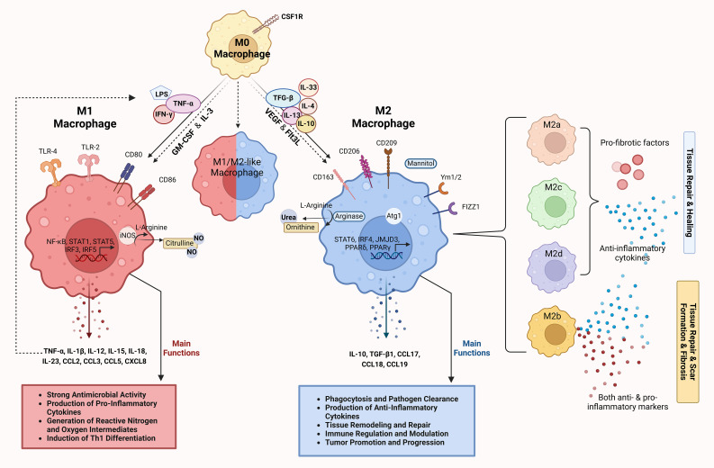
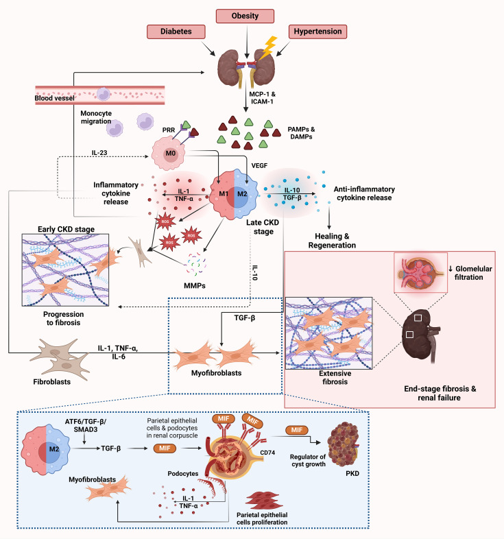
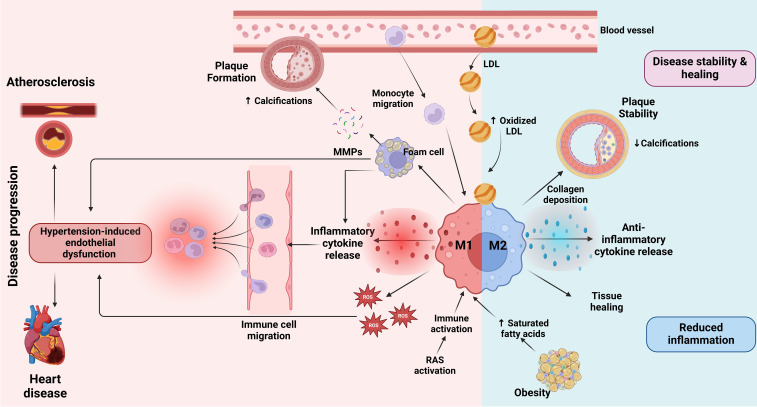
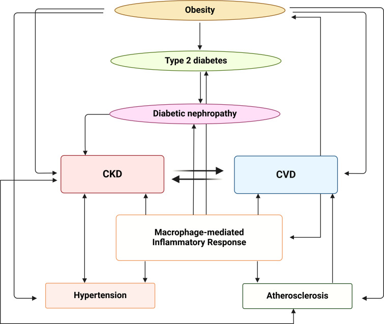
Obesity is a rapidly growing health problem worldwide, affecting both adults and children and increasing the risk of chronic diseases such as type 2 diabetes, hypertension and cardiovascular disease (CVD). In addition, obesity is closely linked to chronic kidney disease (CKD) by either exacerbating diabetic complications or directly causing kidney damage. Obesity-related CKD is characterized by proteinuria, lipid accumulation, fibrosis and glomerulosclerosis, which can gradually impair kidney function. Among the immune cells of the innate and adaptive immune response involved in the pathogenesis of obesity-related diseases, macrophages play a crucial role in the inflammation associated with CKD. In obese individuals, macrophages enter a pro-inflammatory state known as M1 polarization, which contributes to chronic inflammation. This polarization promotes tissue damage, inflammation and fibrosis, leading to progressive loss of kidney function. In addition, macrophage-induced oxidative stress is a key feature of CKD as it also promotes cell damage and inflammation. Macrophages also contribute to insulin resistance in type 2 diabetes by releasing inflammatory molecules that impair glucose metabolism, complicating the management of diabetes in obese patients. Hypertension and atherosclerosis, which are often associated with obesity, also contribute to the progression of CKD via immune and inflammatory pathways. Macrophages influence blood pressure regulation and contribute to vascular inflammation, particularly via the renin-angiotensin system. In atherosclerosis, macrophages accumulate in arterial plaques, leading to chronic inflammation and plaque instability, which may increase the risk of CVD in CKD patients. This review focuses on the involvement of macrophages in CKD and highlights their role as a critical link between CKD and other pathologies. Targeting macrophage polarization and the ensuing macrophage-induced inflammation could be an effective therapeutic strategy for CKD and related diseases and improve outcomes for patients with obesity-related kidney disease.

Keywords: M1; M2; cardiovascular disease; chronic kidney disease; macrophages; obesity.


Conflict of interest statement

The authors declare that the research was conducted in the absence of any commercial or financial relationships that could be construed as a potential conflict of interest. The author(s) declared that they were an editorial board member of Frontiers, at the time of submission. This had no impact on the peer review process and the final decision.

Figures

Figure 1
Figure 1
Differentiation and functions of macrophages along the M1/M2 polarization axis. M0 macrophages represent the undifferentiated or resting state of macrophages. Under the influence of CSF1R, M0 macrophages can differentiate into either M1 or M2 macrophages depending on the cytokine environment and the signaling molecules they encounter. M1 macrophages, induced by stimuli such as LPS and pro-inflammatory cytokines (IFN-γ and TNF-α), express surface markers such as CD80 and CD86 (24). Key signaling pathways involved include TLR-4, NF-κB, STAT1, STAT5 and IRFs, which control the expression of inflammatory genes (25). M1 macrophages produce high levels of pro-inflammatory cytokines (TNF-α, IL-1β, IL-12, IL-15, IL-18, IL-23) and chemokines (CCL2, CCL3, CCL5, CXCL8) (, –31). Through iNOS activity, they generate reactive nitrogen and oxygen species that have a strong antimicrobial effect and promote the differentiation of Th1 cells (28, 32). The main functions of M1 macrophages include pathogen clearance, production of pro-inflammatory cytokines and the generation of reactive oxygen/nitrogen species to fight infections (13, 26, 27, 33, 34). M2 macrophages induced by anti-inflammatory cytokines (IL-4, IL-10, IL-13, IL-33, TGF-β) are involved in tissue repair and immunomodulation. They express surface markers such as CD163, CD206, CD209, Ym1/2 and Fizz1, which are involved in pathways such as STAT6, IRF4, JMJD3, PPARγ and PPARδ, and promote anti-inflammatory and tissue-remodeling functions (, –28). Mannitol serves as a signaling molecule that can enhance M2 macrophage activation and function. M2 macrophages produce anti-inflammatory cytokines and chemokines (IL-10, TGF-β1, CCL17, CCL18, CCL19) and promote tissue repair through arginase activity, which converts L-arginine to urea and ornithine (–31, 35, 36). The main functions of M2 macrophages include phagocytosis, pathogen clearance, tissue remodeling and immune regulation (13, 26, 27, 33, 34). The intermediate or hybrid state between M1 and M2 (M1/M2-like macrophage) represents macrophages with mixed functional properties. These macrophages exhibit features of both M1 and M2 polarization and play a complex role in immune regulation and tissue response. M2 macrophages are further divided into subtypes (M2a, M2b, M2c and M2d), each associated with distinct cytokine profiles and roles in tissue remodeling, as indicated by different colors (26, 35, 37). The dashed arrows show the reversible transitions between the polarization states and illustrate the plasticity of the macrophages. The image was created with Biorender (https://biorender.com).
Figure 2
Figure 2
Role of macrophages in CKD. CKD is triggered by various conditions such as diabetes, obesity and hypertension, leading to renal injury. This initial damage results in the release of DAMPs and PAMPs from injured kidney cells (–67, 79). These molecules activate endothelial cells to release chemokines such as MCP-1 and ICAM-1, which facilitate the recruitment and migration of monocytes from the bloodstream into the kidney, where they differentiate into M0 macrophages (30). PRRs on M0 macrophages recognize DAMPs and PAMPs and initiate further macrophage activation and polarization (–81). After activation by PRRs, macrophages polarize towards the M1 phenotype in the early stages of CKD. M1 macrophages release inflammatory cytokines such as IL-1 and TNF-α together with ROS and MMPs (–70). These molecules exacerbate kidney injury by promoting inflammation and further damaging renal tissue. This release of inflammatory cytokines creates a feedback loop that recruits more immune cells and perpetuates the inflammatory environment in the kidney, as depicted by the arrows in the inflammatory cytokine release cycle. Chronic inflammation due to M1 macrophage activity and the persistent presence of ROS and MMPs leads to fibroblast activation (64, 65). Activated fibroblasts differentiate into myofibroblasts and produce excessive amounts of extracellular matrix proteins such as collagen, leading to tissue scarring (64, 65). As CKD progresses, there is a shift toward the M2 macrophage phenotype to resolve inflammation and promote tissue healing (72). M2 macrophages release anti-inflammatory cytokines, primarily IL-10 and TGF-β, which suppress inflammation and facilitate healing (71, 73). However, despite their role in healing, M2 macrophages also contribute to fibrosis by promoting collagen deposition through the release of VEGF and EGF, which enhances tissue regeneration but can lead to excessive fibrosis (71, 73). The dashed lines highlight the cycle of inflammation and fibrosis driven by IL-10 and IL-23, which perpetuates macrophage polarization and cytokine release (76). IL-23 enhances the M1 macrophage response, while IL-10 in the M2 response can lead to prolonged inflammation and fibrosis, creating a chronic inflammatory environment (76). The ATF6/TGF-β/SMAD3 pathway in M2 macrophages leads to activation of MIF, which plays a central role in renal fibrosis (66, 77). MIF acts via CD74, a receptor expressed on podocytes and parietal epithelial cells in the renal corpuscle (85). The interaction between MIF and CD74 stimulates the production of inflammatory cytokines in podocytes, including IL-1 and TNF-α, and triggers proliferation of parietal epithelial cells (85). This contributes to inflammation, fibrosis, and kidney damage. In the context of polycystic kidney disease (PKD), MIF also acts as a regulator of cyst growth, which further exacerbates disease progression (86). End-stage CKD is characterized by extensive fibrosis and scarring of renal tissue, leading to decreased glomerular filtration rate and loss of kidney function (64). The inflammatory cytokines IL-1, TNF-α and IL-6 contribute to the activation of fibroblasts and differentiation into myofibroblasts and promote the progression of fibrosis (66, 77). Endothelin and TGF-β also play a role in fibroblast activation and collagen deposition, which further drives fibrosis (69). Markers of fibrosis are associated with activated macrophages and myofibroblasts in the fibrotic kidney. This eventually leads to end-stage renal disease, depicted in the figure by the extensive scarring and fibrosis in the kidney tissue. The image was created with Biorender (https://biorender.com).
Figure 3
Figure 3
Role of macrophages in CVD. At the onset of atherosclerosis, LDL particles accumulate in the blood vessel walls, where they undergo oxidation to form oxidized LDL (163, 164). This triggers monocyte migration into the vessel wall, where they differentiate into macrophages. Upon activation by inflammatory stimuli, macrophages transition into the M1 state, releasing inflammatory cytokines and ROS which drive further vascular inflammation (89). Additionally, RAS activation contributes to immune activation and promotes an inflammatory response that exacerbates the progression of atherosclerosis (–99). This inflammation is further amplified by hypertension-induced endothelial dysfunction, which facilitates immune cell migration into the vessel wall and promotes plaque instability (56). Factors such as obesity and saturated fatty acid accumulation may influence macrophage polarization towards M1, contributing to a more unstable plaque environment (138). M1 macrophages contribute to plaque formation by ingesting oxidized LDL and transforming into foam cells that promote the release of matrix MMPs (159). MMPs degrade extracellular matrix components, weakening the plaque and promoting calcifications within it, which can lead to plaque rupture (160, 161). The presence of M1 macrophages is therefore associated with increased inflammation, plaque vulnerability and progression of CVD, which may ultimately lead to heart disease (167, 168). Conversely, M2 macrophages, which are activated in response to anti-inflammatory signals, play a role in disease stability and healing (169). M2 macrophages secrete anti-inflammatory cytokines that attenuate inflammation, promote collagen deposition and stabilize the plaque by reducing calcifications (169). These macrophages support tissue healing and contribute to reduced inflammation in the vessel wall. The balance between M1 and M2 macrophage activation determines the overall inflammatory state of the atherosclerotic plaque and its stability, thus influencing the progression or attenuation of cardiovascular disease. The image was created with Biorender (https://biorender.com).
Figure 4
Figure 4
Interplay between metabolic, inflammatory and vascular pathways in the progression of CKD and CVD. Obesity contributes to the development of type 2 diabetes, which can progress to diabetic nephropathy. Diabetic nephropathy, in turn, is associated with both CKD and CVD that have a bidirectional relationship, with each condition exacerbating the other. The macrophage-mediated inflammatory response plays a central role in this network linking CKD, CVD and atherosclerosis, contributing to the progression of both CKD and hypertension. The image was created with Biorender (https://biorender.com).

Similar articles

Cited by

References

    1. Li H, Meng Y, He S, Tan X, Zhang Y, Zhang X, et al. . Macrophages, chronic inflammation, and insulin resistance. Cells. (2022) 11:3001. doi: 10.3390/cells11193001 - DOI - PMC - PubMed
    1. Wang Y, Chen X, Song Y, Caballero B, Cheskin LJ. Association between obesity and kidney disease: a systematic review and meta-analysis. Kidney Int. (2008) 73:19–33. doi: 10.1038/sj.ki.5002586 - DOI - PubMed
    1. Arabi T, Shafqat A, Sabbah BN, Fawzy NA, Shah H, Abdulkader H, et al. . Obesity-related kidney disease: Beyond hypertension and insulin-resistance. Front Endocrinol (Lausanne). (2023) 13:1095211. doi: 10.3389/fendo.2022.1095211 - DOI - PMC - PubMed
    1. Nawaz S, Chinnadurai R, Al-Chalabi S, Evans P, Kalra PA, Syed AA, et al. . Obesity and chronic kidney disease: A current review. Obes Sci Pract. (2022) 9:61–74. doi: 10.1002/osp4.629 - DOI - PMC - PubMed
    1. Kotsis V, Martinez F, Trakatelli C, Redon J. Impact of obesity in kidney diseases. Nutrients. (2021) 13:4482. doi: 10.3390/nu13124482 - DOI - PMC - PubMed

MeSH terms

LinkOut - more resources