. 2025 Jan 10:15:1450887.
doi: 10.3389/fimmu.2024.1450887. eCollection 2024.

NLRP3 deficiency aggravated DNFB-induced chronic itch by enhancing type 2 immunity IL-4/TSLP-TRPA1 axis in mice

Affiliations

NLRP3 deficiency aggravated DNFB-induced chronic itch by enhancing type 2 immunity IL-4/TSLP-TRPA1 axis in mice

Si-Ting Huang et al. Front Immunol. .

Abstract

Background: The nod-like receptor family pyrin domain-containing 3 (NLRP3) has been implicated in various skin diseases. However, its role in mediating 2, 4-dinitrofluorobenzene (DNFB)-induced chronic itch remains unclear.

Methods: Widetype (WT) and Nlrp3 deletion (Nlrp3-/- )mice, the expression of transient receptor potential (TRP) ankyrin 1 (TRPA1) inhibitor or recombinant mice interleukin-18 (IL-18) were used to establish and evaluate the severity of DNFB-mediated chronic itch. Quantitative real-time PCR, western blotting, immunohistochemistry staining, immunofluorescence staining and enzyme-linked immunosorbent assay (ELISA) was used to examine the expression of NLRP3 inflammasome, type 2 immunity and receptors in dorsal root ganglion (DRG) neurons related with chronic itch. Flow cytometry was performed to quantify the frequency of type 2 immune cells.

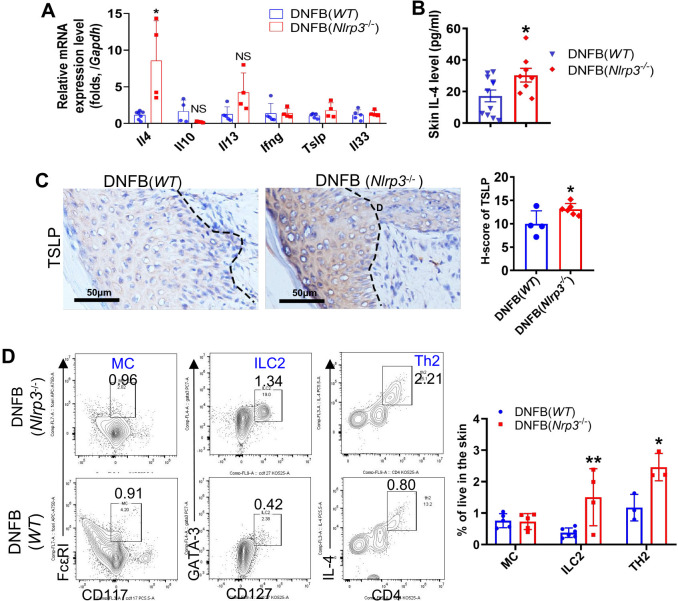
Results: This study revealed that the NLRP3 inflammasome was activated in the skin of DNFB-induced chronic itch mice. Surprisingly, the absence of Nlrp3 exacerbated itch behavior. In Nlrp3-/- mice, IL-18 expression was downregulated, whereas markers of type 2 immunity, such as IL-4 and thymic stromal lymphopoietin (TSLP), were significantly upregulated in the skin. Furthermore, TRPA1 and its colocalization with the IL-4 receptor were increased in the DRG. Inhibition of TRPA1 or administration of recombinant IL-18 significantly reduced DNFB-induced itch behavior in Nlrp3-/- mice. Recombinant IL-18 also decreased the expression of TRPA1, IL-4, and TSLP.

Discussion: These findings suggested that the absence of Nlrp3 aggravated DNFB-induced chronic itch by exacerbating type 2 immunity in the skin and enhancing the IL-4/TSLP-TRPA1 axis, potentially driven by reduced IL-18 levels.

Keywords: DNFB-induced chronic itch; IL-18; Nlrp3 inflammasome; TRPA1; type 2 immunity.


Conflict of interest statement

The authors declare that the research was conducted in the absence of any commercial or financial relationships that could be construed as a potential conflict of interest.

Figures

Figure 1
Figure 1
NLRP3 inflammasome was activated in the skin of mice with DNFB-induced chronic itch. (A) Flowchart illustrating the establishment of the DNFB-induced chronic itch model. (B) The number of scratches within 1 hr was video recorded in normal controls (group NC) and mice with DNFB-induced chronic itch (group DNFB). (C) Relative RNA expression levels of Nlrp3, Il1b, Casp1, Casp11, and Il18 in the skin of indicated mice were detected by RT-qPCR. (D) IL-1β concentration in the skin lysate was analyzed by ELISA. (E) Protein expression levels of NLRP3, IL-18, and ASC (both oligomer and monomer forms) were detected by Western blotting (WB). Representative WB images are shown on the left, and semi-quantitative analysis is presented on the right. (F) Expression of NLRP3 and ASC in skin sections from mice with or without DNFB challenge was visualized by IF staining. Representative IF photographs (400× magnification) are shown on the left, and the percentage of NLRP3 or ASC single positive cells is shown on the right. For each experiment, the sample size was n=5 (NC group) or 5-7 (DNFB), and all experiments were conducted in duplicate. Quantitative data are shown as mean ± SEM. Statistical significance was determined using an unpaired t-test, with *P<0.05, **P<0.01, ***P<0.001, and ****P<0.0001.
Figure 2
Figure 2
Nlrp3 genetic deletion exacerbated DNFB-induced chronic itch. (A) The number of scratching bouts within 1 hour was recorded via video monitoring in WT and Nlrp3-/- mice subjected to DNFB-induced chronic itch. (B) Serum IgE concentrations in WT mice or Nlrp3-/- mice with DNFB-induced chronic itch were detected using an IgE ELISA kit. (C–E) Epidermal thickness and inflammatory cell infiltration in the dermis of the indicated mice were analyzed through H&E staining. (F, G) Avidin staining (representative photographs shown in (F)) and quantitative analysis (G) of avidin-positive cells or the ratio of positive cells to total cells in 200× magnification fields of skin sections were performed. The number of mice in the NC group was 5, and the DNFB (WT) or DNFB (Nlrp3 -/-) groups each included eight mice, with experiments repeated twice. All quantitative data are shown as mean ± SEM. *P<0.05, **P<0.01, ***P<0.001, ****P<0.0001. Two-way ANOVA was used for (A), t-test was used for (B), and one-way ANOVA was applied for (C, E, G).
Figure 3
Figure 3
Nlrp3 deletion diminished IL-18 but not IL-1β in DNFB-induced mice. (A) The protein expression levels of NLRP3 and IL-18 in the skin lysates of DNFB-challenged WT and Nlrp3-/- mice were detected by WB. The representative bands are shown on the left, and the semi-quantitative analysis is presented on the right. (B) Representative photographs of IL-18 positive IF staining in mice skin sections (400×) ware are shown on the left, demonstrating a significant decrease in IL-18 positive cells due to Nlrp3 deletion. (C) The IL-1β concentration in skin lysates of the indicated mice was analyzed by ELISA. (D) Representative photographs of IL-1β (green) or Ly6G (red) single positive and their merged (yellow) IF staining in mice skin sections are shown on the left (400×). Quantification of IL-1β, Ly6G single-positive, and double-positive cells per field (100×) is shown on the right. All quantitative data are presented as mean ± SEM. **P<0.01, unpaired t-test.
Figure 4
Figure 4
Knockout of Nlrp3 enhanced ILC2, Th2 cells and type 2 immunity in the DNFB-induced chronic itch mouse skin. (A) The relative mRNA expression levels of Il4, Il10, Il13, Ifnγ, Tslp, and Il33 in the skin of the indicated mice were detected by RT-qPCR. (B) The IL-4 concentration in the neck skin lysates of the indicated mice was analyzed by ELISA. (C) The expression level of TSLP in neck skin was detected by IHC. Representative photographs are shown on the left, with semi-quantitative analysis shown as H-scores on the right. (D) Frequencies of the indicated cell types isolated from the neck skin of WT mice or Nlrp3 -/- mice with DNFB were analyzed by flow cytometry and FlowJo software. Representative gating plots are shown on the left and their quantifications are displayed on the right. The sample size for the DNFB (WT) or DNFB (Nlrp3 -/-) groups was eight. All quantitative data are shown as mean ± SEM. Statistical significance was determined by unpaired t-test: *P<0.05, **P<0.01.
Figure 5
Figure 5
TRPA1 neurons were involved in severe DNFB-induced chronic itch of Nlrp3-/- mice. (A) RNA expression levels of receptors related to chronic itch in the DRG were analyzed by RT-qPCR. (B) RNA expression levels of Trpa1 and Trpv1 in the DRG of DNFB-challenged mice. (C) IF staining of TRPA1 and TRPV1 in the DRG. Representative photographs are shown on the left, and the quantitative analyses are presented on the right, illustrating the TRPA1 and TRPV1 positive ratios per 100 cells in the DRG sections of mice. (D) IF staining of TRPA1 and IL-4R in the DRG. Representative photographs are shown on the left, and the quantitative analyses are shown on the right, illustrating the average count of TRPA1 and IL-4R double-positive cells per field (400×) in the DRG sections of mice. The sample size in the NC group was five and in DNFB (WT) or DNFB (Nlrp3 -/-) group, it was eight. All quantitative data are shown as mean ± SEM. Statistical significance is indicated as follows: *P<0.05, **P<0.01, ***P<0.001. One-way ANOVA was used for the analyses in (C, D), while an unpaired t-test was applied in (A, B).
Figure 6
Figure 6
IL-18 was associated with the severity of DNFB-induced chronic itch via regulating type 2 immunity and TRPA1 in Nlrp3 -/- mice. (A) Scratching frequency of DNFB-challenged Nlrp3 -/- mice with or without rmIL-18 treatment. (B) Scratching frequency in DNFB-challenged Nlrp3 -/- mice treated with or without TRPA1 antagonist (HC030031). (C) RNA expression levels of Trpa1 in the DRG were analyzed by RT-qPCR. (D) TSLP expression in the neck skin of DNFB-challenged Nlrp3 -/- mice, with or without rmIL-18 treatment, was detected by IHC. Representative photographs are shown on the left, with semi-quantitative analysis (H-score) on the right. (E) IL-4 concentrations in the neck skin lysate of DNFB-challenged Nlrp3 -/- mice, with or without rmIL-18 treatment, were measured by ELISA. All quantitative data are presented as mean ± SEM. Statistical significance is indicated as follows: *P<0.05, **P<0.01, ***P<0.001, ****P<0.0001. A two-way ANOVA was used for (A), and an unpaired t-test was applied for (B–E).

Similar articles

Cited by

References

    1. Wang F, Kim BS. Itch: A paradigm of neuroimmune crosstalk. Immunity. (2020) 52:753–66. doi: 10.1016/j.immuni.2020.04.008 - DOI - PubMed
    1. Agelopoulos K, Pereira MP, Wiegmann H, Ständer S. Cutaneous neuroimmune crosstalk in pruritus. Trends Mol Med. (2022) 28:452–62. doi: 10.1016/j.molmed.2022.03.005 - DOI - PubMed
    1. Gause WC, Rothlin C, Loke P. Heterogeneity in the initiation, development and function of type 2 immunity. Nat Rev Immunol. (2020) 20:603–14. doi: 10.1038/s41577-020-0301-x - DOI - PMC - PubMed
    1. Pirzgalska RM, Veiga-Fernandes H. Type 2 neuroimmune circuits in the shaping of physiology. Immunity. (2023) 56:695–703. doi: 10.1016/j.immuni.2023.03.016 - DOI - PubMed
    1. Oetjen LK, Mack MR, Feng J, Whelan TM, Niu H, Guo CJ, et al. . Sensory neurons co-opt classical immune signaling pathways to mediate chronic itch. Cell. (2017) 171:217–228.e213. doi: 10.1016/j.cell.2017.08.006 - DOI - PMC - PubMed

MeSH terms