. 2024 Dec 20;28(1):111638.
doi: 10.1016/j.isci.2024.111638. eCollection 2025 Jan 17.

Deep learning uncovers histological patterns of YAP1/TEAD activity related to disease aggressiveness in cancer patients

Affiliations

Deep learning uncovers histological patterns of YAP1/TEAD activity related to disease aggressiveness in cancer patients

Benoit Schmauch et al. iScience. .

Abstract

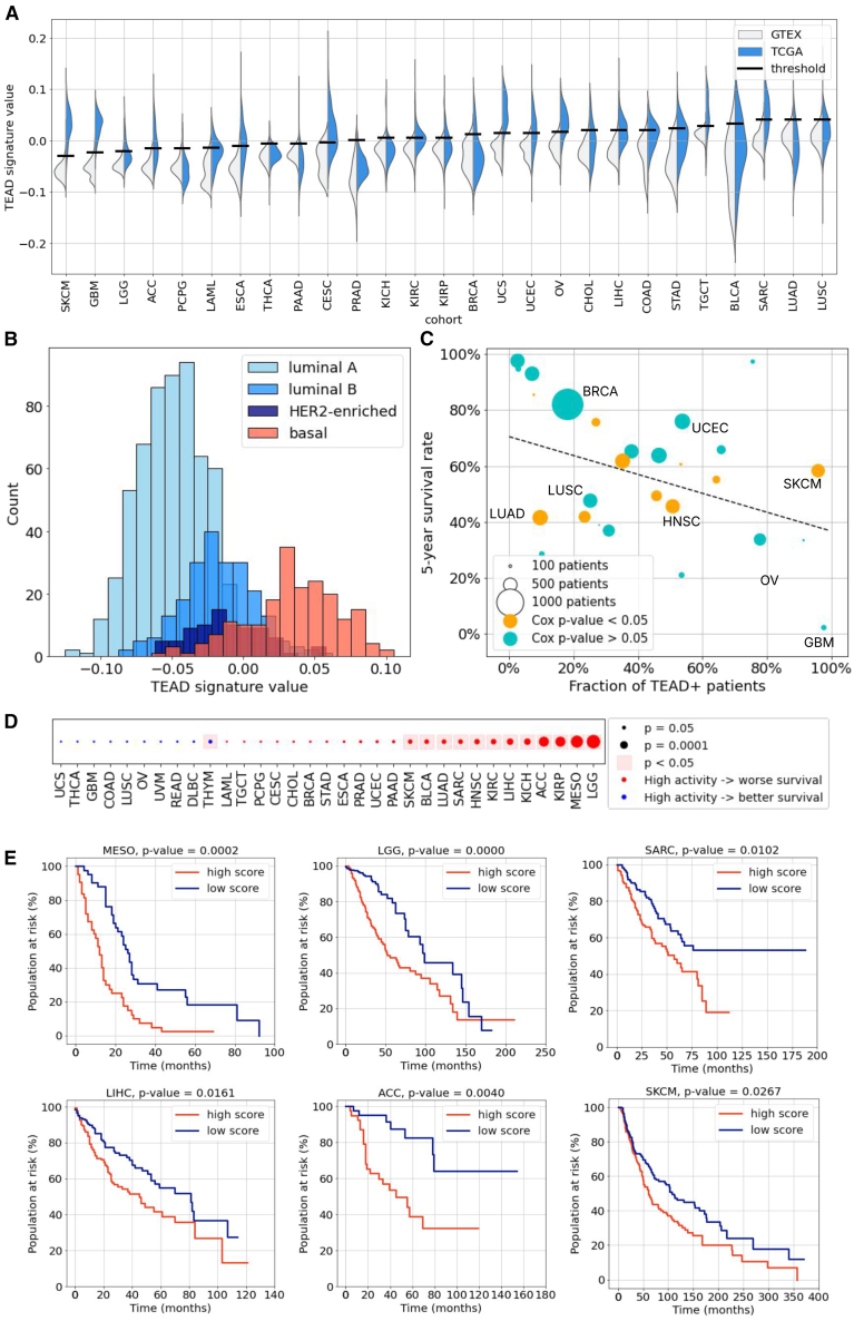
Over the last decade, Hippo signaling has emerged as a major tumor-suppressing pathway. Its dysregulation is associated with abnormal expression of YAP1 and TEAD-family genes. Recent works have highlighted the role of YAP1/TEAD activity in several cancers and its potential therapeutic implications. Therefore, identifying patients with a dysregulated Hippo pathway is key to enhancing treatment impact. Although recent studies have derived RNA-seq-based signatures, there remains a need for a reproducible and cost-effective method to measure the pathway activation. In recent years, deep learning applied to histology slides have emerged as an effective way to predict molecular information from a data modality available in clinical routine. Here, we trained models to predict YAP1/TEAD activity from H&E-stained histology slides in multiple cancers. The robustness of our approach was assessed in seven independent validation cohorts. Finally, we showed that histological markers of disease aggressiveness were associated with dysfunctional Hippo signaling.

Keywords: Applied sciences; Health sciences; Machine learning.


Conflict of interest statement

The authors declare the following competing interests. B.S., V.C., O.D.D, J.E.L.D., A.H., R.B., C.M., A.R., E.D., K.V.L. and E.P. are employed by Owkin, Inc.; E.R., I.V., L.D., E.D.R., F.N., E.S., V.F., M.Cl. and M.Ce. are employed by Sanofi. H.P. received research funds from Owkin for RNA extraction and demographic data. M.E.: Personal fees, travel costs and speaker's honoraria from MSD, AstraZeneca, Janssen-Cilag, Cepheid, Roche, Astellas, Diaceutics, Owkin, BMS; research funding from AstraZeneca, Janssen-Cilag, STRATIFYER, Cepheid, Roche, Gilead, Owkin; advisory role for Diaceutics, MSD, AstraZeneca, Janssen-Cilag, GenomicHealth, Owkin, BMS. A.S.: Honoraria for advisory boards from Amgen, AstraZeneca, Boehringer Ingelheim, Ipsen, Janssen, Lilly, MSD, Pfizer, Roche, Takeda; consulting: AstraZeneca, BMS, Daiichi Sankyo, Janssen, Roche; symposiums: Amgen, AstraZeneca, BMS, Janssen, Pfizer, Sanofi, Takeda; congress: Janssen, Pfizer, Takeda. S.L.: Honoraria for advisory boards from Janssen, MSD, Sanofi, Abbvie; symposiums: MSD, Janssen. N.G.: Honoraria and/or consulting fees from Abbvie, Amgen, AstraZeneca, BMS, Boehringer-Ingelheim, Daiichi Sankyo, Janssen, Lilly, Mirati, MSD, Novartis, Pfizer, Roche, Sanofi, Takeda, and received grants from MSD and AstraZeneca paid to institution outside of the present work. E.S. holds a patent for the TEAD500 signature (WO2023180385A1).

Figures

None
Graphical abstract
Figure 1
Figure 1
Workflow of the study (A) RNA-seq and clinical data from 32 cancer types from The Cancer Genome Atlas (TCGA) were collected. TEAD-500 signature was computed from RNA-seq data. General statistics and association with prognosis were evaluated. (B) Models were trained to predict the signature from Hematoxylin & Eosin (H&E)-stained histology slides. Cross-validation was run on TCGA. (C) Models trained on specific cancer types were transferred to other indications to assess the presence of common histological patterns associated with YAP1/TEAD activity. (D) The best models were further validated on seven external cohorts of non-small cell lung cancer (NSCLC), mesothelioma and head and neck squamous cell carcinoma (HNSC), demonstrating the robustness of our approach. (E) The most predictive tiles were extracted from the model and reviewed by trained pathologists to highlight histological biomarkers associated with YAP1/TEAD activity.
Figure 2
Figure 2
YAP1/TEAD activity is associated with poor prognosis across cancer (A) Distribution of TEAD-500 signature values on TCGA cohorts and on matched healthy tissues from the GTEx database. Bars indicate the 95th percentile value on healthy tissue. (B) Distribution of TEAD-500 signature values in subtypes of breast cancer (BRCA). (C) Correlation between the fraction of patients with a signature value above the GTEx-inferred threshold, and overall survival (OS). In orange, cohorts where the Cox p-value of the signature is significant. To compute this p-value, a univariate Cox model was fitted on the TEAD-500 signature to predict overall survival, and a chi-squared test was used to compute p-values. (D) Summary of the association of the TEAD-500 signature with patient overall survival in different cancer types. Red dots indicate negative associations (higher values associated with a worse prognosis), blue dots indicate positive association. p-values defined as in panel c. (E) Kaplan-Meier curves of OS per cohort, stratified with respect to the GTEx threshold (high score: activity above threshold, low score: activity below threshold). For mesothelioma, since no GTEx data could be matched with cancer data, the threshold is the median value. p-values were obtained with a log-rank test and corrected with Benjamini-Hochberg method to account for multiple-hypothesis testing.
Figure 3
Figure 3
Deep learning can predict YAP1/TEAD activity from histology slides (A) Boxplot of the Pearson correlation values obtained in cross-validation on TCGA cohorts (box: interquartile range (IQR); horizontal line: median; whiskers: 1.5 times IQR, triangle: mean; circles: individual fold values). (B) Correlation between the prognostic power of the RNA-seq signature and that of the histology-based predictions. p-value was computed with a 2-sided t-test. (C) Table of the Pearson correlation obtained by applying a model trained on a given indication to any other indication. Diagonal values are the averages obtained in cross-validation. For readability, negative values were clipped to 0. The blue square indicates indications that were grouped for training the final NSCLC model. (D) Boxplot (defined as in panel a.) of the Pearson correlation values obtained in cross-validation on LUSC, UCEC, ACC, BRCA and LUAD, with models trained individually on each cancer (blue) or with a single model trained simultaneously on the five cohorts (red). p-values were obtained from a Z-test, as described in the STAR Methods section (n.s. nonsignificant, ∗ p-value <0.05, ∗∗ p-value <0.01, ∗∗∗ p-value <0.001).
Figure 4
Figure 4
The HE2TEAD model transfers robustly to external validation cohorts (A) RNA-seq signature values versus histology-based prediction on external NSCLC validation cohorts. Predictions were obtained from a model trained on advanced-stage patients (AJCC stage IV) from five TCGA cohorts: LUAD, LUSC, ACC, UCEC and BRCA. 2-sided p-values, computed with a t-test, are reported. The scale is different on both axes because the model was trained to predict the normalized signature value (zero mean and unit standard deviation). (B) Same as a, on external mesothelioma cohorts from Stanford and NYU. The model was trained on TCGA-MESO and TCGA-LUAD. (C) Id., on the CPTAC-HNSCC cohort (head and neck squamous cell carcinoma). The model was trained on 7 TCGA cohorts: HNSC, SKCM, BLCA, ACC, BRCA, LUAD, LUSC, UCEC.
Figure 5
Figure 5
HE2TEAD identifies areas associated with a high YAP1/TEAD activity (A) Heatmap of the model scores on an NSCLC sample from TCGA. Red indicates regions associated with a high YAP1/TEAD activity value, while blue indicates areas associated with a low activity. Scale bar: 5mm. (B) Top positively predictive (red square) and negatively predictive tiles (blue square) from the TCGA-NSCLC cohort (advanced-stage patients). Scale bar: 50μm. (C) Heatmap of the model scores on an HNSC sample from TCGA. Scale bar: 5mm. (D) Top positively predictive and negatively predictive tiles from the TCGA-HNSC cohort. Scale bar: 50μm.
Figure 6
Figure 6
YAP1/TEAD activity is associated with necrosis and inflammation Distribution of some of the histological patterns in positively and negatively predictive tiles from seven cohorts, as well as in randomly chosen tiles for comparison. p-values were obtained with a chi-square test of contingency between positive and negative tiles, and corrected for multiple-hypothesis testing with the Benjamini-Hochberg method. No tiles were annotated with normal epithelium in the mesothelioma cohorts, and similarly for fibrosis in the head and neck squamous cell carcinoma cohorts. p-values were obtained from a chi-squared test of contingency, as described in the STAR Methods section (n.s. nonsignificant, ∗ p-value <0.05, ∗∗ p-value <0.01, ∗∗∗ p-value <0.001, ∗∗∗∗ p-value <0.0001).

References

    1. Fu M., Hu Y., Lan T., Guan K.L., Luo T., Luo M. The Hippo signalling pathway and its implications in human health and diseases. Signal Transduct. Targeted Ther. 2022;7:376. - PMC - PubMed
    1. Calses P.C., Crawford J.J., Lill J.R., Dey A. Hippo pathway in cancer: aberrant regulation and therapeutic opportunities. Trends Cancer. 2019;5:297–307. - PubMed
    1. Ortega Á., Vera I., Diaz M.P., Navarro C., Rojas M., Torres W., Parra H., Salazar J., De Sanctis J.B., Bermúdez V., Bermúdez V. The YAP/TAZ signaling pathway in the tumor microenvironment and carcinogenesis: Current knowledge and therapeutic promises. Int. J. Mol. Sci. 2021;23:430. - PMC - PubMed
    1. Cunningham R., Hansen C.G. The Hippo pathway in cancer: YAP/TAZ and TEAD as therapeutic targets in cancer. Clin. Sci. 2022;136:197–222. - PMC - PubMed
    1. Bae S.J., Luo X. Activation mechanisms of the Hippo kinase signaling cascade. Biosci. Rep. 2018;38 - PMC - PubMed

LinkOut - more resources