[Preprint]. 2025 Jan 17:2025.01.13.632808.
doi: 10.1101/2025.01.13.632808.

Defining the Polycystin Pharmacophore Through HTS & Computational Biophysics

Affiliations

Defining the Polycystin Pharmacophore Through HTS & Computational Biophysics

Eduardo Guadarrama et al. bioRxiv. .

Update in

Abstract

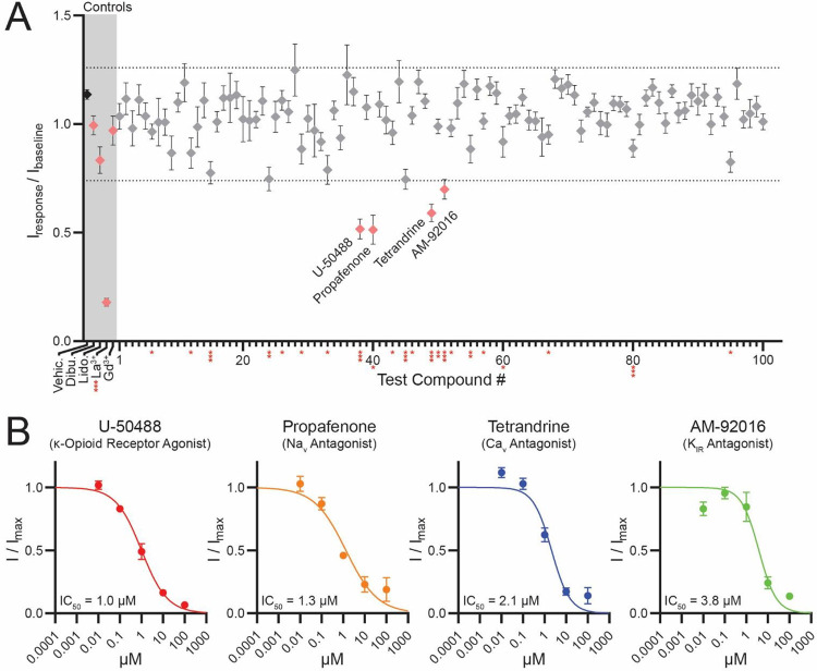
Background and purpose: Polycystins (PKD2, PKD2L1) are voltage-gated and Ca2+-modulated members of the transient receptor potential (TRP) family of ion channels. Loss of PKD2L1 expression results in seizure-susceptibility and autism-like features in mice, whereas variants in PKD2 cause autosomal dominant polycystic kidney disease. Despite decades of evidence clearly linking their dysfunction to human disease and demonstrating their physiological importance in the brain and kidneys, the polycystin pharmacophore remains undefined. Contributing to this knowledge gap is their resistance to drug screening campaigns, which are hindered by these channels' unique subcellular trafficking to organelles such as the primary cilium. PKD2L1 is the only member of the polycystin family to form constitutively active ion channels on the plasma membrane when overexpressed.

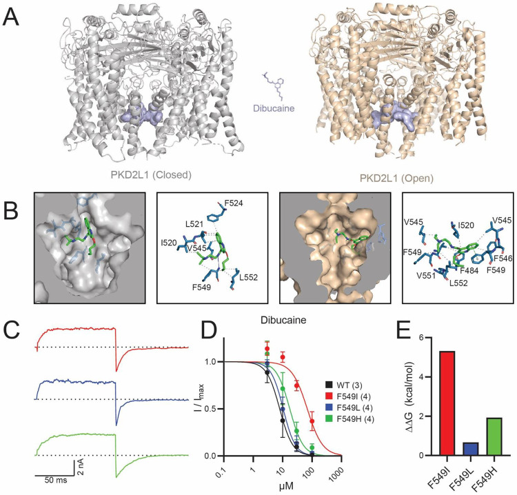
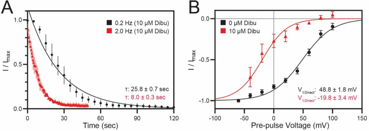
Experimental approach: HEK293 cells stably expressing PKD2L1 F514A were pharmacologically screened via high-throughput electrophysiology to identify potent polycystin channel modulators. In-silico docking analysis and mutagenesis were used to define the receptor sites of screen hits. Inhibition by membrane-impermeable QX-314 was used to evaluate PKD2L1's binding site accessibility.

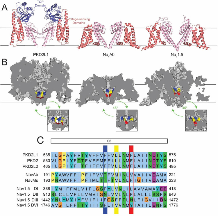
Key results: Screen results identify potent PKD2L1 antagonists with divergent chemical core structures and highlight striking similarities between the molecular pharmacology of PKD2L1 and voltage-gated sodium channels. Docking analysis, channel mutagenesis, and physiological recordings identify an open-state accessible lateral fenestration receptor within the pore, and a mechanism of inhibition that stabilizes the PKD2L1 inactivated state.

Conclusion and implication: Outcomes establish the suitability of our approach to expand our chemical knowledge of polycystins and delineates novel receptor moieties for the development of channel-specific antagonists in TRP channel research.

Keywords: ADPKD; Computational Biophysics; High-Throughput Electrophysiology; Molecular Docking; PKD2; PKD2L1; Pharmacology; TRP channels; autosomal polycystic kidney disease; ion channels; molecular mechanisms; polycystin; primary cilia.


Conflict of interest statement

Conflict of Interest Statement: The authors declare that they have no conflict of interest

Figures

Figure 1.
Figure 1.. High-throughput drug screen identifies four potent PKD2L1 antagonists.
(A) Mean post-treatment F514A tail currents, normalized to pre-treatment amplitude. Asterisks denote compounds with a statistically significant change in amplitude relative to currents from vehicle treated, cells (*p < 0.05, **p < 0.005, ***p < 0.0005, unpaired t-test). Dotted lines denote a ± 25% shift from baseline current. Grey overlay distinguishes control and previously identified PKD2L1 antagonists. (controls DMSO, La3+ n = 7–109 cells; test compounds n = 6–39 cells, Error = SEM) (B) Drug concentration-response relationships for F514A when treated with screen-identified antagonists from A. Data were fit to the Hill equation to calculate IC50 values. (n = 6–17 cells, Error = SEM)
Figure 2.
Figure 2.. In silico docking predicts drug binding within the PKD2L1 pore vestibule.
(A) Clustering histograms of screen hits. Histograms represent the results from 100 runs of docking analysis against PKD2L1 (PDB: 6DU8), grouped by conformational clusters and ranked according to the lowest binding energy in each cluster. Insets present the chemical structure for each screen hit. (B) Surface representations of highest-ranked docking conformation for each screen hit. (D) Stick representations of molecular interactions between screen hits and binding site residues, analyzed via PLIP.
Figure 3.
Figure 3.. Structural homology between PKD2L1 and Nav channels implies shared drug binding properties.
(A) Cartoon representations of two isolated domains from PKD2L1, NavAb, and Nav1.5 high-resolution structures, with channel regions colored to highlight the pore domain (pink), voltage-sensing domains (red), and TOP domain (blue),,. (B) Cross section-view of PKD2L1, NavAb, and Nav1.5 surface representations. Insets provide alternate viewing angle of the pore domain’s interior surface. Colors denote locations NavAb drug-binding residues: T206 blue, M209 yellow, V213 red. (C) Sequence alignment of the S6 transmembrane segments of PKD2L1, PKD2, NavAb, NavMs, and Nav1.5. Sequence residues colored according to ClustalX scheme, with highlights corresponding to colored residues in B.
Figure 4.
Figure 4.. PKD2L1 contains a Nav-like local dibucaine binding site.
(A) Cartoon representations of PKD2L1 in closed (PDB: 6DU8) and open (PDB: 5Z1W) conformations,. Blue surfaces highlight the ten highest ranked docking positions obtained for dibucaine in Autodock4.2. (B) Surface representations of the highest-ranked binding positions for dibucaine. Stick representations of proposed molecular interactions between dibucaine and binding site residues, analyzed with PLIP. (C) Sample current traces from functional F549 mutations. (D) Dose-response curves for dibucaine inhibition of PKD2L1 WT and F549 mutant currents. (n = 4–6 cells, Error = SEM). (E) Change in dibucaine binding affinity for functional F549 mutants, calculated from the change in IC50 values relative to WT PKD2L1.
Figure 5.
Figure 5.. Dibucaine shows use and voltage-dependent inhibition of PKD2L1.
(A) Time course of PKD2L1 inhibition by 10 μM dibucaine at 0.2 and 2 Hz sweep frequencies. Time constants (tau) were calculated by fitting tail currents to a one-phase model of exponential decay. (n = 4 cells, Error = SEM) (B) Normalized current-voltage relationship for PKD2L1 when treated with 0 and 10 μM dibucaine. V1/2 for each condition was estimated by fitting data to a two-state Boltzmann function. (n = 5 cells, Error = SEM)
Figure 6.
Figure 6.. A hypothetical model of drug access to the PKD2L1 lateral fenestration binding site.
(A) LA-AA receptor access to the lateral fenestration binding site preferentially occurs via the inner gate (IG), as evidenced by enhanced inhibition of PKD2L1 during high-frequency opening events. For drugs with high lipid solubility, the lateral fenestrations (LF) present an additional hydrophilic access path to the LF receptor, as in Navs. (B) Membrane-impermeable local anesthetics (QX-314) show bilateral access to the local anesthetic binding site, with intracellular treatment producing near instantaneous inhibition, while extracellular treatment produces exceedingly slow inhibition. Due to QX-314’s inability to traverse through the cell membrane, this extracellular inhibition indicates a path through the tetragonal opening for polycystins (TOP) and selectivity filter (SF) to access the LF receptor. Due to the delayed inhibition produced by extracellular QX-314, this SF pathway is likely much less chemically favorable than the intracellular hydrophilic pathway.

References

    1. Alexander S.P., Kelly E., Marrion N.V., Peters J.A., Faccenda E., Harding S.D., Pawson A.J., Sharman J.L., Southan C., Buneman O.P., et al. (2017). THE CONCISE GUIDE TO PHARMACOLOGY 2017/18: Overview. Br. J. Pharmacol. 174 Suppl 1, S1–S16. 10.1111/bph.13882. - DOI - PMC - PubMed
    1. Esarte Palomero O., Larmore M., and DeCaen P.G. (2023). Polycystin Channel Complexes. Annu. Rev. Physiol. 85, 425–448. 10.1146/annurev-physiol-031522-084334. - DOI - PMC - PubMed
    1. Clapham D.E. (2003). TRP channels as cellular sensors. Nature 426, 517–524. 10.1038/nature02196. - DOI - PubMed
    1. Julius D. (2013). TRP Channels and Pain. Annu. Rev. Cell Dev. Biol. 29, 355–384. 10.1146/annurev-cellbio-101011-155833. - DOI - PubMed
    1. Venkatachalam K., and Montell C. (2007). TRP channels. Annu. Rev. Biochem. 76, 387–417. 10.1146/annurev.biochem.75.103004.142819. - DOI - PMC - PubMed

Publication types

LinkOut - more resources