

This is a preprint.
A High Throughput Assay for Measuring Secreted Protein Based on a de novo Fluorescent Reporter Reveals Regulatory and Structural Insights in Salmonella Type Three Secretion System
- PMID: 39868124
- PMCID: PMC11760704
- DOI: 10.1101/2025.01.17.633628
A High Throughput Assay for Measuring Secreted Protein Based on a de novo Fluorescent Reporter Reveals Regulatory and Structural Insights in Salmonella Type Three Secretion System
Update in
-
A high throughput assay for measuring secreted protein based on a de novo fluorescent reporter reveals regulatory and structural insights in Salmonella type three secretion system.Protein Sci. 2025 Jul;34(7):e70183. doi: 10.1002/pro.70183. Protein Sci. 2025. PMID: 40563197 Free PMC article.
Abstract
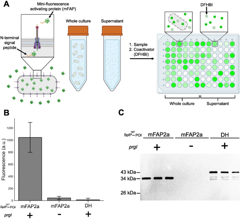
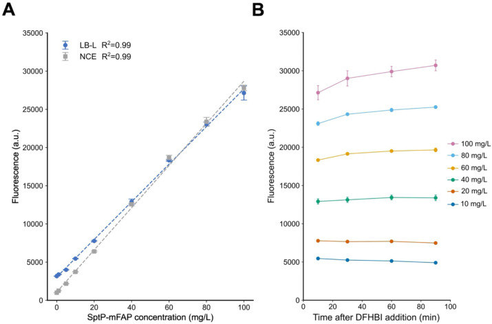
Intracellular protein production in bacteria is limited by the need for lysis and costly purification. A promising alternative is to engineer the host organism for protein secretion. While the Salmonella enterica serovar Typhimurium (S. Typhimurium) Type 3 Secretion System (T3SS) has been utilized for protein secretion, its study and eventual applicability for recombinant protein production is constrained by the lack of high-throughput assays to quantitatively measure secretion titer. Developing such assays is challenging, as proteins must remain unfolded for secretion, limiting the use of several common reporter proteins. In this work, we develop a high-throughput secretion assay using mini-Fluorescent Activating Protein (mFAP). mFAP forms a chromophore only upon addition of an exogenous substrate, allowing secretion and subsequent fluorescence detection. We demonstrate mFAP secretion via the T3SS with an N-terminal secretion tag and show that the fluorescent signal in the secreted fraction is rapid and linear over three orders of magnitude. Using this assay, we screen S. Typhimurium strains with secretion-enhancing mutations, identifying a constitutively active strain and reveal temporally controlled secretion dynamics. We also show that this assay may be applicable to other secretion systems, providing a universal tool for tracking heterologous protein secretion.
Keywords: fluorescence assay; protein secretion; recombinant protein production; type III secretion system (T3SS).
Conflict of interest statement
Conflict of interest statement: DTE has a financial interest in Opera Bioscience, which is commercializing bacterial protein production and secretion. DTE’s conflict of interest is reviewed and managed by Northwestern University in accordance with their conflict-of-interest-policies.
Figures


Similar articles
-
A high throughput assay for measuring secreted protein based on a de novo fluorescent reporter reveals regulatory and structural insights in Salmonella type three secretion system.Protein Sci. 2025 Jul;34(7):e70183. doi: 10.1002/pro.70183. Protein Sci. 2025. PMID: 40563197 Free PMC article.
-
Signs and symptoms to determine if a patient presenting in primary care or hospital outpatient settings has COVID-19.Cochrane Database Syst Rev. 2022 May 20;5(5):CD013665. doi: 10.1002/14651858.CD013665.pub3. Cochrane Database Syst Rev. 2022. PMID: 35593186 Free PMC article.
-
Active body surface warming systems for preventing complications caused by inadvertent perioperative hypothermia in adults.Cochrane Database Syst Rev. 2016 Apr 21;4(4):CD009016. doi: 10.1002/14651858.CD009016.pub2. Cochrane Database Syst Rev. 2016. PMID: 27098439 Free PMC article.
-
Salmonella exploits host- and bacterial-derived β-alanine for replication inside host macrophages.Elife. 2025 Jun 19;13:RP103714. doi: 10.7554/eLife.103714. Elife. 2025. PMID: 40536105 Free PMC article.
-
Single-incision sling operations for urinary incontinence in women.Cochrane Database Syst Rev. 2017 Jul 26;7(7):CD008709. doi: 10.1002/14651858.CD008709.pub3. Cochrane Database Syst Rev. 2017. Update in: Cochrane Database Syst Rev. 2023 Oct 27;10:CD008709. doi: 10.1002/14651858.CD008709.pub4. PMID: 28746980 Free PMC article. Updated.
References
-
- Global Bioengineered Protein Drugs Market Growth Analysis Report. https://www.bccresearch.com/market-research/biotechnology/bioengineered-....
-
- Lilie H, Schwarz E, Rudolph R (1998) Advances in refolding of proteins produced in E. coli. Curr Opin Biotechnol 9:497–501. - PubMed
Publication types
Grants and funding
LinkOut - more resources
Full Text Sources