[Preprint]. 2025 Mar 12:2025.01.17.633618.
doi: 10.1101/2025.01.17.633618.

Systematic Determination of the Impact of Structural Edits on Peptide Accumulation into Mycobacteria

Affiliations

Systematic Determination of the Impact of Structural Edits on Peptide Accumulation into Mycobacteria

Rachita Dash et al. bioRxiv. .

Update in

Abstract

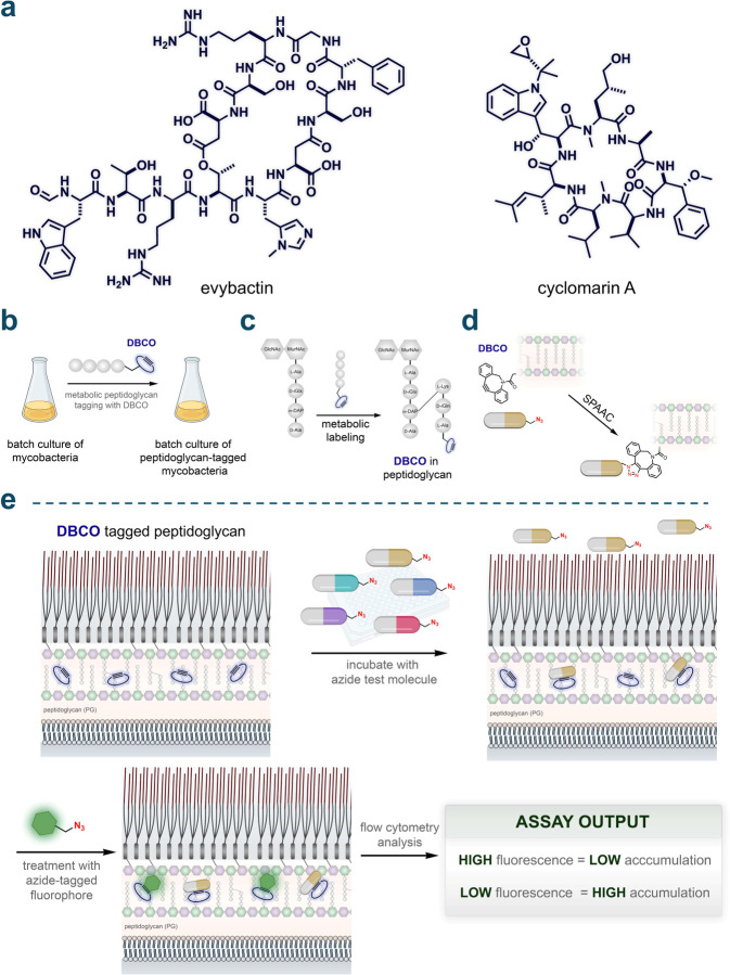
Understanding the factors that influence the accumulation of molecules beyond the mycomembrane of Mycobacterium tuberculosis (Mtb) - the main barrier to accumulation - is essential for developing effective antimycobacterial agents. In this study, we investigated two design principles commonly observed in natural products and mammalian cell-permeable peptides: backbone N-alkylation and macrocyclization. To assess how these structural edits impact molecule accumulation beyond the mycomembrane, we utilized our recently developed Peptidoglycan Accessibility Click-Mediated Assessment (PAC-MAN) assay for live-cell analysis. Our findings provide the first empirical evidence that peptide macrocyclization generally enhances accumulation in mycobacteria, while N-alkylation influences accumulation in a context-dependent manner. We examined these design principles in the context of two peptide antibiotics, tridecaptin A1 and griselimycin, which revealed the roles of N-alkylation and macrocyclization in improving both accumulation and antimicrobial activity against mycobacteria in specific contexts. Together, we present a working model for strategic structural modifications aimed at enhancing the accumulation of molecules past the mycomembrane. More broadly, our results also challenge the prevailing belief in the field that large and hydrophilic molecules, such as peptides, cannot readily traverse the mycomembrane.


Figures

Fig. 1.
Fig. 1.
(a) Chemical structures of the peptides evybactin and cyclomarin A. (b) General schematic illustrating the labeling of mycobacteria with DBCO. Mycobacteria were grown in batches and metabolically labeled with DBCO using a modified exogenous stem peptide probe. (c) Schematic depicting site-specific tagging of the peptidoglycan layer with DBCO. An exogenous stem peptide modified with a DBCO moiety on its N-terminus is crosslinked onto the existing peptidoglycan framework. This occurs via the installation of a covalent link between the meso-α,ε-diaminopimelic acid (m-DAP) residue on a stem peptide of the existing peptidoglycan framework and the Lys residue on the exogenously added stem peptide, by transpeptidases. (d) Schematic illustration of strain-promoted azide-alkyne cycloaddition (SPAAC) occurring between the azide-tagged test molecules and the installed DBCO landmarks within the PG. (e) Schematic illustration of PAC-MAN. Incubation of azide-tagged molecules with the DBCO-labeled mycobacteria allows the registration of their arrival past the mycomembrane onto the PG through SPAAC. A subsequent incubation step with an azide-tagged fluorophore in a secondary SPAAC reveals the unoccupied DBCO landmarks. Cellular fluorescence is then measured by flow cytometry where high fluorescence values correspond to low accumulation and vice versa.
Fig. 2
Fig. 2
(a) Schematic illustration and chemical structure of TetD. (b) Chemical structure of the peptide Nmet0. (c) Dose-response analysis showing the apparent accumulation of Nmet0 across the mycomembrane in Msm. (d) Mass spectrometry analysis of sacculi fragments isolated from TetD treated cells that were subsequently treated with the dye Fl-az. (e) Dose-response analysis showing the apparent accumulation of Nmet0 across the mycomembrane in Msm labeled with varying concentrations of TetD. (f) Dose-response analysis showing the apparent accumulation of Nmet0 across the mycomembrane in Msm treated with different azide-tagged fluorophores, and their chemical structures. (g) Dose-response analysis showing the apparent accumulation of Nmet0 across the mycomembrane in Msm treated with varying concentrations of Fl-az. (h) Dose-response analysis showing the apparent accumulation of Nmet0 across the mycomembrane in Msm with varying Fl-az treatment durations. (i) Dose-response analysis showing the apparent accumulation of Nmet0 across the mycomembrane in Msm with varying Nmet0 treatment durations. (j) Schematic illustration of optimized assay conditions. For the rest of experiments, TetD labeled mycobacterial cells were incubated with 50 μM azide-peptides for 1 h or 2 h and washed with PBST (phosphate buffered saline with 0.05% tween80) twice. Cells were then treated with 50 μM azide-tagged fluorophore for 1 h and washed with PBST twice and fixed in 4% formaldehyde (in PBS) before high throughput analysis on flow cytometry. All dose-response analyses involving Nmet0 were performed with a 1 h incubation period with the peptide. Data are represented as mean ± SD (n= 3). For dose-response curves, Boltzmann sigmoidal curves were fitted to the data using GraphPad Prism.
Fig. 3
Fig. 3
(a) Chemical structures of the N-methylation sub-library with varying degrees of backbone N-methylation (Nmet0-5). (b) Apparent accumulation of the N-methylation sub-library with varying degrees of backbone N-methylation across the mycomembrane in Msm and Mtb following 1 h of treatment with 50 μM compound. (c) Chemical structures of the N-methylation sub-library with varying positions of backbone N-methylation (Nmet6–9 and Nmet1). (d) Apparent accumulation of the N-methylation sub-library with varying positions of backbone N-methylation across the mycomembrane in Msm and Mtb following 1 h of treatment with 50 μM compound. Data are represented as mean ± SD (n= 3). P-values were determined by a two-tailed t-test (ns = not significant, *p < 0.1, **p < 0.01, ***p < 0.001, ****p < 0.0001).
Fig. 4
Fig. 4
(a) Schematic illustration of a macrocyclic and a linear peptide interacting with the surrounding water molecules in an aqueous solution. Hydrogen bonds are indicated by dashed lines. Macrocyclization can promote intramolecular hydrogen bonding resulting in the reduction of hydrogen bonds between the backbone amides and the solvent, thereby potentially reducing membrane permeation associated desolvation penalty. (b) Chemical structures of the linear vs macrocyclic sub-library members containing macrocyclic peptides of increasing sizes (Cyc0-3) and their linear counterparts (Lin0–3). (c) Apparent accumulation of the linear vs macrocyclic sub-library across the mycomembrane in Msm and Mtb following 2 h of treatment with 50 μM compound. (d) Chemical structures of the ring size (lariat) sub-library members containing macrocyclic peptides of increasing ring sizes (Lar1-5) and their linear counterpart (Lar0). (e) Apparent accumulation of the ring size sub-library across the mycomembrane in Msm and Mtb following 2 h of treatment with 50 μM compound. (f) Chemical structures of the cyclization chemistry sub-library members containing a disulfide bonded macrocyclic peptide (Dit1), a bis-electrophilic linker based macrocyclic peptide (Dit2) and their linear counterpart (Dit0). (g) Apparent accumulation of the cyclization chemistry sub-library across the mycomembrane in Msm and Mtb following 2 h of treatment with 50 μM compound. Data are represented as mean ± SD (n= 3). P-values were determined by a two-tailed t-test (ns = not significant, *p < 0.1, **p < 0.01, ***p < 0.001, ****p < 0.0001).
Fig. 5
Fig. 5
(a) Schematic illustration of the hydrogen-deuterium exchange (HDX) analysis of peptides. Peptides are incubated in a D2O buffer, allowing the exchange of amide protons (N-H) with deuterium, with the exchange rate reflecting the solvent accessibility of the N-H bonds. The HDX process is tracked using 1H-NMR spectroscopy. (b) 1H-NMR spectra illustrating the time-dependent HDX of the backbone amide protons in Lin0 and Cyc0 over 1 h. (c) Fraction of Kaz1 N-H protons of Lin0 and Cyc0 exchanged to N-D over time during HDX. (d) Fraction of L2 N-H protons of Lin0 and Cyc0 exchanged to N-D over time during HDX. (e) 3-D representation and chemical structure of Cyc0 showing hydrogen bonding observed through molecular dynamics simulations. (f) Comparison of serum stability of Lin0 and Cyc0 incubated in mouse serum at 37 °C over 1 h. For serum stability analyses, one-phase decay curves were fitted to the data using GraphPad Prism.
Fig. 6
Fig. 6
(a) Chemical structure of the peptide tri, the tridecaptin parent analog in our series. (b) Chemical structures of the peptides triMe1, triMe2, triMe3, the N-methylated tridecaptin analogs in our series. (c) Dose-response analysis showing the apparent accumulation of the N-methylated tridecaptin analogs across the mycomembrane in Msm. (d) MIC values of the N-methylated tridecaptin analogs against Msm. (e) Chemical structure of triCyc, the macrocyclic tridecaptin analog in our series. (f) Dose-response analysis showing the apparent accumulation of the macrocyclic tridecaptin analog triCyc in comparison to the parent tridecaptin analog tri, across the mycomembrane in Msm. (g) MIC values of the macrocyclic and linear tridecaptin analogs triCyc and triLin, in comparison to the parent tridecaptin analog tri, against Msm. (h) Chemical structures of the peptides grisLin, grisCyc, grisMeLin, and grisMeCyc, the griselimycin analogs in our study. (i) Apparent accumulation of the griselimycin analogs across the mycomembrane in Msm following 6 h of treatment with 50 μM compound. (j) MIC values of the of the griselimycin analogs against Msm. All MIC values were obtained from three biological replicates which showed identical results. Other data are represented as mean ± SD (n= 3). For dose-response curves, Boltzmann sigmoidal curves were fitted to the data using GraphPad Prism. All dose-response analyses involving the tridecaptin series were performed with a 1 h incubation period with the peptides. P-values were determined by a two-tailed t-test (ns = not significant, * p < 0.1, ** p < 0.01, *** p < 0.001, **** p < 0.0001).

References

    1. Global Tuberculosis Report 2023 - TB Incidence. https://www.who.int/teams/global-tuberculosis-programme/tb-reports/globa... (accessed 2024-06-03).
    1. Williams P. M. Tuberculosis — United States, 2023. MMWR Morb Mortal Wkly Rep 2024, 73. 10.15585/mmwr.mm7312a4. - DOI - PMC - PubMed
    1. Chiaradia L.; Lefebvre C.; Parra J.; Marcoux J.; Burlet-Schiltz O.; Etienne G.; Tropis M.; Daffé M. Dissecting the Mycobacterial Cell Envelope and Defining the Composition of the Native Mycomembrane. Sci Rep 2017, 7 (1), 12807. 10.1038/s41598-017-12718-4. - DOI - PMC - PubMed
    1. Dulberger C. L.; Rubin E. J.; Boutte C. C. The Mycobacterial Cell Envelope — a Moving Target. Nat Rev Microbiol 2020, 18 (1), 47–59. 10.1038/s41579-019-0273-7. - DOI - PubMed
    1. Bansal-Mutalik R.; Nikaido H. Mycobacterial Outer Membrane Is a Lipid Bilayer and the Inner Membrane Is Unusually Rich in Diacyl Phosphatidylinositol Dimannosides. Proc Natl Acad Sci U S A 2014, 111 (13), 4958–4963. 10.1073/pnas.1403078111. - DOI - PMC - PubMed

Publication types

LinkOut - more resources