[Preprint]. 2024 Dec 16:2024.12.16.628748.
doi: 10.1101/2024.12.16.628748.

Identification of a minimal Alu domain required for retrotransposition

Affiliations

Identification of a minimal Alu domain required for retrotransposition

John B Moldovan et al. bioRxiv. .

Update in

Abstract

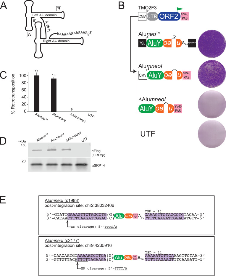
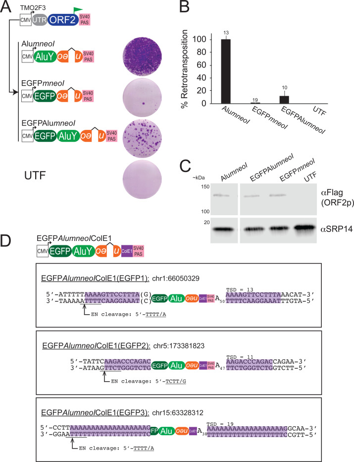
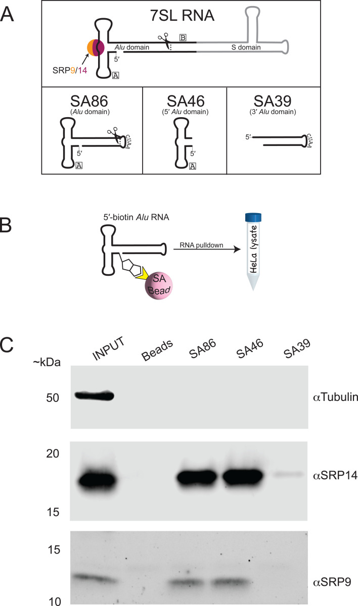
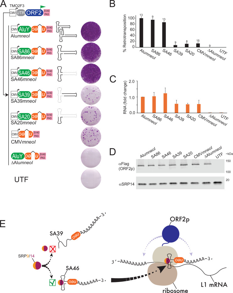
Alu elements are primate-specific retrotransposon sequences that comprise ~11% of human genomic DNA. Alu sequences contain an internal RNA polymerase III promoter and the resultant Alu RNA transcripts mobilize by a replicative process termed retrotransposition. Alu retrotransposition requires the Long INterspersed Element-1 (LINE-1) open reading frame 2-encoded protein (ORF2p). Current models propose that Alu RNA binds to signal recognition particle proteins 9 and 14 (SRP9/14) and localizes to ribosomes, which allows Alu to 'hijack' L1 ORF2p. Here, we used HeLa cell-based retrotransposition assays to define a minimal Alu domain necessary for retrotransposition. We demonstrate that Alu transcripts expressed from a cytomegalovirus (CMV) RNA polymerase II promoter can efficiently undergo retrotransposition. The use of an external CMV promoter to express Alu RNA allowed us to construct separation-of-function mutations to examine the effects of large deletions within the Alu sequence on retrotransposition. Deletion mutagenesis demonstrated that a 46 nucleotide (nt) domain located at the 5' end of the Alu RNA transcript is necessary for Alu retrotransposition. Consistent with current models, the 46 nt 5' Alu domain associates with SRP9/14 in HeLa-HA cell extracts and can promote a single round of retrotransposition in HeLa-HA cells. We propose that the 46 nt 5' Alu domain forms a discrete structure that allows for SRP 9/14 binding and ribosomal association, thereby allowing the Alu poly(A) tract to compete with the L1 poly(A) tail for ORF2p RNA binding to mediate its retrotransposition.


Conflict of interest statement

COMPETING INTERESTS J.V.M. is an inventor on patent US6150160, is a paid consultant for Gilead Sciences, serves on the scientific advisory board of Tessera Therapeutics Inc. (where he is paid as a consultant and has equity options), and has licensed reagents to Merck Pharmaceutical. He also recently served on the American Society of Human Genetics Board of Directors. The other authors do not declare competing interests.

Figures

Figure 1:
Figure 1:. Retrotransposition of an AluY element transcribed by an RNA pol II CMV promoter.
(A) Schematic of a consensus human AluY element. AluY is ~282 nt in length and consists of two 7SL RNA Alu domain monomers (left and right) connected by an A-rich linker and ends in a variable length 3′ encoded poly(A) tract. The approximate locations of the pol III A and B boxes are indicated. (B) Results of AluY retrotransposition assays. HeLa-HA cells were co-transfected with TMO2F3 and the indicated AluY plasmid. TMO2F3 expresses ORF2p with a carboxyl terminus 3XFLAG tag (green flag). ORF2p expression is augmented by a CMV promoter (white square), the native L1 5′ UTR (grey oval), and an SV40 PAS (pink square). The AluneoTet retrotransposition marker (orange oval) contains a backwards copy of the neomycin phosphotransferase gene interrupted by a self-splicing group I intron (loop) that is in the same transcriptional orientation as the Alu sequence. A 44 bp encoded poly(A) tract follows the neoTet sequence. AluneoTet expression is augmented by a 7SL gene enhancer (black square) and a sequence of four consecutive thymidine residues (black square) located downstream of the 44 bp poly(A) tract. The AlumneoI retrotransposition marker (orange oval) contains a backward copy of the neomycin phosphotransferase gene interrupted by a γ-globin intron (^) that is in the same transcriptional orientation as the Alu sequence. AlumneoI expression is augmented by a CMV promoter (white square) and an SV40 PAS (pink square) located after the mneoI indicator. Displayed next to each plasmid construct is a single well of a representative six-well tissue culture plate from retrotransposition assays. UTF = untransfected. (C) Quantification of AluY retrotransposition assays. The X-axis indicates Alu expressing construct co-transfected with TMO2F3 and the Y-axis indicates average percent (%) retrotransposition normalized to AluneoTet from (n) independent biological replicates for each transfection condition. Error bars indicate standard deviation; (n) number of biological replicates indicated above the error bars. (D) ORF2p-Flag expression levels. Western blots of HeLa-HA lysates co-transfected with TMO2F3 and the Alu retrotransposition construct (top of lane). The antibody used for detection is indicated on right. MW markers are indicated on the left. Western blot experiments were repeated twice with similar results. (E) Structure of de novo CMVAlumneoI insertions (c1983, top) and (c2177, bottom). Genomic insertion site locations are based on human reference genome sequence T2T CHM13v2.0/hs1. The AlumneoI insertions contain typical TPRT structural hallmarks indicative of L1 ORF2p-mediated retrotransposition (see main text). Target site duplications are highlighted in magenta. The ORF2p EN target site is underlined; the arrow and “/” indicates the L1 EN cleavage site. Parentheses indicate untemplated nucleotides.
Figure 2:
Figure 2:. Embedded Alu sequences promote the retrotransposition of RNA pol II transcripts.
(A) Results of retrotransposition assays. HeLa-HA cells were co-transfected with TMO2F3 and the indicated embedded Alu construct. Displayed to the right of construct are single wells of a representative six-well tissue culture plate from retrotransposition assays. (B) Quantification of retrotransposition assays. The X-axis indicates the Alu construct co-transfected with TMO2F3, and the Y-axis indicates average percent (%) retrotransposition normalized to AlumneoI for each transfection condition; error bars indicate standard deviation; (n) number of biological replicates indicated above error bars. (C) ORF2p-Flag expression levels. Western blots of HeLa-HA lysates co-transfected with TMO2F3 and the Alu retrotransposition construct (top of lane). The antibodies used for detection are indicated on the right and MW markers are indicated on the left of the blot image. Western blot experiments were repeated twice with similar results. (D) Top panel: Diagram of the EGFPAluColE1 rescue vector. Insertions were characterized from G418-resistant HeLa-HA cells that were co-transfected with TMO2F3 and EGFPAluColE1 using the rescue method (see Methods). Bottom panel: Structure of three EGFPAluColE1 insertions (EGFP1, EGFP2, EGFP3). Genomic insertion site locations are based on human reference genome sequence T2T CHM13v2.0/hs1. The EGFPAluColE1 insertions contain typical TPRT structural hallmarks indicative of L1 ORF2p-mediated retrotransposition (see main text). Target site duplications are highlighted in magenta. The L1 ORF2p EN target site is underlined; the arrow and “/” indicates the L1 EN cleavage site. Parentheses indicate an untemplated nucleotide.
Figure 3:
Figure 3:. 5′ Alu domain RNA binds to SRP9/14 in HeLa-HA cells.
(A) Diagram of Alu domain RNAs. The Alu domain (black) of the 7SL RNA (top) binds SRP9/14. SA86 (7SL RNA Alu domain) is an 86 nt fragment derived from the 7SL RNA Alu domain where the scissors indicate the approximate location of SA86 in relation to the 7SL RNA. The 3′ stem of SA86 is joined by a “CUAA” sequence. SA46 (5′ Alu domain) is composed of nts 1–46 of SA86. SA39 (3′ Alu domain) is composed of the last 39 nts of SA86. (B) Schematic of Alu domain RNA pulldown experiments. Alu domain RNAs containing a 5′ biotin moiety were pre-bound to streptavidin (SA) coated magnetic beads. Alu RNA-SA bead complexes were incubated in HeLa-HA cell lysates and western blotting was used to detect SRP9/14 eluted from Alu RNA-SA bead complexes. (C) SRP9/14 binds to minimal Alu RNAs. Western blots from RNA pulldown experiments. Alu RNA domain is indicated top of each lane. Input = ~2% of WCL; Beads = empty beads (no RNA). The antibody used for detection is indicated on the right of the gel and MW markers indicated on the left of blot image. RNA pulldowns experiments were repeated three times with similar results.
Figure 4:
Figure 4:. SA46 is necessary to promote efficient retrotransposition.
(A) Results of retrotransposition assays. HeLa-HA cells were co-transfected with TMO2F3 and the indicated Alu-containing pol II transcribed RNA construct with a schematic diagram of the respective Alu domain RNA. Displayed next to each plasmid are single wells of a representative six-well tissue culture plate from retrotransposition assays. (B) Quantification of retrotransposition assays. The X-axis indicates the Alu RNA expressing construct co-transfected with TMO2F3, and the Y-axis indicates average percent (%) retrotransposition normalized to AlumneoI for each transfection condition; error bars indicate standard deviations; (n) number of biological replicates are indicated above the error bars. (C) Quantification of Alu domain RNA by RT-qPCR. The X-axis indicates the Alu RNA expressing construct co-transfected with TMO2F3. The Y-axis indicates the RNA fold change normalized to AlumneoI from three independent experiments for each transfection condition with the exception of ΔCMVAlumneoI, which is based on two independent experiments; error bars indicate the standard deviations. (D) ORF2p-Flag expression levels. Western blots of HeLa-HA lysates co-transfected with TMO2F3 and the Alu retrotransposition construct (indicated top of each lane). Western blot experiments were done twice with similar results. The antibody used for detection is indicated on right of the blot image and MW markers are indicated on the left of blot image. (E) Model of SA46 retrotransposition. Alu RNAs that contain an intact 5′ Alu domain (i.e., AluY, SA86, SA46) bind SRP9/14 to form a stable Alu RNP that can localize to a ribosome where the Alu encoded poly(A) tract can compete with the L1 poly(A) tail to bind ORF2p. Polyadenylated RNA transcripts that do not contain a stable Alu domain (i.e., SA39, SA20, CMVmneoI) cannot efficiently bind SRP9/14, may be less stable compared to 5′ Alu domain containing RNAs, and likely do not associate with ribosomes. Thus, SA39 and SA20 have a much lower propensity for retrotransposition when compared to AluY, SA86 and SA46.

References

    1. Deininger P. (2011) Alu elements: know the SINEs. Genome Biol, 12, 236. - PMC - PubMed
    1. Ullu E. and Tschudi C. (1984) Alu sequences are processed 7SL RNA genes. Nature, 312, 171–172. - PubMed
    1. Boeke J.D., Garfinkel D.J., Styles C.A. and Fink G.R. (1985) Ty elements transpose through an RNA intermediate. Cell, 40, 491–500. - PubMed
    1. Dewannieux M., Esnault C. and Heidmann T. (2003) LINE-mediated retrotransposition of marked Alu sequences. Nat Genet, 35, 41–48. - PubMed
    1. Cordaux R., Hedges D.J., Herke S.W. and Batzer M.A. (2006) Estimating the retrotransposition rate of human Alu elements. Gene, 373, 134–137. - PubMed

Publication types