[Preprint]. 2025 Jan 18:2025.01.15.633151.
doi: 10.1101/2025.01.15.633151.

Temporal Microenvironment Mapping (μMap) of Intracellular Trafficking Pathways of Cell-Penetrating Peptides Across the Blood-Brain Barrier

Affiliations

Temporal Microenvironment Mapping (μMap) of Intracellular Trafficking Pathways of Cell-Penetrating Peptides Across the Blood-Brain Barrier

Danielle C Morgan et al. bioRxiv. .

Abstract

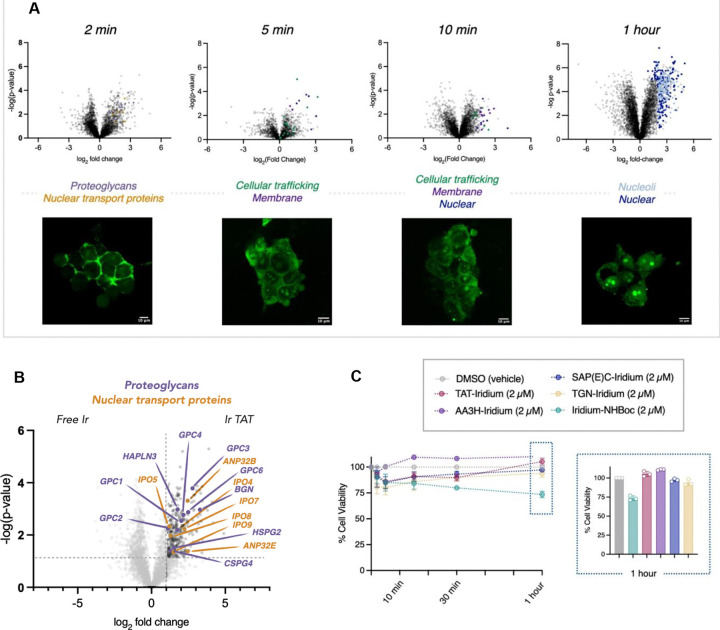
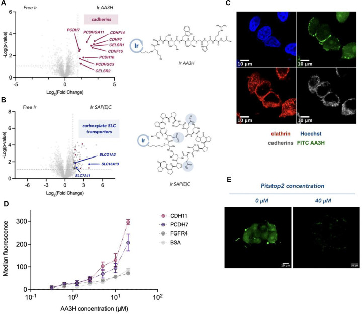
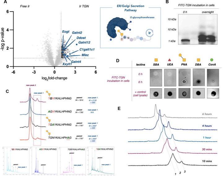
Peptides play critical roles in cellular functions such as signaling and immune regulation, and peptide-based biotherapeutics show great promise for treating various diseases. Among these, cell-penetrating peptides (CPPs) are particularly valuable for drug delivery due to their ability to cross cell membranes. However, the mechanisms underlying CPP-mediated transport, especially across the blood-brain barrier (BBB), remain poorly understood. Mapping intracellular CPP pathways is essential for advancing drug delivery systems, particularly for neurological disorders, as understanding how CPPs navigate the complex environment of the BBB could enable the development of more effective brain-targeted therapies. Here, we leverage a nanoscale proximity labeling technique, termed μMap, to precisely probe the peptide-receptor interactions and intracellular trafficking mechanisms of photocatalyst-tagged CPPs. The unique advantage of the μMap platform lies in the ability to control the timing of light exposure, which enables the collection of time-gated data, depending on when the blue light is applied to the cells. By harnessing this spatiotemporal precision, we can uncover key peptide-receptor interactions and cellular processes, setting the stage for new innovations in drug design and brain-targeted therapies.


Figures

Fig 1 |
Fig 1 |. The peptide μMap platform.
The spatiotemporal control of μMap enables time-resolved labeling of cellular trafficking events. HIV-1 TAT was used to validate the platform, capturing cell-surface entry receptors at 2 minutes and intracellular interactors at 1 hour, thus providing insights into dynamic, time-dependent trafficking processes.
Fig 2 |
Fig 2 |. Spatiotemporal profiling of HIV-1 TAT subcellular trafficking.
A. Parallel microscopy and μMap analysis were employed to examine the intracellular trafficking of the Iridium-HIV-1 TAT peptide, providing both visual and proteomic insights. B. Early interactome analysis identified cell surface receptors and proteoglycans (HSPG2, CSPG4, GPC1-4, GPC6, BGN, HAPLN3, purple) known to facilitate internalization. Additionally, nuclear import factors (importins IPO4, IPO5, IPO7, IPO8, IPO9, yellow) were detected. C. Toxicity assessments were conducted on all peptide-Iridium conjugates used in this study, evaluating their effects at a concentration of 2 μM over the maximum incubation time (1 hour) in cells.
Fig 3 |
Fig 3 |. Identifying the entry receptors for neutral AA3H and anionic SAP(E)C CPPs.
A. μMap proteomic analysis of AA3H entry identified cadherins (red) and B. SLC transporters (blue) as interactors for SAP(E)C. C. Immunofluorescence microscopy displays colocalization of FITC-AA3H with cadherins and clathrin-coated pits (scale bar = 10 μm). D. ELISA binding studies demonstrate significant interaction between AA3H and cadherins, with minimal binding to control proteins. E. Microscopic evaluation of AA3H entry is disrupted via inhibition of clathrin-mediated endocytosis (scale bar = 10 μm).
Fig 4 |
Fig 4 |. Entry receptor mapping of BBB penetrating PepTGN identified through in vivo HTS.
A. PepTGN, a brain-penetrating peptide identified via in vivo phage display, has an unknown BBB entry mechanism. We utilized μMap to investigate its transcytosis trafficking pathway. B. μMap experiments were performed on ice for 15 minutes in two different cell lines (hCMEC/d3, shown, and bEND.3 cells) to identify potential entry receptors. One of the top hits identified in both volcano plots was the calcitonin receptor-like receptor (CALCRL, pink), a G-protein coupled receptor (GPCR) involved in peptide signaling. The experiments also revealed clathrin (GAK and NECAP2, maroon) and dynamin (DNM2, maroon) proteins. C. Blocking experiments were conducted to validate CALCRL as the entry receptor for PepTGN. These studies showed a significant reduction in peptide uptake when cells were pre-incubated with either the native ligand CGRP or the monoclonal antibody Erenumab. In contrast, incubation with a control isotype antibody had no effect on internalization.
Fig 5 |
Fig 5 |. Transcytosis mapping of BBB penetrating PepTGN identified through in vivo HTS
A. μMap profiling of PepTGN intracellular interactors in bEND.3 cell line after 1 hour incubation. 30% of the top hits in the volcano plot relate to ER and Golgi secretory proteins (blue), suggesting a potential exocytosis mechanism of PepTGN. B. Fluorescence microscopy of secretion kinetics of FITC PepTGN. C. FITC-PepTGN (green) colocalization in the ER (red, ERp57/ERp60 antibody) (scale bar = 100 μm).
Fig 6 |
Fig 6 |. PepTGN is glycosylated during transcytosis.
A. μMap proteomic analysis of PepTGN in the bEND.3 cell line identifies several O-glycosyltransferases (blue), suggesting that PepTGN may get glycosylated during transcytosis. B. Gel electrophoresis of fluorescein labelled PepTGN after incubation with cells displayed multiple new products. C. PepTGN analogs were synthesized, replacing potential O- or N-glycosylation sites (Thr-12, Asn-10, Asn-2), identifying Thr-12 as a key residue for glycosylation. D. To further confirm glycosylation, we used a biotinylated lectin dot-blot, which showed positive binding for DBA and PNA lectins in PepTGN cellular samples, indicating O-linked glycans. E. UPLC-MS analysis of the purified peptides samples revealed a cleavage fragment and a second product with a +322 Da mass shift, suggesting a glycosylation event (1 = cleavage product, 2 = parent peptide & 3 = glycosylated product).

References

    1. Wu D., Chen Q., Chen X., Han F., Chen Z. and Wang Y., Signal Transduct Target Ther, 2023, 8, 217. - PMC - PubMed
    1. Abbott N. J., Patabendige A. A., Dolman D. E., Yusof S. R. and Begley D. J., Neurobiol Dis, 2010, 37, 13–25. - PubMed
    1. Patabendige A. and Janigro D., Biochem Soc Trans, 2023, 51, 613–626. - PMC - PubMed
    1. Mitragotri S., Burke P. A. and Langer R., Nat Rev Drug Discov, 2014, 13, 655–672. - PMC - PubMed
    1. Pirhaghi M., Mamashli F., Moosavi-Movahedi F., Arghavani P., Amiri A., Davaeil B., Mohammad-Zaheri M., Mousavi-Jarrahi Z., Sharma D., Langel U., Otzen D. E. and Saboury A. A., Mol Pharm, 2024, 21, 2097–2117. - PubMed

Publication types

LinkOut - more resources