[Preprint]. 2025 Jan 17:2025.01.15.633247.
doi: 10.1101/2025.01.15.633247.

Oral prodrug of a novel glutathione surrogate reverses metabolic dysregulation and attenuates neurodegenerative process in APP/PS1 mice

Affiliations

Oral prodrug of a novel glutathione surrogate reverses metabolic dysregulation and attenuates neurodegenerative process in APP/PS1 mice

Swetha Pavani Rao et al. bioRxiv. .

Update in

Abstract

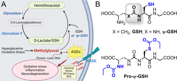
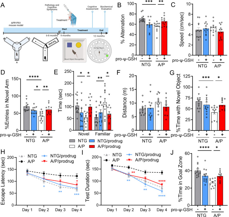
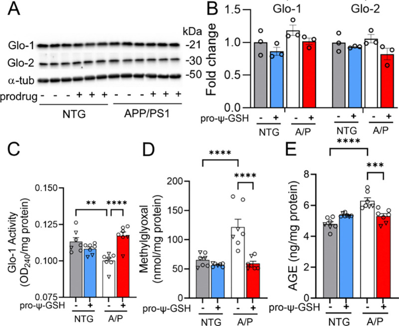
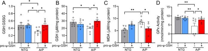
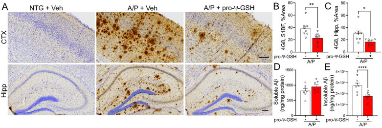
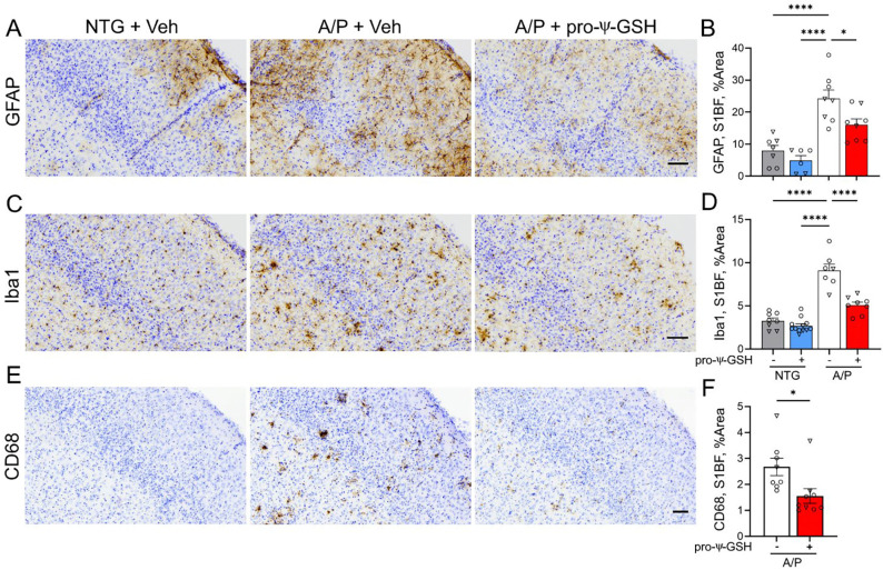
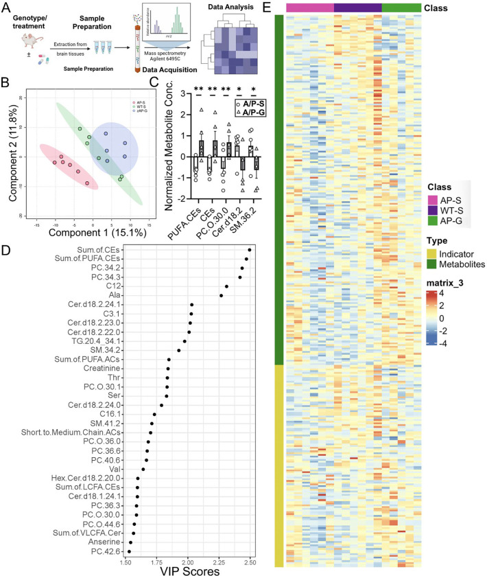
Glycation-induced oxidative stress underlies the numerous metabolic ravages of Alzheimer's disease (AD). Reduced glutathione levels in AD lead to increased oxidative stress, including glycation-induced pathology. Previously, we showed that the accumulation of reactive 1,2-dicarbonyls such as methylglyoxal, the major precursor of non-enzymatic glycation products, was reduced by the increased function of GSH-dependent glyoxalase-1 enzyme in the brain. In this two-pronged study, we evaluate the therapeutic efficacy of an orally bioavailable prodrug of our lead glyoxalase substrate, pro-ψ-GSH, for the first time in a transgenic Alzheimer's disease mouse model. This prodrug delivers pharmacodynamically relevant brain concentrations of ψ-GSH upon oral delivery. Chronic oral dosing of pro-ψ-GSH effectively reverses the cognitive decline observed in the APP/PS1 mouse model. The prodrug successfully mirrors the robust effects of the parent drug i.e., reducing amyloid pathology, glycation stress, neuroinflammation, and the resultant neurodegeneration in these mice. We also report the first metabolomics study of such a treatment, which yields key biomarkers linked to the reversal of AD-related metabolic dysregulation. Collectively, this study establishes pro-ψ-GSH as a viable, disease-modifying therapy for AD and paves the way for further preclinical advancement of such therapeutics. Metabolomic signatures identified could prove beneficial in the development of treatment-specific clinically translatable biomarkers.

Keywords: Alzheimer’s disease; Glyoxalase-1; advanced glycation end products; metabolomics; neuroinflammation; oxidative stress.


Conflict of interest statement

Competing interests S.S.M. and R.V. are co-inventors on the patent applications relating to ψ-GSH and its analogs as treatment options for neurodegenerative disorders and liver diseases. The funders had no role in the design of the study; in the collection, analyses, or interpretation of data; in the writing of the manuscript; or in the decision to publish the results.

Figures

Figure 1.
Figure 1.
(A) Glyoxalase enzymatic pathway involved in detoxification of methylglyoxal. Formation of advanced glycation end products (AGE) by methylglyoxal with biological macromolecules results in oxidative stress and inflammatory cascade. (B) Chemical structures of the glyoxalase substrate glutathione (GSH), ψ-GSH, and the prodrug, pro-ψ-GSH.
Figure 2.
Figure 2.
Oral administration of pro-ψ-GSH in symptomatic APP/PS1 mice reverses cognitive impairment. (A) Schematic of the experimental timeline in aged APP/PS1 (A/P) mice. (B) Percent spontaneous alternations in Y-maze of animals treated for 12 weeks. Significant improvement in the alternation behavior was observed between prodrug and saline treated APP/PS1 mice. (C) Average speed of the mice during the Y-maze alternation test were unaffected by the genotype and the prodrug treatment. (D-F) In Y-maze spatial recognition test, the prodrug treated APP/PS1 group spent more time in the previously unexplored (novel) arm compared to the familiar arm, as evident from significantly higher number of entries (D) in the novel arm, and corollary, more time spent (E) in the novel arm. The total distance traveled (F) was unchanged within the genotypes and treatment groups. (G) In novel object recognition test, prodrug-treated APP/PS1 mice exhibited higher exploratory behavior with the novel object compared to corresponding vehicle controls. (H-I) Escape latency and test duration for identification of target hole during the training period in Barnes maze. Prodrug-treated APP/PS1 mice learned to find the escape hole faster than saline-treated APP/PS1 mice over the test duration. (J) Proportion of time spent in the target quadrant over rest of the maze area during the probe trial conducted 24 h after the last training session. Oral pro-ψ–GSH group performed significantly better in the probe trial, and spent larger proportion of total time in the target zone compared to the controls. Circles are males and triangles are females. Data are represented as mean ± SEM. One-way ANOVA or two-way ANOVA (for E) followed by Tukey’s or Holm-Sidak’s post-hoc multiple comparisons test *p<0.05, **p<0.01, ***p<0.001, and ****p<0.0001.
Figure 3.
Figure 3.
Effect of pro-ψ-GSH supplementation on Glo-1 related pathway markers in aged APP/PS1 mice. (A) Glo-1 and Glo-2 protein expression analyses by western blot and (B) their respective quantification. (C) Glo-1 enzyme activity was significantly compromised in APP/PS1 (A/P) mice compared to the corresponding non-transgenic controls (NTG), which was restored by the oral prodrug treatment. (D) Reduced Glo-1 function resulted in increased MG levels in the APP/PS1-saline group. The MG levels were normalized to NTG-saline group levels by the prodrug. (E) Increased AGEs content, corresponding to higher MG levels, in untreated APP/PS1 mice was reduced significantly by pro-ψ-GSH treatment. Circles are males and triangles are females. Data are presented as the mean ± S.E.M and is representative of two (for A & B) or three (C–E) independent experiments. Pro-ψ-GSH and saline-treated APP/PS1 and NTG groups were compared with one-way ANOVA with Tukey’s post-hoc multiple comparison test for statistical analysis. ** p < 0.01, *** p < 0.001, **** p < 0.0001.
Figure 4.
Figure 4.
Mitigation of increased oxidative stress by pro-ψ-GSH treatment in APP/PS1 mice brain. (A-B) Improved redox ratio (GSH/GSSG, A) was apparent in pro-ψ-GSH treated APP/PS1 mice. Quantification of reduced GSH (B) showed lower cytoplasmic GSH content in the APP/PS1 brains compared to NTG mice, which were restored by the prodrug treatment. (C) Lipid peroxidation levels as calculated from the levels of MDA were attenuated by the prodrug in the APP/PS1 group. (D) Compromised GPx activity in aged APP/PS1 saline group was restored to NTG levels by pro-ψ-GSH. Circles are males and triangles are females. Data are shown as the mean ± SEM. Data are representative of three independent experiments. Statistical significance was examined by a one-way ANOVA with Tukey’s or Sidak’s post-hoc multiple comparison test. * p < 0.05, ** p < 0.01.
Figure 5.
Figure 5.
Oral pro-ψ-GSH effectively reduces Aβ burden in symptomatic APP/PS1 mice. (A) Representative images of Aβ immunoreactivity (using 4G8 antibody) in the S1BF cortex (CTX) and hippocampus (Hipp) from non-Tg littermates (NTG) and APPswe/PS1ΔE9 Tg mice (A/P). Animals were treated with either Vehicle (Veh) or pro-ψ-GSH for 3 months prior to tissue collection. Scale bar, 100 μm. (B, C) Quantitative analysis of amyloid pathology was done by determining the percent area covered by 4G8 immunoreactivity in S1BF (B) and hippocampus (C). Compared to the vehicle (Veh) treated mice, pro-ψ-GSH-treated mice showed significantly lower 4G8 immunoreactivity (n = 8 APP/PS1 + Saline; n = 9 APP/PS1 + pro-ψ-GSH). There was no Aβ immunoreactivity in NTG mice. (D, E) ELISA assay quantitation of the β-amyloid load in A/P mice. Levels of insoluble Aβx–42 levels (E) in the brain homogenate of A/P mice treated with pro-ψ-GSH were reduced significantly compared to the Veh treated A/P mice. Circles are males and triangles are females. Unpaired Student’s t-test was used for all statistical comparisons of saline and ψ-GSH-treated A/P cohorts. For comparisons between saline and pro-ψ-GSH-treated APP/PS1 groups, a one-way ANOVA with Tukey’s or Sidak’s post-hoc multiple comparison test was performed for statistical analysis. * p < 0.05, ** p < 0.01, *** p < 0.005. * p < 0.05, ** p < 0.01, *** p < 0.005.
Figure 6.
Figure 6.
Oral pro-ψ-GSH treatment reduces cortical reactive astrocytosis and microglial activation in the APP/PS1 mouse model of AD. (A, C, E) Representative images of brain sectioned stained for astrocytes (GFAP), total microglia (Iba1) and activated microglia (CD68) in S1BF of NTG and APPswe/PS1ΔE9 (A/P) mouse cohorts. Animals were treated with either Vehicle (Veh) or pro-ψ-GSH for 3 months prior to tissue collection. Scale bar, 100 μm. (B, D. F) Glial reaction in S1BF was quantified by defining the percent area covered by GFAP (B), Iba1(D), or CD68 (F) immunoreactivity. The results show the expected increase in glial activation in the Veh treated A/P mice compared to NTG mice. Consistent with reduced amyloid pathology, oral pro-ψ-GSH treatment significantly reduced astrocyte and microglial activation. Circles are males and triangles are females. Data are shown as mean ± S.E.M. Statistical comparisons between group were performed by one-way ANOVA with Tukey’s post-hoc multiple comparison test. * p < 0.05, ** p < 0.01, *** p < 0.005, **** p < 0.001.
Figure 7.
Figure 7.
Cytokine array analysis of mice based on genotype and treatment. (A) Representative membranes showing the change in the inflammatory cytokines in brain homogenates from APP/PS1 mice treated with either saline or pro-ψ-GSH. The intensities of the spots were quantified densitometrically by ImageJ and compared to the density of the internal standards. (B) The data is normalized with respect to NTG saline controls. Cytokine analysis showed a significant increase in pro-inflammatory cytokines (IL-1β, IL5, IFN-γ, MIP-α, TNF-α, VCAM-1) in saline treated APP/PS1 mice compared to age-matched NTG controls. Treatment with pro-ψ-GSH reversed the effect of AD pathology in aged APP/PS1 mice, and offered cytokine profile similar to the NTG controls. Data are expressed as mean ± SEM. (C) Pathway analysis using Enrichr online tool displayed modulation of signaling pathways involved in neurodegeneration, specifically Alzheimer’s processes. (D) Display of pathways impacted by pro-ψ-GSH and the involved cytokines. Data are shown as mean ± S.E.M and is representative of two independent experiments. Statistical significance in (B) was determined by two-way ANOVA analysis using Fisher’s LSD multiple comparison or Tukey’s post-hoc test. (* p < 0.05, ** p < 0.01, *** p < 0.001).
Figure 8.
Figure 8.
Oral pro-ψ-GSH treatment attenuates the loss of cortical TH+ afferents and atrophy of TH+ neurons in Locus Coeruleus (LC) in the APP/PS1 model. (A) Representative images of TH+ afferents in S1BF of Vehicle (Veh) and pro-ψ-GSH-treated APPswe/PS1ΔE9 (A/P) subjects. Also shown is Veh treated NTG mice. Scale bar, 50 μm. (B) Quantitative analysis of TH+ afferent density show that the TH+ afferent density is reduced in Veh treated A/P animals compared to NTG subjects. Oral pro-ψ-GSH treatment of A/P mice leads to a significant increase in TH+ afferent density compared to Veh treated A/P subjects. (C, E) Representative low (C) and high (E) magnification images of TH+ neurons of the LC. (D) Consistent with prior studies, stereological counting of TH+ neurons in the LC do not show significant neuronal loss in A/P mice. (F) Analysis of TH+ neuron volume in LC show that, consistent with the loss of TH+ afferents in A/P + Veh mice, TH+ neurons are significantly smaller compared to NTG mice. Further, oral pro-ψ-GSH treatment of A/P mice show significant attenuation of neuronal atrophy. Circles are males and triangles are females. Data are mean ± S.E.M. One-way ANOVA with Tukey’s post-hoc test. **p < 0.01, *** p < 0.005, **** p < 0.001.
Figure 9.
Figure 9.
Metabolite alterations caused by AD pathology in APP/PS1 mice were reversed by pro-ψ-GSH treatment. (A) Schematic of the metabolomics study. (B) PLS-DA plot classifying the hippocampal metabolites from NTG and APP/PS1 (AP) mice treated with pro-ψ-GSH (AP and AP/G, resp.). (C) Bar graph showing top metabolites that are affected by the prodrug treatment in APP/PS1 (A/P-G) compared to corresponding vehicle treated mice (A/P-S) with established role in AD pathology. PUFA-CEs = polyunsaturated fatty acid cholesteryl esters, CEs = sum of cholesteryl esters, PC.O = phosphatidylcholine, cer = ceramide, SM = sphigomyelins. (D) Metabolite profiling analysis by VIP scores (> 1.5) provided top metabolites contributing significantly to variations within groups. The relative contribution of the metabolites is proportional to the VIP score. (E) Heat maps showing changes in top 50 metabolites in NTG and APP/PS1 groups treated with either saline or pro-ψ-GSH. Each column represents an individual animal within the group. Hierarchical clustering found NTG-saline group closer to the A/P + pro-ψ-GSH group, suggesting beneficial effect of the treatment. Data are represented as the mean ± SEM. Statistical significance was derived by Student t-test, * p < 0.05, ** p < 0.01, n = 5–6.

Similar articles

References

    1. DeTure M. A.; Dickson D. W. The neuropathological diagnosis of Alzheimer’s disease. Molecular Neurodegeneration 2019, 14 (1), 32. DOI: 10.1186/s13024-019-0333-5. - DOI - PMC - PubMed
    1. Cummings J.; Osse A. M. L.; Cammann D.; Powell J.; Chen J. Anti-Amyloid Monoclonal Antibodies for the Treatment of Alzheimer’s Disease. BioDrugs 2024, 38 (1), 5–22. DOI: 10.1007/s40259-023-00633-2 From NLM. - DOI - PMC - PubMed
    1. Osborne O. M.; Naranjo O.; Heckmann B. L.; Dykxhoorn D.; Toborek M. Anti-amyloid: An antibody to cure Alzheimer’s or an attitude. iScience 2023, 26 (8), 107461. DOI: 10.1016/j.isci.2023.107461. - DOI - PMC - PubMed
    1. Carreiras M. C.; Mendes E.; Perry M. J.; Francisco A. P.; Marco-Contelles J. The multifactorial nature of Alzheimer’s disease for developing potential therapeutics. Curr Top Med Chem 2013, 13 (15), 1745–1770. DOI: 10.2174/15680266113139990135 From NLM. - DOI - PubMed
    1. Trejo-Lopez J. A.; Yachnis A. T.; Prokop S. Neuropathology of Alzheimer’s Disease. Neurotherapeutics 2022, 19 (1), 173–185. DOI: 10.1007/s13311-021-01146-y From NLM. - DOI - PMC - PubMed

Publication types