[Preprint]. 2025 Jan 17:2025.01.17.633277.
doi: 10.1101/2025.01.17.633277.

ASSOCIATIONS BETWEEN EPILEPSY-RELATED POLYGENIC RISK AND BRAIN MORPHOLOGY IN CHILDHOOD

Alexander Ngo  1   2 Lang Liu  1   3 Sara Larivière  4 Valeria Kebets  1   2 Serena Fett  1   2 Clara F Weber  1   5   6 Jessica Royer  1   2 Eric Yu  1   3 Raúl Rodríguez-Cruces  1   2 Zhiqiang Zhang  7 Leon Qi Rong Ooi  8   9   10 B T Thomas Yeo  8   9   10 Birgit Frauscher  11   12 Casey Paquola  13 Maria Eugenia Caligiuri  14 Antonio Gambardella  15 Luis Concha  16 Simon S Keller  17   18 Fernando Cendes  19 Clarissa L Yasuda  19 Leonardo Bonilha  20 Ezequiel Gleichgerrcht  21 Niels K Focke  22 Raviteja Kotikalapudi  23 Terence J O'Brien  24   25 Benjamin Sinclair  24   25 Lucy Vivash  24   25 Patricia M Desmond  25 Elaine Lui  25 Anna Elisabetta Vaudano  26   27 Stefano Meletti  26   27 Reetta Kälviäinen  28   29 Hamid Soltanian-Zadeh  30   31 Gavin P Winston  32   33   34 Vijay K Tiwari  35 Barbara A K Kreilkamp  22 Matteo Lenge  36   37 Renzo Guerrini  36 Khalid Hamandi  38   39 Theodor Rüber  40   41   42 Tobias Bauer  40   41   42 Orrin Devinsky  43 Pasquale Striano  44   45 Erik Kaestner  46 Sean N Hatton  47 Lorenzo Caciagli  33   48 Matthias Kirschner  49   50 John S Duncan  33   34 Paul M Thompson  51 ENIGMA Consortium Epilepsy Working GroupCarrie R McDonald  46   52 Sanjay M Sisodiya  33   34 Neda Bernasconi  1   2 Andrea Bernasconi  1   2 Ziv Gan-Or  1   2   3 Boris C Bernhardt  1   2
Affiliations

ASSOCIATIONS BETWEEN EPILEPSY-RELATED POLYGENIC RISK AND BRAIN MORPHOLOGY IN CHILDHOOD

Alexander Ngo et al. bioRxiv. .

Update in

  • Associations between epilepsy-related polygenic risk and brain morphology in childhood.
    Ngo A, Liu L, Larivière S, Kebets V, Fett S, Weber CF, Royer J, Yu E, Rodríguez-Cruces R, Zhang Z, Ooi LQR, Yeo BTT, Frauscher B, Paquola C, Caligiuri ME, Gambardella A, Concha L, Keller SS, Cendes F, Yasuda CL, Bonilha L, Gleichgerrcht E, Focke NK, Kotikalapudi R, O'Brien TJ, Sinclair B, Vivash L, Desmond PM, Lui E, Vaudano AE, Meletti S, Kälviäinen R, Soltanian-Zadeh H, Winston GP, Tiwari VK, Kreilkamp BAK, Lenge M, Guerrini R, Hamandi K, Rüber T, Bauer T, Devinsky O, Striano P, Kaestner E, Hatton SN, Caciagli L, Kirschner M, Duncan JS, Thompson PM; ENIGMA Consortium Epilepsy Working Group; McDonald CR, Sisodiya SM, Bernasconi N, Bernasconi A, Gan-Or Z, Bernhardt BC. Ngo A, et al. Brain. 2025 Aug 14:awaf259. doi: 10.1093/brain/awaf259. Online ahead of print. Brain. 2025. PMID: 40811581

Abstract

Temporal lobe epilepsy with hippocampal sclerosis (TLE-HS) is associated with a complex genetic architecture, but the translation from genetic risk factors to brain vulnerability remains unclear. Here, we examined associations between epilepsy-related polygenic risk scores for HS (PRS-HS) and brain structure in a large sample of neurotypical children, and correlated these signatures with case-control findings in in multicentric cohorts of patients with TLE-HS. Imaging-genetic analyses revealed PRS-related cortical thinning in temporo-parietal and fronto-central regions, strongly anchored to distinct functional and structural network epicentres. Compared to disease-related effects derived from epilepsy case-control cohorts, structural correlates of PRS-HS mirrored atrophy and epicentre patterns in patients with TLE-HS. By identifying a potential pathway between genetic vulnerability and disease mechanisms, our findings provide new insights into the genetic underpinnings of structural alterations in TLE-HS and highlight potential imaging-genetic biomarkers for early risk stratification and personalized interventions.

Keywords: Imaging-genetics; brain structure; childhood; genetic risk; temporal lobe epilepsy.


Conflict of interest statement

POTENTIAL CONFLICTS OF INTEREST All authors declare no competing conflicts of interest.

Figures

Figure 1 |
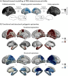
Figure 1 |. PRS-HS associations with cortical thickness (ABCD).
(A) Distribution of genetic risk effects on morphology across the different lobes (in order from top to bottom: all, frontal, limbic, occipital, parietal, temporal). (B) Regional imaging-genetic correlations between PRS-HS and thickness. Blue and red colours represent negative and positive correlations, respectively. White outline indicates pFDR < 0.05. L, left; PRS-HS; polygenic risk score for epilepsy-related hippocampal sclerosis; R, right.
Figure 2 |
Figure 2 |. Network epicentres of morphological changes associated with PRS-HS.
(A) Schematic representation of epicentre mapping approach using seed-based cortico- and subcortico-cortical connectivity. PRS-HS, polygenic risk score for epilepsy-related hippocampal sclerosis. (B) Correlation coefficients indexing spatial similarity between imaging-genetic effects and seed-based functional (top) and structural (bottom) connections for every cortical and subcortical region. Red and blue colours represent negative associations, while grey depicts positive correlations. White outline indicates pspin < 0.05. L, left; R, right.
Figure 3 |
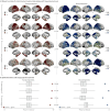
Figure 3 |. Comparison between PRS-HS effects and epilepsy case-control atrophy.
(A) Case-control differences in left and right TLE-HS from ENIGMA-Epilepsy (top) and from MICs and NKG (bottom). Blue and red colours point to atrophy and hypertrophy in patients relative to healthy controls, respectively. Outline in white represents pFDR < 0.05. L, left; R, right; TLE-HS, temporal lobe epilepsy with hippocampal sclerosis. (B) Spatial correlations between epilepsy-related atrophy (top: ENIGMA-Epilepsy; bottom: MICs and NKG) and imaging-genetic effect maps (ABCD) are compared against permutation-based null correlations. Points represent the empirical correlation (with significance defined as pspin < 0.05). In the boxplots, the ends of boxes represent the first (25%) and third (75%) quartiles, the centre line (median) represents the second quartile of the null distribution (n = 5,000 permutations), the whiskers represent the non-outlier endpoints of the distribution.
Figure 4 |
Figure 4 |. Comparison between imaging-genetic and epilepsy-related disease epicentres.
(A) Functional and structural disease epicentres in left and right TLE-HS from ENIGMA-Epilepsy (top) and from MICs and NKG (bottom). Red and blue colours represent negative associations, while grey depicts positive correlations. Outline in white represents pspin < 0.05. L, left; R, right; TLE-HS, temporal lobe epilepsy with hippocampal sclerosis. (B) Spatial correlations between epilepsy-related epicentres (top: ENIGMA-Epilepsy; bottom: MICs and NKG) and imaging-genetic effect maps (ABCD) are compared against permutation-based null correlations. Points represent the empirical correlation (with significance defined as pspin < 0.05). In the boxplots, the ends of boxes represent the first (25%) and third (75%) quartiles, the centre line (median) represents the second quartile of the null distribution (n = 5,000 permutations), the whiskers represent the non-outlier endpoints of the distribution.

References

    1. Feigin V. L. et al. The global burden of neurological disorders: translating evidence into policy. Lancet Neurol. 19, 255–265 (2020). - PMC - PubMed
    1. Epilepsy C. by H.-G W. for the I. C. on N. of. Mesial Temporal Lobe Epilepsy with Hippocampal Sclerosis. Epilepsia 45, 695–714 (2004). - PubMed
    1. Lin J. J. et al. Reduced Neocortical Thickness and Complexity Mapped in Mesial Temporal Lobe Epilepsy with Hippocampal Sclerosis. Cereb. Cortex 17, 2007–2018 (2007). - PubMed
    1. Keller S. S. & Roberts N. Voxel-based morphometry of temporal lobe epilepsy: An introduction and review of the literature. Epilepsia 49, 741–757 (2008). - PubMed
    1. McDonald C. R. et al. Regional neocortical thinning in mesial temporal lobe epilepsy. Epilepsia 49, 794–803 (2008). - PubMed

Publication types