[Preprint]. 2025 Jan 27:2025.01.09.632077.
doi: 10.1101/2025.01.09.632077.

High-throughput Mucus Microrheology for Phenotyping and Disease Modeling

Affiliations

High-throughput Mucus Microrheology for Phenotyping and Disease Modeling

Feng Ling et al. bioRxiv. .

Abstract

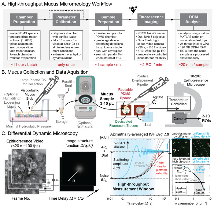
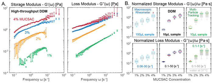
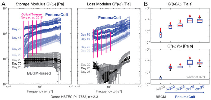
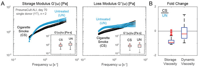
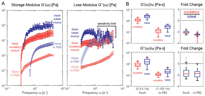
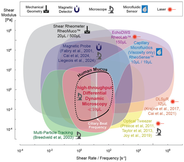
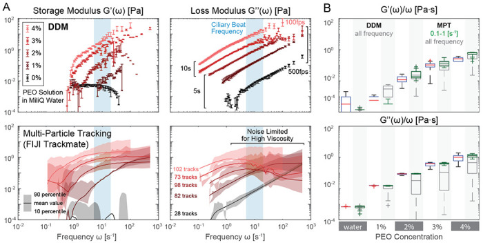
Mucus plays an integral role for the barrier function of many epithelial tissues. In the human airways, mucus is constantly secreted to capture inhaled microbes and pollutants and cleared away through concerted ciliary motion. Many important respiratory diseases exhibit altered mucus flowability and impaired clearance, contributing to respiratory distress and increased risk of infections. Understanding how mucus rheology changes during disease progression and in response to treatments is thus of great interest for subtyping patients and tailoring treatments, probing disease mechanisms, and tailoring therapies; however, basic research of mucus rheology is greatly hampered by the lack of scalable and user-friendly rheometry assays for the small volumes of mucus typically produced by in vitro respiratory models and in clinical ex vivo settings. To address this challenge, we developed a streamlined, high-throughput protocol leveraging Differential Dynamic Microscopy (DDM) to reliably measure the frequency-dependent microrheology of minuscule (3-10 μL) mucus samples using standard epifluorescence microscopy. Our method does not require time-consuming user-interventions common in particle tracking routines and measures microrheology at the time scale of mucus relaxation (1-20s), hence greatly reducing assay time. We demonstrate the successful application of our method in mucus samples harvested from state-of-the-art air-liquid-interface (ALI) human respiratory cultures to assess mucus rheology in airway disease models and different culture conditions. To show that our approach equally applies to other types and sources of human mucus, we also validated our method with clinical samples of cervical mucus. We envision that our method can be seamlessly adopted by non-expert users, without the need for specialized equipment or extensive training, to study diseases and their treatments in the respiratory, intestinal, reproductive and other mucosal organ systems. This advancement opens up new avenues for large-scale studies, providing new insights into the role of mucus rheology which was previously limited by data accessibility and resource constraints.


Conflict of interest statement

Competing interests: The authors declare that they have no competing interests.

Figures

Figure 1:
Figure 1:. High-throughput mucus microrheology workflow.
A. Our streamlined workflow can measure the viscoelastic moduli of each 3-10 μL mucus sample at a total time cost of ca. 30 min, with only a few minutes requiring sustained human attention. In a trial run, 60 samples were prepared in 62 min, and 235 regions of interest were imaged in 123 min. B. Mucus from sterile in vitro culture were extracted from the apical side of transwell cultures and sealed in parafilm wrapped Eppendorf tube for analysis. PDMS spacers with small holes (3 mm or 6 mm diameter) were cut and put on glass slides to form capillary chambers. Dilute fluorescent bead solution was filled to each chamber and let dry in a dark box at room temperature until the liquid had fully evaporated. After verifying bead integrity with a one-time hydrodynamic radius calibration using a pure water sample, the mucus samples were transferred to one chamber each, gently mixed and sealed for epifluorescence microscopy inside a temperature controlled incubator. C. Differential dynamic microscopy (DDM) works by finding the azimuthally-averaged image structure function based on the Fourier transform of video frame differences at different time delay Δt. To increase throughput capacity, we restricted measurement time to <20 s and estimated the scattering amplitude A(q) using periodically shifted images instead of direct observation. Insets visualize particle motion due to diffusion versus such shifts. Bottom panel compares results of this shift-based estimation strategy (blue and black dashed line) with shortened video (light gray dashed line) of tracers in water to values obtained from original long video (dark gray dashed line) taken with 10x (0.3 NA) objectives. Transwell and centrifuge illustration created with BioRender https://BioRender.com/d80d467.
Figure 2:
Figure 2:. Viscoelastic properties of reconstituted MUC5AC gels.
Porcine gastric mucin (MUC5AC) was reconstituted in acidic buffer (pH 4) to simulate mucus gels with physiologically-relevant viscoelastic character. A. Frequency response of the storage and loss moduli of 1 to 4% w/w MUC5AC gel are clearly separable using our high-throughput DDM protocol. Error bar indicates 10 to 90th percentile calculation based on 5, 512×512 px ROIs. B. Microrheology output from DDM (middle panels) and Multi-Particle Tracking (MPT, via FIJI Trackmate, right panels) can deviate from macrorheology (left panels). DDM microrheology at all frequency range (0.1–50 Hz) closely matches MPT results at restricted frequency (0.1–1 Hz, green). MPT results at full frequency ranges (0.1–50 Hz, dark gray) produce significantly higher variance especially for high viscosity samples. Light gray line indicates the dynamic viscosity of pure water at measurement temperature. Measurement performed on n = 2 technical replicates using GeneFrame 25 μL capillary chambers. Slides were placed on a temperature-controlled sample holder for additional thermal stability.
Figure 3:
Figure 3:. Impact of differentiation medium and culture age.
A. Storage and loss moduli derived from mucus extracted from in vitro primary airway epithelial Air-Liquid-Interface (ALI) cultures in PneumaCult (PC, blue markers) versus BEGM-based medium (grayscale markers) at different time points (days numbered after air lift at day 0). Pink markers are reproduced optical tweezer measurement of mucus extracted from PC ALI culture [68]. B. Frequency-normalized frequency normalized viscoelastic moduli shows that viscoelasticity of PC culture mucus saturates over time. In contrast, BEGM-based mucus consistently showed a low viscosity near that of the water / culture medium. Measurements performed from samples of n = 2 to 3 ALI insert cultures per condition from N=1 donor using 6 mm capillary chambers. Slides were placed on a temperature-controlled sample holder for additional thermal stability.
Figure 4:
Figure 4:. Rheological stability of sterile in vitro mucus collection.
A. Storage and loss moduli of mucus extracted from ALI culture maintained in PneumaCult from two separate donors: donor 7783 (green shaded markers) is of bronchial origin, and donor 8938 (magenta shaded markers) is from a small airway source. B. Left panels show the fresh and stored absolute viscosity (|G*|/ω) before (lighter shade) and after 7 days of storage (darker shade). Right panel shows that fold changes in absolute viscosity between the two donors remain stable after 7 days of storage inside the sealed capillary chambers. Measurements performed on pooled n = 3 12-well plate inserts with specified N = 2 donors using 3 mm capillary chambers.
Figure 5:
Figure 5:. Rheological changes of in vitro airway mucus in response to cigarette smoke exposure.
A. Storage and loss moduli of mucus extracted from ALI cultures maintained in PneumaCult and exposed to cigarette smoke extract twice a week over 8 weeks resembling a subchronic condition (blue, CS) and untreated conditions (black, UN). A reduction in viscoelastic moduli induced by cigarette smoke is observed for a single proof-of-concept donor and time point (day 70 after air lift), hinting a potential compensation due to irritation. B. Fold change of the storage (left) and dynamic (right) viscosity from CS treated sample and untreated one. Measurements performed on n = 2 technical replicates from N = 1 donor samples per condition using 6 mm capillary chambers at approximately 1:4 v/v dilution in PBS. Slides were placed on a temperature-controlled sample holder for additional thermal stability.
Figure 6:
Figure 6:. Rheological stability of mucus after osmotic swelling.
A. Storage and loss moduli derived from clinically extracted cervical mucus at luteal (blue shaded markers) and ovulatory phase (red shaded markers), both from fresh collection (darker shade) and after exposure to PBS without homogenization (lighter shade). The luteal phase viscosity at higher frequencies are most likely outside the sensitivity limit of our methodology and thus discarded in later statistical analyses. B. frequency normalized viscoelastic moduli (left panels) and fold change in absolute viscosity (right panels) of the luteal and ovulatory mucus before and after exposure to PBS solution. Different frequency range (axis label) are used for normalized moduli to avoid sensitivity limited zone (0.3-5 Hz before swelling) and minimize effects of sample drift (1-100 Hz after swelling). Measurements performed on sample per condition from N = 1 donor with 6 mm capillary chambers. Slides were placed on a temperature-controlled sample holder for additional thermal stability.
Figure 7:
Figure 7:. DDM is uniquely suitable for user-friendly, high-throughput assessment of low volume mucus samples.
Accessible shear modulus and shear rate / frequency range of common rheology methods compared to human mucus modulus at frequencies relevant for biomedical research (black line) [15,16]. Ciliary beat frequency range based on our in vitro ALI culture measurements, with white line indicating median [14]. Symbols indicate type of equipment required, and where applicable, we also indicate the required minimum sample volume for each method. Multi-Particle Tracking (light green) and Differential Dynamic Microscopy (pink) ranges are based on [43] and our reported setup. Dynamic Light Scattering (orange), Magnetic Probe (dark purple), and Optical Tweezer (yellow) ranges are based on numbers reported in [, –78, 81, 82, 86]. Mechanical shear rheometer (gray) values are based on our experience with Aanton Paar devices. Sample volume for RheoMuco device are minimum ALI mucus volume reported in online FAQ and recommended sputum volume in [70]. Capillary microfluidic viscometer (light blue) and Diffusing Wave Spectroscopy (light purple) ranges are based on values reported by RheoSense m-VROC II and VROC Initium 1+ and LS Instruments RheoLab devices [74], respectively.
Figure 8:
Figure 8:. High-throughput DDM validation with PEO solutions.
A. storage and loss moduli from DDM and particle tracking microrheology. DDM results show a small variance (error bar 10-90 percentile from 5 ROI average) that easily distinguishes PEO solution at different concentrations by weight, especially near typical ciliary beat frequency ranges (light blue). B. Comparison of frequency normalized viscoelastic moduli derived from DDM and particle tracking. Particle tracking results closely match that of DDM but only if the noisy frequency range is ignored. DDM box plots are based on all measured frequencies, green MPT box plots are restricted to data measured between 0.1 to 1 Hz, and dark gray for all measured MPT frequencies.

Similar articles

References

    1. Thornton DJ, Sheehan JK. From mucins to mucus: toward a more coherent understanding of this essential barrier. Proceedings of the American Thoracic Society. 2004;1(1):54–61. - PubMed
    1. Thornton DJ, Rousseau K, McGuckin MA. Structure and function of the polymeric mucins in airways mucus. Annu Rev Physiol. 2008;70(1):459–86. - PubMed
    1. Vasquez PA, Forest MG. Complex fluids and soft structures in the human body. In: Complex fluids in biological systems: Experiment, theory, and computation. Springer; 2014. p. 53–110.
    1. Ma J, Rubin BK, Voynow JA. Mucins, mucus, and goblet cells. Chest. 2018;154(1):169–76. - PubMed
    1. Denton R, Forsman W, Hwang S, Litt M, Miller C. Viscoelasticity of mucus: Its role in ciliary transport of pulmonary secretions. American Review of Respiratory Disease. 1968;98(3):380–91. - PubMed

Publication types

LinkOut - more resources