[Preprint]. 2025 Jan 19:2025.01.15.633105.
doi: 10.1101/2025.01.15.633105.

IL-9 as a naturally orthogonal cytokine with optimal JAK/STAT signaling for engineered T cell therapy

Affiliations

IL-9 as a naturally orthogonal cytokine with optimal JAK/STAT signaling for engineered T cell therapy

Hua Jiang et al. bioRxiv. .

Abstract

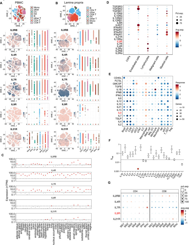
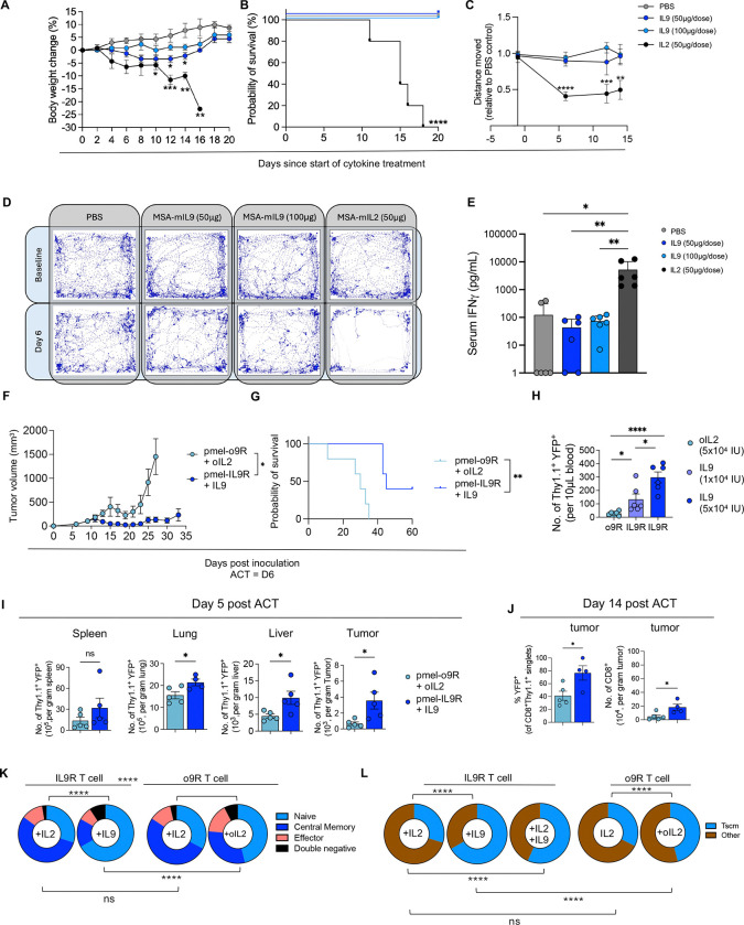
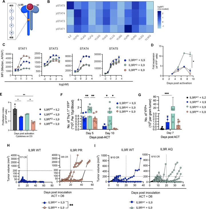
Arming T cells with a synthetically orthogonal IL-9 receptor (o9R) permits facile engraftment and potent anti-tumor functions. We considered whether the paucity of natural IL-9R expression could be exploited for T cell immunotherapy given that, in mice, high doses of IL-9 were well-tolerated without discernible immune modulation. Compared to o9R, T cells engineered with IL-9R exhibit superior tissue infiltration, stemness, and anti-tumor activity. These qualities are consistent with a stronger JAK/STAT signal, which in addition to STAT1/3/5, unexpectedly includes STAT4 (canonically associated with IL-12 but not common γ-chain cytokines). IL-9R T cells are exquisitely sensitive to perturbations of proximal signaling, including structure-guided attenuation, amplification, and rebalancing of JAK/STAT signals. Biased IL-9R mutants uncover STAT1 as a rheostat between proliferative stem-like and terminally differentiated effector states. In summary, we identify native IL-9/IL-9R as a natural cytokine-receptor pair with near-orthogonal qualities and an optimal JAK/STAT signaling profile for engineered T cell therapy.


Conflict of interest statement

Declaration of interests A.K. serves on the advisory board and holds stock for Dispatch Therapeutics and Certis Oncology and consults for Sastra Cell Therapy. K.C.G. is the founder of Synthekine and co-founder of Dispatch, which are developing cytokine receptor-based therapeutics. The use IL-9 and L-9R signaling composition and methods are claimed in a patent application (PCT/US2023/070251). E.J.M. has served as a consultant for GLG and Guidepoint.

Figures

Figure 1.
Figure 1.. Nomination of IL-9 as a naturally orthogonal cytokine for T cell therapy.
(A) Expression of common γc cytokines receptors in single cell RNA sequencing data of PBMC from normal healthy donors. tSNE plots with cell annotations are shown in the top panel as a reference. For each cytokine receptor, a feature plot colored by scaled expression level (left panel) and a scatter plot summarizing expression for each cell type (right panel) are shown. (B) As in (A), but for lamina propria lymphocytes from the uninflamed ileum of patients with Crohn’s disease. (C) Expression scatterplot of normalized expression of receptors for γc cytokines in publicly available RNA sequencing data of 37 normal tissues from the GTEx database. Dashed line indicates a strict nTPM threshold of 0.7, below which expression is not detected or negligible. (D) Dot plot of intratumoral expression of a panel of cytokine receptors across malignant and non-malignant cell types, based on single cell RNA-sequencing data from soft tissue sarcoma (n=65,945 cells across 15 patients). (E) Differential expression of genes (number of genes, magnitude of response) among cell types within lymph nodes of mice treated with one of several cytokines versus PBS control (based on publicly available data). (F) Reported median Shet values for γc and other cytokines. Shet quantifies the impact of predicted heterozygous loss-of-function variants on evolutionary fitness, with higher scores associated with greater impact on evolutionary fitness. Error bars represent 95% highest posterior density. The dashed line at 0.02 represents the median Shet value for all canonical transcripts. (G) Expression dot plot of receptors for γc cytokines in sorted CD19 CAR T cells (n=66,042) from 15 pediatric patients at various timepoints after infusion. Data presented separately for CD4 (left) and CD8 (right) CAR T cells. PBMC: peripheral blood mononuclear cells; TPM: transcripts per million; GTEX: Genotype-Tissue Expression; CAR: chimeric antigen receptor
Figure 2.
Figure 2.. Exogenous IL-9 is well-tolerated and results in superior tissue infiltration, stemness and anti-tumor activity of IL-9R-engineered antigen-specific T cells.
(A) Body weight (percentage change from baseline) of mice (n=8 mice/group) treated with PBS, IL-9 (50 μg or 100 μg), or IL-2 (50 μg) i.p. every other day. (B) Survival of mice from (A). (C) Sum of distance traveled over 30 second period in mice from (A), plotted relative to the average distance traveled by mice in the PBS group. (D) Graphical representation of mouse movement (n=8 mice/group) over a period of 30 seconds at baseline (top panel) and six days after starting treatment (bottom panel). (E) Serum IFNγ levels measured by ELISA six days after initiating treatment (n=3 mice/group) as described in A-C. (F) B16-F10 tumor growth (mean ± SEM, n=5 mice/group) after ACT with pmel T cells engineered with o9R or IL-9R (0.4×10⁶ transduced cells, i.v.), and cytokine treatment with oIL-2 or IL-9, respectively (5×10⁴ IU i.p., daily for 5 days starting with ACT). Data is representative of three independent experiments. (G) Survival of mice from (F). (H) Quantification of transduced pmel T cells in the blood five days after ACT (n=5 mice/group). (I) Quantification of transduced pmel T cells (Thy1.1+YFP+) across different tissues five days after ACT (n=5 mice/group). (J) Transduced (YFP+) pmel T cells as percentage of all pmel T cells within the tumor 14 days after ACT (left panel) and total number of CD8+ T cells per gram of tumor 14 days after ACT (right panel) (n=5 mice per group). (K) o9R or IL9R transduced C57BL/6 T cells stimulated for 24h with IL-2 (10nM), IL-9 (10nM), oIL-2 (10μM), or a combination as noted. The proportion of naïve (CD62L+CD44−), central memory (CD62L+CD44+), effector (CD44+CD62L−) or double negative (CD62L−CD44−) T cells. Data from two independent experiments, with 4 technical replicates per condition. (L) The proportion of TSCM cells (CD44−, CD62L+, Sca-1+) from (K). For in vivo experiments, cytokines were tagged with mouse serum albumin (MSA) for half-life extension. *P < 0.05, **P < 0.005, ***P < 0.0005, ****P < 0.0001 (one-way ANOVA for A-C and E-F; Mantel-Cox for G; unpaired t test for H and J, two-way ANOVA for K-L).
Figure 3.
Figure 3.. IL-9R signaling results in phosphorylation of STAT4, in addition to STAT1, STAT3, and STAT5.
(A) Dose-response curves of STAT1, STAT3, and STAT5 phosphorylation in IL-9R or o9R transduced (YFP+) pmel T cells stimulated with either oIL-2 or IL-9 for 20 minutes (shown are technical duplicates; representative of at least three independent experiments). (B) Volcano plots depicting differential gene expression based on RNA-sequencing of IL-9R or o9R transduced C57BL/6 T cells and treated with IL-9 (10 nM), IL-2 (10 nM) or oIL2 (10 μM) for 48 hours. Comparisons for each volcano plot are shown below the x-axis. Significance (red) indicates adjusted p < 10−5 and absolute fold change ≥2. (C) Differentially phosphorylated proteins between IL-9R transduced C57BL/6 T cells stimulated for 20’ with either IL-2 (10nM) or no cytokine (left) or IL-9 (10nM) versus no cytokine (right). Significance (red) indicates adjusted p<0.05 and log2(fold change)≥ 0.5. (D) Dose-response curve for STAT4 phosphorylation among IL-9R transduced (YFP+) pmel T cells treated for 20 minutes with IL-9 or IL-12, or o9R transduced T cells treated with oIL-2 (shown are technical duplicates; representative of two independent experiments). (E) Waterfall plot summarizing transcription factor enrichment scores based on RNA-seq data of IL-9R T cells treated with IL-9 or IL-2 (top 15 for each). Enrichment score is inferred by fitting a linear model that predicts observed gene expression based on prior knowledge of a curated set of transcription factors and their target genes. (F) Heat map of the expression of the Biocarta IL-12 Pathway gene set based on RNA-sequencing from (B). Samples and genes clustered hierarchically without supervision. (G) In vitro expansion of IL-9R (YFP+) transduced pmel T cells and treated with 10nM cytokine starting on day 3 after activation (n=3 technical replicates/group). Data is representative of three biological replicates and two independent experiments. (H) Representative contour plots of CD44 and CD62L expression of T cells from (G) after 24h treatment with cytokine. (I) Quantification of naïve CD62L+CD44 (left) and stem-like CD62L+CD44Sca-1+ (right) T cells from (H). Data from two independent experiments. *P < 0.05, **P < 0.005, ***P < 0.001, ****P < 0.0001 (one-way ANOVA for G; two-way ANOVA for G, I).
Figure 4.
Figure 4.. Structure-based attenuation or amplification of IL-9/IL-9R signaling diminishes the anti-tumor qualities of T cells signaling through the native receptor complex.
(A) Structural prediction of the interleukin-9 (IL-9) receptor complex based on AlphaFold2. The complex consists of the IL-9 receptor (IL-9R, green), IL-9 (blue), and the γc (pink). The inset demonstrates the interaction of the glutamine at amino acid position 115 of IL-9 (Q115) with the γc, which we subsequently mutated (IL-9Q115T) to generate an attenuated cytokine. (B) Dose-response curves of STAT1, STAT3, STAT4, and STAT5 phosphorylation in IL-9R transduced pmel T cells stimulated with IL-9WT (blue) or IL-9Q115T (light gray) for 20 minutes. Error bars represent SEM of technical duplicates; Data is representative of two independent experiments. (C) Tumor growth after ACT with IL-9R pmel T cells seven days after B16-F10 tumor inoculation. Mice were treated with either IL-9WT (n=7 mice) or IL-9Q115T (n=9 mice) (10 doses, every other day). Data is representative of two independent experiments. (D) Peripheral blood quantification of IL-9R transduced pmel T cells over time in mice treated with ACT and either IL-9WT or IL-9Q115T cytokine (each cytokine dose indicated by a vertical dashed line). In the IL-9Q115T group, IL-9R pmel T cells were not observed in the blood at day 41 or after three additional cytokine doses on days 44–48, and thus only the IL-9WT group received doses beyond day 48. Data is representative of two independent experiments. (E) Schematic of IL-9R variants with either three (IL-9R3x) or five (IL-9R5x) repeated phosphotyrosine (pY) elements within the intracellular domain of the IL-9 receptor (created with Biorender.com). Sequence of the phosphotyrosine element shown in the legend. (F) Phosphorylation of indicated STATs at Emax (100nM) for IL-9RWT, IL-9R3x, and IL-9R5x transduced pmel T cells stimulated with IL-2 or IL-9 for 20 minutes. Data is representative of three biological experiments (mean ± SEM). (G) B16-F10 tumor growth after ACT with either IL-9RWT (n=7 mice) or IL-9R3x transduced pmel T cells (n=8 mice). Mice were also treated with IL-9 (5×10⁴ IU i.p., every other day for 5 doses starting with ACT). Shown are individual tumor growth curves (left and middle) and mean ± SEM (right). Data is representative of two independent experiments. For in vivo experiments, cytokines were tagged with mouse serum albumin (MSA) for half-life extension. *P < 0.05, **P < 0.005, ***P < 0.001, ****P < 0.0001. (two-way ANOVA for C; Welch’s t-test for D, G; one-way ANOVA for F). See also Figure S4.
Figure 5.
Figure 5.. IL-9R intracellular domain mutants skew STAT phosphorylation and alter in vivo proliferative capacity and anti-tumor efficacy.
(A) Schematic of the IL-9 receptor complex, highlighting the phosphotyrosine residue within the IL-9R intracellular domain (ICD) and three adjacent amino acids. A panel of ten single amino acid mutations were generated within the ICD at the proline or glutamine residues (created with Biorender.com). (B) Heat map of MFI (log-scaled and row-scaled) at Emax for phosphorylation of STAT1, STAT3, STAT4, and STAT5 for C57BL/6 T cells transduced with wildtype IL-9R or one of ten IL-9R mutants. Transduced pmel T cells (technical duplicates) were stimulated with IL-9 for 20 minutes. Data is representative of two independent experiments. (C) Dose-response curves of STAT1, STAT3, STAT4, and STAT5 phosphorylation among transduced IL-9RWT, IL-9RAQ, and IL-9RPR pmel T cells (YFP+) stimulated with recombinant IL-9 for 20 minutes. Error bars represent SEM of technical duplicates. Data is representative of three biological replicates. (D) Relative in vitro expansion of YFP+ IL-9RWT, IL-9RAQ, IL-9RPR pmel T cells cultured with IL-9 on day 3 post-activation (10 nM; n=3 technical replicates/group). Data is representative of 3 biological replicates. (E) In vitro proliferation index of C57BL/6 T cells engineered with IL-9RWT, IL-9RAQ, and IL-9RPR over the course of nine days post-activation (transduced on day 1), as measured by dilution of CellTrace Violet dye (n=6 replicates/group). Cells were treated with either recombinant IL-2 or IL-9 (10 nM) on day 3 after T cell activation. Proliferation index was quantified using FlowJo software based on values of histogram peaks (see Figure S5B). (F) Peripheral blood quantification of IL-9RWT, IL-9RAQ, or IL-9RPR transduced (Thy1.1+YFP+) pmel T cells in B16-F10 tumor-bearing mice (n=6–7 mice/group) on the indicated days after ACT. IL-9 treatment (5×10⁴ IU i.p., every other day) was started with ACT and continued for 5 doses. Data is representative of at least three independent experiments. (G) Quantification of IL-9RWT, IL-9RAQ, or IL-9RPR transduced (YFP+) pmel T cells within the tumors (B16-F10) of mice (n=6–7 mice/group) seven days post adoptive cell transfer (ACT). Data is representative of two independent experiments. (H) B16-F10 tumor growth in mice treated with IL-9RWT (n=7 mice) or IL-9RPR (n=6 mice) pmel T cells. Data is representative of at least three independent experiments. (I) B16-F10 tumor growth in mice treated with IL-9RWT or IL-9RAQ pmel T cells (n=10 mice/group). IL-9 treatment as per (F). Data is representative of at least 4 independent experiments. For in vivo experiments, cytokines were tagged with mouse serum albumin (MSA) for half-life extension. *P < 0.05, **P < 0.005, ***P < 0.001, ****P < 0.0001 (two-way ANOVA for D-F; Welch’s t-test for G-I).
Figure 6.
Figure 6.. IL-9R variants reveal STAT1 as a rheostat skewing T cells from a stem and memory state toward a terminal effector state.
(A) Schematic for in vivo experiment to generate single cell RNAseq data. IL-9RWT, IL-9RAQ, or IL-9RPR engineered pmel T cells are adoptively transferred into B16-F10 tumor-bearing mice seven days after tumor inoculation. Mice (n=7–8 mice/group) are treated with IL-9 (5×10⁴I.U. every other day) starting on the day of ACT (day 7) until tumors are harvested on day 15. Transduced pmel T cells (Thy1.1+YFP+) are sorted by FACS prior to library preparation and single cell RNAseq. See related Figure S6A – S6B. (B) UMAP plots based on scRNAseq of n=6,706 cells pmel T cells from (A). Ten major clusters colored according to annotation. See related Figure S6C–E. (C) Propeller plots demonstrating the relative proportion of each cluster from (A), split by treatment group (IL-9RWT, IL-9RAQ, or IL-9RPR). (D) Violin plots summarizing single cell expression of a gene set differentially expressed between KLRG1hi effector and naïve mouse T cells (GSE10239). Gene sets are summarized as a module score in Seurat and plotted by treatment group. (E) Violin plots summarizing single cell expression of a gene set differentially expressed between mouse T cells eight days (D8) after encounter with acute infection versus malignancy (GSE60501). Gene sets are summarized as a module score in Seurat and plotted by treatment group. (F) Ridgeplot of pseudotime scores for single cell data from (A) organized by treatment group. (G) UMAP plot of scRNA-sequencing data from (A) annotated with pseudotime trajectories. Cells within the Tscm-like (Tcf7) cluster were selected as the root for the pseudotime analysis (white circles with black outlines). Black circles with white outlines represent nodes of the differentiation trajectory. (H) Schematic for linking phosphoflow data with RNA-sequencing data. o9R, IL-9RWT, IL-9RAQ, IL-9RPR, or IL-9R5x pmel T cells were treated with cytokines for either 20 mins (phosphoflow) or 48h (RNA-sequencing). MFIs at Emax for pSTAT1, pSTAT3, pSTAT4 and pSTAT5 were merged with RNA-sequencing data to identify genes highly correlated with phosphorylation of each STAT protein. (I) Histograms of phosphorylation of STAT1, STAT3, STAT4 and STAT5 for transduced (YFP+) T cells for each condition after 20 minutes of cytokine stimulation at Emax: IL-2 (10nM), IL-9 (10nM) and oIL-2 (10μM). (J) Venn diagram of the top 100 genes most correlated with the phosphorylation of each STAT. (K) Scatterplot depicting the relationship between the pSTAT1 module score (y-axis) and pSTAT1 phosphorylation levels (x-axis) in vitro. Module score was calculated from the expression of 100 genes most strongly correlated with STAT1 phosphorylation in vitro. Shown are biological triplicates for the gene expression data, colored by sample condition. For STAT phosphorylation data, shown are the average of technical replicates. The gray line indicates the linear regression fit. (L) Violin plot depicting projection of pSTAT1 module score from (J)-(K) onto scRNA-seq data from (A), organized by treatment group. (M) Projection of pSTAT1 module score from (J)-(K) onto UMAP of scRNA-seq data from (A). (N) Waterfall plot summarizing transcription factor (TF) enrichment scores when comparing in vitro RNAseq data of IL-9RWT vs IL-9RAQ groups (left) and IL-9RPR vs IL-9RWTgroups (right). Gene expression changes across IL-9RAQ → IL-9RWT → IL-9RPR are captured by increasing activity of STAT1 and related TFs (shown in red). (O) Ridgeplots summarizing STAT1, STAT3, STAT4, and STAT5a regulon activity as an AUC score calculated using SCENIC. Results are presented by treatment group, with pairwise statistical comparisons (Wilcoxon Rank-Sum Test).
Figure 7.
Figure 7.. The balance of JAK/STAT signaling through IL-9R and IL-9R variants, and their impact on anti-tumor activity, is conserved in human CAR T cells.
(A) Dose-response curves of STAT1, STAT3, STAT4, and STAT5 phosphorylation in human T cells transduced with human IL-9R or human o9R (YFP+) and stimulated with either human IL-9 or human oIL-2 for 20 minutes. Data are representative of two independent donors. (B) Schematic of Nalm6 human leukemia orthotopic model in NSG mice treated with second generation human CAR T cells targeting CD19 (CD19-BBz) and co-transduced with human IL-9R. Mice were also treated with either human IL-2 or IL-9 (or no cytokine). (C) Weight of NSG mice (n=5 mice/group) in response to treatment schema shown in (B). Mice were weighed twice per week. Mice were euthanized at day 10 in the group receiving IL-2 due to toxicity. (D) Bioluminescence (photons/second) of tumors in mice treated as described in (B). (E) Survival curves of mice from (D). (F) Schematic of 143B human osteosarcoma orthotopic solid tumor model in NSG mice treated with second generation human CAR T cells targeting Her2 (Her2-BBz). CAR T cells were cotransduced with human IL-9R (or not) and treated with human IL-9 (or not). (G) Tumor growth as measured by leg volume of mice (n=5 mice/group) as treated per (F). (H) Survival curves of mice from (G). (I) Dose-response curves of STAT1, STAT3, STAT4, and STAT5 phosphorylation in human T cells transduced with human IL-9R, IL-9RPR or IL-9RAQ and stimulated with IL-9 for 20 minutes. Shown are technical duplicates (bars represent SEM) and representative of two independent donors. (J) In vivo expansion and enrichment of CAR T cells from (K). Panel shows absolute number of (YFP+) CAR T cells per 10 microliters of blood twelve days after tumor inoculation (8 days after ACT). (K) Tumor growth as measured by leg volume of mice (n=5 mice/group) treated as per schematic in (F). Tumor volume was measured three times per week. (L) In vivo survival curves of mice from (K). For all experiments in Figure 7, cytokines were tagged with mouse serum albumin for half-life extension.*P < 0.05, **P < 0.005, ***P < 0.0005, ****P < 0.0001 (two-way ANOVA for D, G, J; Mantel-Cox test for E, H, K; one-way ANOVA for L).

References

    1. Leonard W.J., Lin J.X., and O’Shea J.J. (2019). The γ(c) Family of Cytokines: Basic Biology to Therapeutic Ramifications. Immunity 50, 832–850. 10.1016/j.immuni.2019.03.028. - DOI - PubMed
    1. Dwyer C.J., Knochelmann H.M., Smith A.S., Wyatt M.M., Rangel Rivera G.O., Arhontoulis D.C., Bartee E., Li Z., Rubinstein M.P., and Paulos C.M. (2019). Fueling Cancer Immunotherapy With Common Gamma Chain Cytokines. Frontiers in Immunology 10. 10.3389/fimmu.2019.00263. - DOI - PMC - PubMed
    1. Siegel J.P., and Puri R.K. (1991). Interleukin-2 toxicity. Journal of Clinical Oncology 9, 694–704. 10.1200/jco.1991.9.4.694. - DOI - PubMed
    1. Gilleece M.H., Scarffe J.H., Ghosh A., Heyworth C.M., Bonnem E., Testa N., Stern P., and Dexter T.M. (1992). Recombinant human interleukin 4 (IL-4) given as daily subcutaneous injections – a phase I dose toxicity trial. British Journal of Cancer 66, 204–210. 10.1038/bjc.1992.243. - DOI - PMC - PubMed
    1. Davis I.D., Skrumsager B.K., Cebon J., Nicholaou T., Barlow J.W., Moller N.P., Skak K., Lundsgaard D., Frederiksen K.S., Thygesen P., and McArthur G.A. (2007). An open-label, two-arm, phase I trial of recombinant human interleukin-21 in patients with metastatic melanoma. Clin Cancer Res 13, 3630–3636. 10.1158/1078-0432.Ccr-07-0410. - DOI - PubMed

Publication types