[Preprint]. 2025 Jan 15:2025.01.15.633208.
doi: 10.1101/2025.01.15.633208.

Structural mechanism for recognition of E2F1 by the ubiquitin ligase adaptor Cyclin F

Affiliations

Structural mechanism for recognition of E2F1 by the ubiquitin ligase adaptor Cyclin F

Peter Ngoi et al. bioRxiv. .

Update in

Abstract

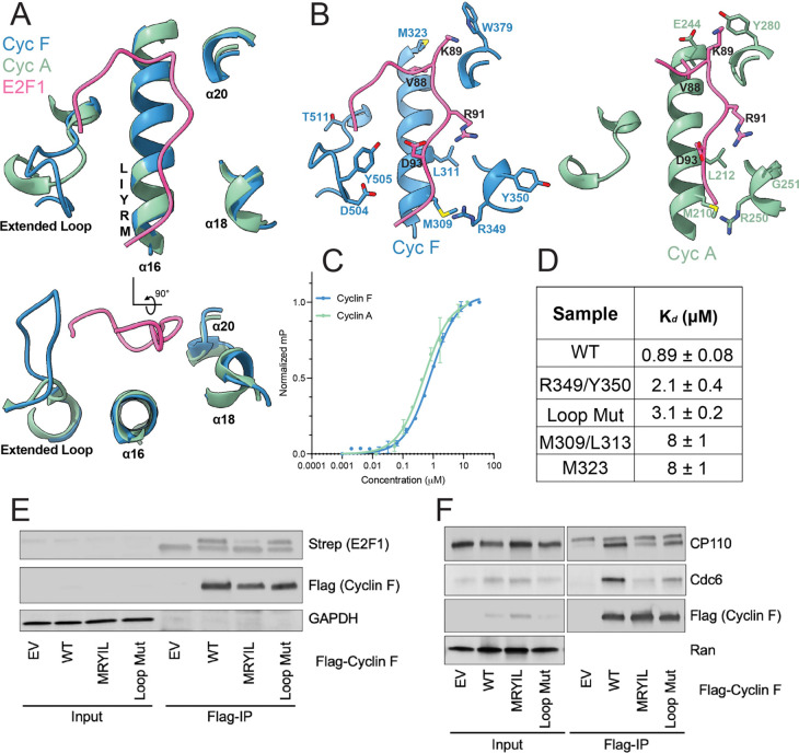
Cyclin F, a non-canonical member of the cyclin protein family, plays a critical role in regulating the precise transitions of cell-cycle events. Unlike canonical cyclins, which bind and activate cyclin-dependent kinases (CDKs), Cyclin F functions as a substrate receptor protein within the Skp1-Cullin-F box (SCF) E3 ubiquitin ligase complex, enabling the ubiquitylation of target proteins. The structural features that distinguish Cyclin F as a ligase adaptor and the mechanisms underlying its selective substrate recruitment over Cyclin A, which functions in complex with CDK2 at a similar time in the cell cycle, remain largely unexplored. We utilized single-particle cryo-electron microscopy to elucidate the structure of a Cyclin F-Skp1 complex bound to an E2F1 peptide. The structure and biochemical analysis reveal important differences in the substrate-binding site of Cyclin F compared to Cyclin A. Our findings expand on the canonical cyclin-binding motif (Cy or RxL) and highlight the importance of electrostatics at the E2F1 binding interface, which varies for Cyclin F and Cyclin A. Our results advance our understanding of E2F1 regulation and may inform the development of inhibitors targeting Cyclin F.


Figures

Figure 1.
Figure 1.
A) Model for E2F1 ubiquitylation by SCFCyclinF and proteasomal degradation. Cyclin F binds and activates the Skp1-Cul1-F box (SCF) E3 ligase to E2F1 and other substrates. B) Domain architecture for Cyclin F and Cyclin A. C) Final electron density map of E2F1-Cyclin F-Skp1 complex with local resolution from 3.0–4.0 Å, visualized by the color-coded surface. D) Final model of the E2F1-Cyclin F-Skp1 complex (PDB: 9CB3). E2F1 (pink), Skp1 (green), and Cyclin F (domains colored according to figure B) are shown. E) Overlay of E2F1-Cyclin F (9CB3) and E2F1-CDK2-Cyclin A (PDB: 1H24) cyclin domains. E2F1 peptide (pink), Cyclin A (green), CDK2 (yellow), Cyclin F cyclin domain (blue), and Cyclin F CBD (orange) are shown.
Figure 2.
Figure 2.
A) Overlay of the E2F1-binding interface in Cyclin F and Cyclin A. The E2F1 peptide is from the E2F1-Cyclin F structure. B) Location of amino acids that are not conserved between Cyclin F and Cyclin A are rendered as sticks. C) Fluorescence polarization (FP) binding curves for Cyclin F-Skp1-GST and GST-Cyclin A for the TAMRA-E2F1 peptide. D) FP binding affinity values of alanine mutant Cyclin F proteins for TAMRA-E2F1 peptide. The Loop Mut contains alanine substitutions in the extended loop at all residues between D504-T511. E) HEK293T cells were co-transfected with plasmids encoding Flag-Cyclin F wild type (WT), a mutant M309A/L311A (MRYIL) mutant, or a mutant containing D504-T511 all changed to alanine (Loop Mut) along with a plasmid encoding Strep-E2F1. Anti-Flag antibodies were used for coimmunoprecipitation, and Western blots were performed with anti-Strep and anti-Flag antibodies to detect Strep-E2F1 and Flag-Cyclin F, respectively. F) HEK293T cells were transfected with plasmids encoding Flag-Cyclin F WT, the MRYIL, or Loop Mut. Coimmunoprecipitation with anti-Flag antibodies was followed by Western blot analysis to detect endogenous CP110 and Cdc6.
Figure 3.
Figure 3.
A) SPOT peptide array of E2F1 and p130 peptide probed for GST-Cyclin F (25–546)-GST-Skp1 binding with an anti-GST antibody. B) Sequence alignment of RxL motifs from Cyclin F substrates. C) Fluorescence polarization competition assay with E2F1 RxL mutant peptides. D) Coulombic electrostatic surface potential for Cyclin F and Cyclin A at the E2F1 peptide binding interface. Numbers represent the fold change increase KD associated with the mutation in E2F1 peptides (from panel C).
Figure 4.
Figure 4.
Model of SCFCyclinF E3 ubiquitin ligase complex bound to E2F1-DP1. E2F1 (pink), DP1 (light blue) is positioned by Cyclin F (blue). Skp1 (green), Cul1 (yellow), and Rbx1 (grey) are shown.

References

    1. D’Angiolella V., Esencay M., Pagano M., A cyclin without cyclin-dependent kinases: cyclin F controls genome stability through ubiquitin-mediated proteolysis. Trends Cell Biol 23, 135–140 (2013). - PMC - PubMed
    1. Galper J. et al. , Cyclin F: A component of an E3 ubiquitin ligase complex with roles in neurodegeneration and cancer. Int J Biochem Cell Biol 89, 216–220 (2017). - PubMed
    1. Clijsters L. et al. , Cyclin F Controls Cell-Cycle Transcriptional Outputs by Directing the Degradation of the Three Activator E2Fs. Mol Cell 74, 1264–1277 e1267 (2019). - PMC - PubMed
    1. Enrico T. P. et al. , Cyclin F drives proliferation through SCF-dependent degradation of the retinoblastoma-like tumor suppressor p130/RBL2. Elife 10 (2021). - PMC - PubMed
    1. D’Angiolella V. et al. , Cyclin F-mediated degradation of ribonucleotide reductase M2 controls genome integrity and DNA repair. Cell 149, 1023–1034 (2012). - PMC - PubMed

Publication types

LinkOut - more resources