Review
. 2025 Feb;14(3):e70599.
doi: 10.1002/cam4.70599.

Nanoparticle-Mediated Cuproptosis and Photodynamic Synergistic Strategy: A Novel Horizon for Cancer Therapy

Affiliations
Review

Nanoparticle-Mediated Cuproptosis and Photodynamic Synergistic Strategy: A Novel Horizon for Cancer Therapy

Junrui Zhang et al. Cancer Med. 2025 Feb.

Erratum in

Abstract

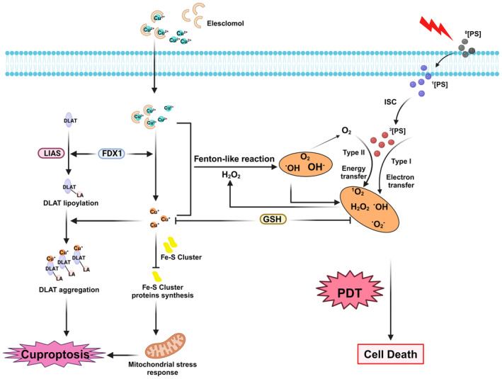
Background: Photodynamic therapy (PDT) is a noninvasive cancer treatment that works by using light to stimulate the production of excessive cytotoxic reactive oxygen species (ROS), which effectively eliminates tumor cells. However, the therapeutic effects of PDT are often limited by tumor hypoxia, which prevents effective tumor cell elimination. The oxygen (O2) consumption during PDT can further exacerbate hypoxia, leading to post-treatment adverse events.

Objectives: This review aims to explore the potential of cuproptosis, a recently discovered copper-dependent form of programmed cell death, to enhance the anticancer effects of PDT. Cuproptosis is highly dependent on mitochondrial respiration, specifically the tricarboxylic acid (TCA) cycle, and can increase O2 and ROS levels or decrease glutathione (GSH) levels, thereby improving PDT outcomes.

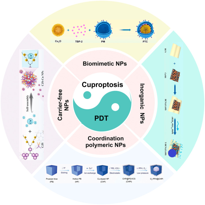
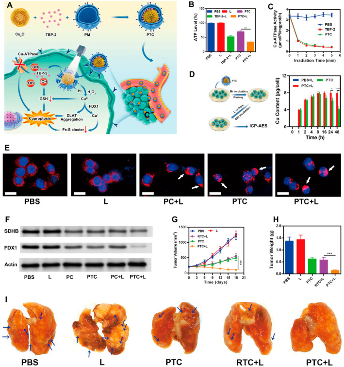
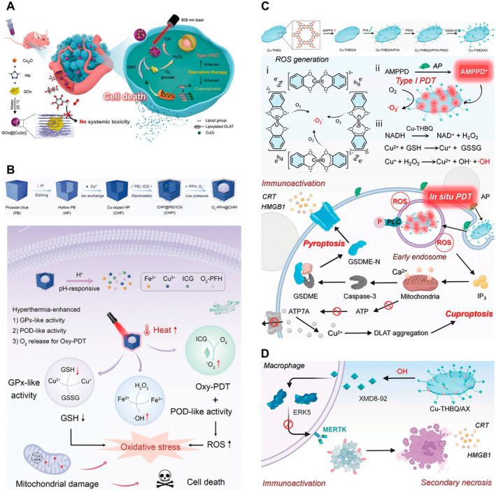
Methods: The review discusses the latest research advancements in the field, detailing the mechanisms that regulate cuproptosis and PDT. It also explores how nanoparticle (NP)-based strategies can be used to exploit the synergistic potential between cuproptosis and PDT. The article examines the prospects of synergistic anticancer activity guided by nanodelivery systems, which could overcome the challenges associated with hypoxia in cancer treatment.

Conclusions: The combination of cuproptosis and PDT, facilitated by NP-based delivery systems, presents a promising approach to enhance the effectiveness of cancer therapy. The review concludes by discussing the challenges and future research directions for this combination therapy, highlighting the need for further investigation into the mechanisms and optimization of treatment strategies to improve outcomes in cancer treatment.

Keywords: PDT; cancer; cuproptosis; therapy nanomaterials.


Conflict of interest statement

The authors declare no conflicts of interest.

Figures

FIGURE 1
FIGURE 1
Schematic mechanism of synergistic cancer therapy of cuproptosis and PDT. Type I and Type II reactions occur when a 0PS absorbs a photon and becomes excited to the 1PS. The excited PS can undergo ISC to form a triplet‐state 3PS. Type I involves electron transfer; Type II involves direct energy transfer. The Cu2+ released by elesclomol (ES) is reduced to Cu+ by the mitochondrial enzyme FDX1, leading to oligomerization of fatty acylated DLAT and instability of the Fe–S cluster protein, ultimately resulting in cuproptosis. GSH can act as a Cu‐chelating agent to remove excess Cu ions and as a reducing agent to remove ROS, significantly affecting the efficacy of cuproptosis and PDT. Cu ions can produce ROS and O2 through a Fenton‐like reaction. 0PS, singlet ground state photosensitizer; 1PS, singlet excited state photosensitizer; 3PS, excited triplet state photosensitizer; DLAT, dihydrolipoamide S‐acetyltransferase; FDX1, ferredoxin 1; GSH, glutathione; ISC, intersystem crossing; LA‐DLAT, lipoylated DLAT; LIAS, lipoyl synthase; ROS, reactive O2 species. Created with BioRender.com.
FIGURE 2
FIGURE 2
Schematic illustration of emerging NP‐assisted cuproptosis and PDT for synergistic cancer therapy. Created with BioRender.com.
FIGURE 3
FIGURE 3
(A) Schematic illustration of biomimetic system PTC for tumor cuproptosis/PDT synergistic therapy. (B) Intracellular ATP levels of 4T1 after different treatment. (C) Cu‐ATPase activity changes after specified treatment under white light irradiation. (D) Schematic illustration and results of measuring intracellular Cu content during the treatment period. (E) DLAT fluorescence images of cancer cells after the indicated treatment. (F) Fe–S cluster protein expression in cancer cells after the indicated treatments. (G, H) Evolution of the tumor volume/weight in mice with different treatments. (I) The collected lungs in mice with different treatments. 4T1, mouse‐derived breast cancer cell line; ATP, adenosine triphosphate; DLAT, dihydrolipoamide S‐acetyltransferase; FDX1, ferredoxin 1; GSH, glutathione; ICP‐AES, inductively coupled plasma—atomic emission spectroscopy; PC, PM‐coated Cu2O without TBP‐2; PM, platelet membrane; PTC, platelet membrane‐coated Cu2O/TBP‐2 biomimetic cuproptosis sensitization system; RTC, red cell membrane‐coated Cu2O/TBP‐2 biomimetic cuproptosis sensitization system; TBP‐2, thioflavin B‐Photosensitizer 2. Reprinted from Ref. [55] with permission. Copyright (2023) the American Chemical Society. *** p‐value < 0.001, which is considered to be highly significant.
FIGURE 4
FIGURE 4
Coordination polymer as nanocarriers for synergistic cancer therapy. (A) Schematic illustration of the synthesis of nonporous GOx@[Cu(tz)] and their mechanism (Gox, glucose oxidase; Htz, 1,2,4‐triazole). Reprinted from Ref [22] with permission. Copyright (2022) WILEY. (B) Schematic illustration of the synthesis of O2‐PFH@CHPI nanozymes and their mechanism (GPx‐like, glutathione peroxidase‐like; GSH, glutathione; GSSG, oxidized glutathione; Oxy‐PDT, oxygen self‐enriching PDT; POD‐like, peroxidase‐like; ROS, reactive oxygen species). Reprinted from Ref [58] with permission. Copyright (2024) WILEY. (C) Schematic illustration of the synthesis of Cu‐THBQ/AX and the mechanism of cuproptosis/PDT synergistic cancer therapy. (D) Release of XMD8‐92 triggered by •OH effectively inhibits macrophage phagocytosis of apoptotic cancer cells, converting immunosuppressive cell death into proinflammatory secondary necrosis (AMPPD, adenosine 5′‐monophosphate propyl disulfide; AP, antioxidant protein; ATP7A, Cu‐transporting ATPase 7A; CRT, calreticulin; Cu‐THBQ/AX, Cu‐tetrahydroxybenzoquinone nanosized metal–organic framework; PVA, poly(vinyl alcohol); Cu‐THBQ, Cu‐tetrahydroxybenzoquinone; ERK5, extracellular signalregulated kinase 5; GSDME, cleavage of gasdermin‐E; GSDME‐N, gasdermin E N‐terminal domain; HMGB1, high mobility group box 1 protein; IL‐10, immunosuppressive cytokine interleukin 10; IP3, intracellular 1,4,5‐triphosphate; MERTK, MER protooncogene tyrosine kinase; NADH, nicotinamide adenine dinucleotide; PSDA, 2,2′‐[propane‐2,2‐diylbis(thio)]diacetic acid; XMD8‐92, extracellular signalregulated kinase 5 inhibitor). Reprinted from Ref [59] with permission. Copyright (2024) the American Chemical Society.
FIGURE 5
FIGURE 5
(A) Schematic illustration of the synthesis of T‐HCN@CuMS Nanoagent and their mechanism. (B) ROS produced by photocatalytic reaction was tested by Rhodamine B degradation assay in vitro. (C) ESR analysis of three typical ROS generation performance of different NPs under light irradiation. (D) High‐resolution XPS spectra of Cu 2p in HCN@CuMS and HCN@MS nanoheterojunction. (E) Live/dead staining of 143B cells after different treatments. (CuMS, Cu‐loaded molybdenum bisulfide nanosheets; ESM1, endothelial cellspecific molecule 1; ESR, electron spin resonance; HCN, heterogeneous carbon nitride nanosheets; HCN@CuMS, heterogeneous carbon nitride nanosheets @ Cu‐loaded metallic molybdenum bisulfide nanosheets; HCN@MS, heterogeneous carbon nitride nanosheets @ metallic molybdenum bisulfide nanosheets; NIR, near‐infrared; RhB, rhodamine B; ROS, reactive oxygen species; T‐HCN@CuMS, targeting nanoheterojunction; XPS, X‐ray photoelectron spectroscopy). Reprinted from Ref. [70] with permission. Copyright (2023) the American Chemical Society.
FIGURE 6
FIGURE 6
(A) Schematic illustration of the synthesis of CJS‐Cu NPs and their mechanism. (B) ROS production pathway of CJS‐Cu NPs. (C) The time‐dependent fluorescence intensity of the CJS‐Cu NPs under light irradiation indicates the generation of •O2−. (D) The cell proliferation was evaluated after treatment with different concentrations of CJS‐Cu NPs under various lighting conditions, and exhibited high cell‐killing efficacy under photoirradiation. (E) The relative TMB absorbance upon the addition of CJS‐Cu NPs and Cu+ with different lighting conditions. (F) Western blot analysis on the expressions of LIAS, ACO‐2, and FDX1. (G) Relative GSH contents in 4T1 cells after different treatments. ACO‐2, aconitase 2; DLAT, dihydrolipoamide S‐acetyltransferase; FDX1, ferredoxin 1; GSH, glutathione; HGF, hepatocyte growth factor/scatter factor; LIAS, lipoic acid synthase; MTA2, metastasis‐associated protein 2; ROS, reactive oxygen species; S0, singlet ground state; S1, singlet first excited state; SDHB, Succinate Dehydrogenase Subunit B; T1, triplet first excited state; TMB, 3,3,5,5′‐Tetramethylbenzidine; VCAM‐1, vascular cell adhesion molecule‐1. Reprinted from Ref. [72] with permission. Copyright (2023) WILEY. *** p‐value < 0.001, which is considered to be highly significant.
FIGURE 7
FIGURE 7
(A) Schematic illustration of the fabrication of CCNAs and their mechanism. (B) Fluorescence images of DCFHDA‐stained PC‐3 cells after different treatments in normoxia and hypoxia. (C) H2O2 staining images of PC‐3 cells after CCNA treatment. (D) The viability of PC‐3 cells following various treatments. (E) Immunofluorescence staining of DLAT aggregation and FDX1 in PC‐3 cells in different groups. (F) Western blot analysis of FDX1 expression after different treatments. (G) The mean fluorescence intensity (MFI) of CRT surface exposure on PC‐3 cells after the indicated treatments. (H) The mean fluorescence intensity (MFI) of CRT surface exposure on PC‐3 cells after the indicated treatments. (I) The MFI of HMGB1 released after the indicated treatments in PC‐3 cells. CCNAs, Cu‐coordinated nanoassemblies; CTLs, cytotoxic T lymphocytes; CRT, calreticulin; CFHDA, 2′,7′‐dichlorodihydrofluorescein diacetate; DLAT, dihydrolipoamide s‐acetyltransferase; DOX, doxorubicin; FDXI, ferredoxin 1; GAPDH, glyceraldehyde 3‐phosphate dehydrogenase; ICD, immunogenic cell death; IDO, ndoleamine 2,3‐dioxygenase; PC‐3, prostate cancer cell line 3; TK, thioketal; Trp/Kyn, tryptophan/kynurenine ratio; ZnPc, zinc phthalocyanine; ZnPc‐TK‐DOX, a prodrug composed of the photosensitizer ZnPc and the chemotherapy drug DOX linked by thioketone TK). Reprinted from Ref. [24] with permission. Copyright (2024) ELSEVIER. A single asterisk (*) is used to denote a p‐value of less than 0.05, which is considered to be statistically significant. This means that there is less than a 5% probability that the observed difference occurred by chance. A double asterisk (**) is used to denote a p‐value of less than 0.01, which is considered to be highly statistically significant. This indicates that there is less than a 1% probability that the observed difference is due to random variation.

Similar articles

Cited by

References

    1. Bray F., Laversanne M., Sung H., et al., “Global Cancer Statistics 2022: GLOBOCAN Estimates of Incidence and Mortality Worldwide for 36 Cancers in 185 Countries,” CA: A Cancer Journal for Clinicians 74, no. 3 (2024): 229–263. - PubMed
    1. Kit O. I., Katelnitskaya O. V., Maslov A. A., et al., “Combined Surgeries for Retroperitoneal Tumors,” Journal of Clinical Oncology 38, no. 15_suppl (2020): e23579.
    1. Chen H., Han Z., Luo Q., et al., “Radiotherapy Modulates Tumor Cell Fate Decisions: A Review,” Radiation Oncology 17, no. 1 (2022): 196. - PMC - PubMed
    1. D'alterio C., Scala S., Sozzi G., et al., “Paradoxical Effects of Chemotherapy on Tumor Relapse and Metastasis Promotion,” Seminars in Cancer Biology 60 (2020): 351–361. - PubMed
    1. Wang Y., Wang D., Zhang Y., et al., “Tumor Microenvironment‐Adaptive Nanoplatform Synergistically Enhances Cascaded Chemodynamic Therapy,” Bioactive Materials 22 (2023): 239–253. - PMC - PubMed