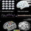
White matter connections within the central sulcus subserving the somato-cognitive action network
- PMID: 39869456
- PMCID: PMC12073987
- DOI: 10.1093/brain/awaf022
White matter connections within the central sulcus subserving the somato-cognitive action network
Abstract
The somato-cognitive action network (SCAN) consists of three nodes interspersed within Penfield's motor effector regions. The configuration of the somato-cognitive action network nodes resembles the one of the 'plis de passage' of the central sulcus: small gyri bridging the precentral and postcentral gyri. Thus, we hypothesize that these may provide a structural substrate of the somato-cognitive action network. Using microdissections of 16 human hemispheres, we consistently identified a chain of three distinct plis de passage with increased underlying white matter in locations analogous to the somato-cognitive action network nodes. We mapped localizations of plis de passage into standard stereotactic space to seed functional MRI connectivity across 9000 resting-state functional MRI scans, which demonstrated the connectivity of these sites with the somato-cognitive action network. Intraoperative recordings during direct electrical central sulcus stimulation further identified inter-effector regions corresponding to plis de passage locations. This work provides a critical step towards an improved understanding of the somato-cognitive action network in both structural and functional terms. Furthermore, our work has the potential to guide the development of refined motor cortex stimulation techniques for treating brain disorders and operative resective techniques for complex surgery of the motor cortex.
Keywords: SCAN; motor cortex; plis de passage; somato-cognitive action network; white matter connectivity.
© The Author(s) 2025. Published by Oxford University Press on behalf of the Guarantors of Brain.
Conflict of interest statement
N.U.F.D. has a financial interest in Turing Medical Inc. and may financially benefit if the company is successful in marketing FIRMM motion monitoring software products. E.M.G. and N.U.F.D. may receive royalty income based on FIRMM technology developed at Washington University School of Medicine and licensed to Turing Medical Inc. N.U.F.D. is a co-founder of Turing Medical Inc. These potential conflicts of interest have been reviewed and are managed by Washington University School of Medicine. A.H. reports lecture fees for Boston Scientific and is a consultant for FxNeuromodulation and Abbott.
Figures


Comment in
-
Shedding light on a novel circuit within primary motor cortex as a target for neuromodulation.Brain. 2025 May 13;148(5):1454-1455. doi: 10.1093/brain/awaf128. Brain. 2025. PMID: 40261808 No abstract available.
References
-
- Piotrowska N, Winkler PA. Otfrid Foerster, the great neurologist and neurosurgeon from Breslau (Wrocław): His influence on early neurosurgeons and legacy to present-day neurosurgery. J Neurosurg. 2007;107:451–456. - PubMed
-
- Penfield W, Boldrey E. Somatic motor and sensory representation in the cerebral cortex of man as studied by electrical stimulation1. Brain. 1937;60:389–443.
-
- Gray GW. The great ravelled knot. Sci Am. 1948;179:26–39. - PubMed
-
- Kandel ER, Koester JD, Mack SH, Siegelbaum SA. Principles of neural science. 6th edition. McGraw Hill; 2021.

