. 2025 Feb 17;482(4):BCJ20240608.
doi: 10.1042/BCJ20240608.

Characterisation of RNA guanine-7 methyltransferase (RNMT) using a small molecule approach

Affiliations

Characterisation of RNA guanine-7 methyltransferase (RNMT) using a small molecule approach

Lesley-Anne Pearson et al. Biochem J. .

Abstract

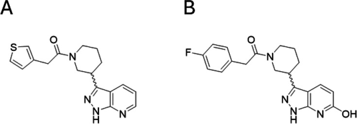
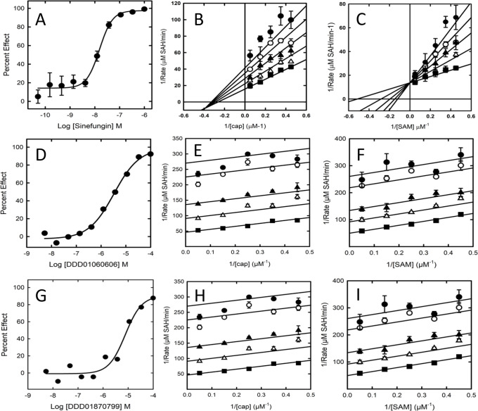
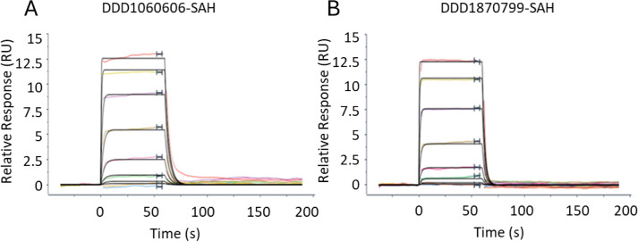
The maturation of the RNA cap involving guanosine N-7 methylation, catalyzsed by the HsRNMT (RNA guanine-7 methyltransferase (HsRNMT)-RAM (RNA guanine-N7 methyltransferase activating subunit (RAM) complex, is currently under investigation as a novel strategy to combat PIK3CA -mutant breast cancer. However, the development of effective drugs is hindered by a limited understanding of the enzyme's mechanism and a lack of small molecule inhibitors. Following the elucidation of the HsRNMT-RAM molecular mechanism, we report the biophysical characterizsation of two small molecule hits. Biophysics, biochemistry and structural biology confirm that both compounds bind competitively with cap and bind effectively to HsRNMT-RAM in the presence of the co-product SAH, with a binding affinity (KD) of approximately 1 μM. This stabilisation of the enzyme--product complex results in uncompetitive inhibition. Finally, we describe the properties of the cap pocket and provided suggestions for further development of the tool compounds.

Keywords: RNMT; Sinefungin; breast cancer; methyl transferase.


Conflict of interest statement

The Authors declare that there are no competing interests associated with this manuscript.

Figures

Figure 1
Figure 1. Diagram of enzymatic reaction and structures of key substrate and analogues.
(A) Diagram of enzymatic reaction carried ofout by RNMT. The N7 nitrogen on the cap and the methyl group are highlighted in red. (B) The cap structure minus RNA as used in the present paperstudy. (C) GMP-PnP as used as a cap surrogate in the crystallography experiments and (D) Sinefungin, a non-selective methyltransferase inhibitor acting as a SAM mimetic. Abbreviations: GMP-PnP, guanosine 5’-[β,γ-imido]triphosphate; RNMT, RNA guanine-7 methyltransferase; SAM, S-adenosylmethionine.
Figure 2
Figure 2. Diagram comparing the two assay formats developed.
Both measure SAH. The MTase-Glo™ assay uses coupling enzymes to convert SAH to a light readout. The RapidFire™ mass spectrometry assay measures SAH levels directly. Abbreviation: SAH, S-adenosylhomocysteine.
Figure 3
Figure 3. Sensograms derived from SPR-binding experiments. All experiments were conducted with eight concentrations.
SAM: - top concentration 5 mM, 1 in 3 dilutions (A), SAH: – top concentration 5 mM, 1 in 3 dilutions (B), Sinefungin: – top concentration 0.5 mM, 1 in 2 dilutions (C), or G(5′)ppp(5′)G-Sinefungin: – top concentration 0.37 mM, 1 in 3 dilutions were flowed over immobilizsed RNMT-RAM complexes. Data isare representative of 2two experiments. Data wasere processed using Langmuir 1:1 model and isare summarizsed in Table 1. Abbreviations: RAM, RNMT-activating miniprotein; RNMT, RNA guanine-7 methyltransferase; SAH, S-adenosylhomocysteine; SAM, S-adenosylmethionine; SPR, surface plasmon resonance.
Figure 4
Figure 4. Michaelis–Menten plots for HsRNMT-RAM substrates.
cap (40–0.078 µM) (A) and SAM (40–0.078 µM) (B). Plots shown are representative examples from three experiments. Error bars are ± standard deviation from four technical replicates. Abbreviations: RAM, RNMT-activating miniprotein; RNMT, RNA guanine-7 methyltransferase; SAM, S-adenosylmethionine.
Figure 5
Figure 5. Chemical structures of (A) DDD1060606 and (B) DDD1870799. DDD1060606 and DDD1870799 showed >80% inhibition of HsRNMT-RAM activity with determined pIC50s (negative logarithm of half-maximal inhibitory concentration [molar]) of 5.5 ± 0.2 (IC50 3 mM; Figure 6D and 5.0 ± 0.2 (IC50 10 mM; Figure 6G), respectively. Abbreviations: RAM, RNMT-activating miniprotein; RNMT, RNA guanine-7 methyltransferase.
Figure 6
Figure 6. Characterisation of inhibition by Sinefungin, DDD1060606 and DDD1870799.
(A) is tThe inhibition curve for Sinefungin (10 point 1:3 dilutions starting at 1 μM, duplicates on assay plate). (B) The Sinefungin ((0 (■), 40 (△), 80 (▲), 120 (◯) and 160 nM (●)) Lineweaver–-Burk plot against cap. and (C) The Sinefungin ((0 (■), 8 (△), 16 (▲), 24 (◯) and 32 μM (●)) Lineweaver-–Burk plot against SAM. (D–F) The inhibition curve (10 point 1:3 dilutions starting at 100 μM, singlicates on assay plate) and Lineweaver-–Burk plots against cap and SAM, respectively, for DDD1060606 (0 (■), 1.25 (△), 2.5 (▲), 5 (◯) and 6.25 μM (●)). (G–I) The inhibition curve (10 point 1:3 dilutions starting at 100 μM, singlicates on assay plate) and Lineweaver-–Burk plots against cap and SAM, respectively, for DDD1870799 (0 (■), 1.25 (△), 2.5 (▲), 5 (◯) and 6.25 μM (●)). Inhibition curves and Lineweaver-–Burk plots are representative of 3three experiments. Error bars in the Lineweaver-–Burk plots are standard deviation from three technical replicates. Abbreviation: SAM, S-adenosylmethionine.
Figure 7
Figure 7. Sensograms derived from SPR binding experiments.
All experiments were conducted with an eight concentration in a 1:3 serial dilution. DDD1060606: – top concentration 30 mM (A) or DDD1870799: – top concentration 30 mM (B) were flowed over immobilizsed RNMT-RAM complexes in the presence of SAH (5 mM). Data isare representative of 2two experiments. Data wasere processed using Langmuir 1:1 model and are summarizsed in Table 3. Abbreviations: RAM, RNMT-activating miniprotein; RNMT, RNA guanine-7 methyltransferase; SAH, S-adenosylhomocysteine; SPR, surface plasmon resonance.
Figure 8
Figure 8. Structural analysis of the HsRNMT cap binding site.
(A) GMP-PnP in complex with Sinefungin, (B) DDD1060606 in complex with SAH and (C) DDD1870799 in complex with SAH. Residues within 4 Å are drawn in sticks. Dash lines represent hydrogen bonds (black), salt bridges (red) and aromatic H-bonds (cyan). Abbreviations: GMP-PnP, guanosine 5′-[β,γ-imido]triphosphate; RNMT, RNA guanine-7 methyltransferase; SAH, S-adenosylhomocysteine.
Figure 9
Figure 9. Cap, DDD1060606 and DDD1870799 titrations into RNMT-RAM.
(A) Raw data and integration of data, corrected for the heat of dilution. The line represents the least-squares fit to the single-site binding model by the ORIGIN program. (B) Thermodynamic signatures (ΔG° in blue, ΔH° in green, − TΔS° in red, all data in Kkcal/mol). Abbreviations: RAM, RNMT-activating miniprotein; RNMT, RNA guanine-7 methyltransferase.
Figure 10
Figure 10. Potential compound development routes informed by MIF-based structures.
The GRID MIFs calculated for the RNMT polypeptide chain (with corresponding energy cut-offs in brackets) are contoured yellow (hydrophobic: −1.5 kcal/mol), blue (hydrogen-bond donor: −5.5 kcal/mol) and red (hydrogen-bond acceptor: −5.0 kcal/mol). (A) Sinefungin, (B) cap analogue with amine modified to methyl group, (C) SAH-DDD1060606 and (D) SAH-DDD1870799 crystal structures. Abbreviations: MIF, molecular interaction field; RNMT, RNA guanine-7 methyltransferase; SAH, S-adenosylhomocysteine.
Figure 11
Figure 11. Diagram summarising the ordered bi-bi mechanism of enzymatic activity of RNMT and the enzyme-product stabilisation driven by binding of the compounds resulting in the observed uncompetitive inhibition. Abbreviation: RNMT, RNA guanine-7 methyltransferase.

Similar articles

Cited by

References

    1. Galloway A., Cowling V.H. mRNA cap regulation in mammalian cell function and fate. Biochim. Biophys. Acta Gene Regul. Mech. 2019;1862:270–279. doi: 10.1016/j.bbagrm.2018.09.011. - DOI - PMC - PubMed
    1. Pelletier J., Schmeing T.M., Sonenberg N. The multifaceted eukaryotic cap structure. Wiley Interdiscip. Rev. RNA. 2021;12:e1636. doi: 10.1002/wrna.1636. - DOI - PubMed
    1. Inesta-Vaquera F., Cowling V.H. Regulation and function of CMTR1-dependent mRNA cap methylation. Wiley Interdiscip. Rev. RNA. 2017;8:e1450. doi: 10.1002/wrna.1450. - DOI - PMC - PubMed
    1. Liang S., Almohammed R., Cowling V.H. The RNA cap methyltransferases RNMT and CMTR1 co-ordinate gene expression during neural differentiation. Biochem. Soc. Trans. 2023;51:1131–1141. doi: 10.1042/BST20221154. - DOI - PMC - PubMed
    1. Furuichi Y. Discovery of m(7)G-cap in eukaryotic mRNAs. Proc. Jpn. Acad., Ser. B, Phys. Biol. Sci. 2015;91:394–409. doi: 10.2183/pjab.91.394. - DOI - PMC - PubMed

Publication types

MeSH terms

LinkOut - more resources