. 2025 Jan 27;11(1):27.
doi: 10.1038/s41420-025-02304-3.

METTL1 coordinates cutaneous squamous cell carcinoma progression via the m7G modification of the ATF4 mRNA

Affiliations

METTL1 coordinates cutaneous squamous cell carcinoma progression via the m7G modification of the ATF4 mRNA

Xinru Zhang et al. Cell Death Discov. .

Abstract

Methyltransferase-like 1 (METTL1)-mediated m7G modification is a common occurrence in various RNA species, including mRNAs, tRNAs, rRNAs, and miRNAs. Recent evidence suggests that this modification is linked to the development of several cancers, making it a promising target for cancer therapy. However, the specific role of m7G modification in cutaneous squamous cell carcinoma (cSCC) is not well understood. In this study, we observed conspicuously elevated levels of METTL1 in cSCC tumors and cell lines. Inhibiting METTL1 led to reduced survival, migration, invasion, and xenograft tumor growth in cSCC cells. Mechanistically, through a combination of RNA sequencing, m7G methylated immunoprecipitation (MeRIP)-qPCR, and mRNA stability assays, we discovered that METTL1 is responsible for the m7G modification of ATF4 mRNA, leading to increased expression of ATF4. Importantly, we demonstrated that this modification is dependent on the methyltransferase activity of METTL1. Additionally, we observed a positive association between ATF4 expression and METTL1 levels in cSCC tumors. Intriguingly, restoring ATF4 expression in cSCC cells not only promoted glycolysis but also reversed the anti-tumor effects of METTL1 knockdown. In conclusion, our results underscore the critical role of METTL1 and m7G modification in cSCC tumorigenesis, suggesting a promising target for future cSCC therapies.


Conflict of interest statement

Competing interests: The authors declare no competing interests. Ethical approval and consent to participate: All the experiments related to human tissues were conducted in accordance with Helsinki criteria and were approved by the Ethics Committees of Shanghai Outdo Biotech Co. Ltd (approval number SHYJS-CP-2001009). All mice studies are conducted according to protocols approved by the Animal Ethics Committee of Anhui Medical University with the approval number LLSC20240910.

Figures

Fig. 1
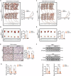
Fig. 1. METTL1 is overexpressed in cSCC tumors and cell lines.
A The protein levels of METTL1 in normal specimens (n = 7) and cSCC tumors (n = 36) with different tumor grade were detected by IHC. B The samples highlighted in red in A were magnified. The intensity optical density (IOD) for each sample was calculated. C The mRNA levels of METTL1 in normal specimens and cSCC tumors were detected by qRT-PCR. D, E The expression of METTL1 in HaCaT keratinocytes and cSCC cell lines (HSC-1 and A431) were detected by western blot and qRT-PCR, respectively. β-Tubulin was used for the normalization control. The relative protein level of METTL1 was calculated. F The m7G methylation levels in total RNA and decapped mRNA from cell lines were detected by dot blot assay. Data are shown as mean ± SEM. ***P < 0.001, n = 6. MB methylene blue.
Fig. 2
Fig. 2. METTL1 knockdown significantly represses cSCC cells survival.
A, B Western blot and qRT-PCR showed METTL1 was successfully knocked down by shMETTL1 at protein and mRNA levels. β-Tubulin was used for the normalization control. C The m7G methylation levels in METTL1-knockdown cSCC cell lines were detected by dot blot assay. D Measurements of cell viability by CCK8 assay (n = 6). E EdU staining assay was used to assess cell proliferation (n = 3). The relative ratio of EdU+ cells were calculated. F The proportions of EdU+ cells were detected by flow cytometry (n = 3). G Measurements of cell proliferation by colony formation assay (n = 3). The colony number was calculated. H The cell apoptosis was determined by Annexin V/PI double staining (n = 6). I, J The protein and mRNA levels of BCL-2 and BAX were detected by western blot and qRT-PCR (n = 6), respectively. β-Tubulin was used for the normalization control. K, L TUNEL staining of HSC-1 and A431 cells was performed (n = 3). Data are shown as mean ± SEM. *P < 0.05, **P < 0.01, ***P < 0.001.
Fig. 3
Fig. 3. METTL1 knockdown inhibits migration and invasion of cSCC cells.
A Migration of HSC-1 and A431 cells was evaluated by wound-healing assay (n = 3). The migration area was calculated. B The mobility was assessed by trans-well migration assay (n = 5). C The invasiveness of HSC-1 and A431 cells was evaluated by Matrigel invasiveness measurement (n = 5). Data are shown as mean ± SEM. ***P < 0.001.
Fig. 4
Fig. 4. METTL1 deficiency reduces xenograft tumor growth in vivo.
Knockdown of METTL1 inhibited HSC-1 xenograft tumor growth (A) and reduced tumor weight (B). Knockdown of METTL1 inhibited A431 xenograft tumor growth (C) and reduced tumor weight (D). E, F The protein levels of PCNA in tumor tissues were detected by IHC and western blot. The IOD of PCNA was calculated. β-Tubulin was used for the normalization control. The relative protein level was calculated. G The mRNA level of Pcna was evaluated by qRT-PCR. Data are shown as mean ± SEM. n = 5. **P < 0.01, ***P < 0.001.
Fig. 5
Fig. 5. METTL1 induces the m7G modification of ATF4 mRNA.
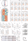
A Volcano plot of significantly altered mRNA in METTL1-knockdown HSC-1 cell compared to control cell. B Differentially expressed genes (DEG) were clustered and shown in a heat map of RNA sequencing. C, D qRT-PCR was performed to detect the downregulated DEG in HSC-1 and A431 cells (n = 6). E Suppressed m7G methylated ATF4 mRNA in METTL1-knockdown cells was detected by MeRIP-qPCR (n = 3). F qRT-PCR showing ATF4 mRNA transcripts stability in ActD-treated HSC-1 and A431 cells transfected with or without shMETTL1 (n = 3). G The protein levels of ATF4 in HSC-1 and A431 cells with or without shMETTL1 were detected by western blot (n = 3). β-Tubulin was used for the normalization control. H The mRNA levels of ATF4 in cells transfected with WT METTL1 or Mut METTL1 were assessed by qRT-PCR (n = 6). I qRT-PCR showing ATF4 mRNA transcripts stability in ActD-treated HSC-1 and A431 cells transfected with WT METTL1 or Mut METTL1 (n = 3). J The protein levels of ATF4 in cells transfected with WT METTL1 or Mut METTL1 were assessed by western blot (n = 3). β-Tubulin was used for the normalization control. K The protein levels of METTL1 and ATF4 in normal specimens (n = 7) and cSCC tumors (n = 36) with different tumor grade were detected by IHC on continuous sections. The intensity optical density (IOD) for each sample was calculated. L Scatter plots showing the positive correlation between METTL1 and ATF4 expression in cSCC tumors. Data are shown as mean ± SEM. *P < 0.05, **P < 0.01, ***P < 0.001.
Fig. 6
Fig. 6. ATF4 overexpression reverses the anti-tumor effect of METTL knockdown.
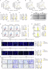
A, B Metabolic phenotypes of HSC-1 and A431 cells were determined by the both OCR and ECAR assays (n = 5). C Glucose uptake in HSC-1 and A431 cells were detected glucose assay (n = 5). D The ATP production was evaluated through ATP assay kit (n = 5). E Lactate levels in the medium were assessed (n = 5). F The protein levels of GLUT1, HK2 and LDHA were detected by western blot (n = 5). β-Tubulin was used for the normalization control. G MMP was measured by flow cytometry (n = 3). H The mitochondrial mass was measured by MitoTracker (n = 3). I EdU staining assay was used to assess cell proliferation (n = 3). J TUNEL staining of HSC-1 and A431 cells was performed (n = 3). K The mobility and invasiveness were assessed by trans-well migration assay and matrigel invasiveness measurement (n = 5), respectively. Data are shown as mean ± SEM. *P < 0.05, ***P < 0.001.
Fig. 7
Fig. 7. ATF4 overexpression counteracted the inhibitory effects of METTL knockdown on tumor growth in vivo.
ATF4 overexpression promoted METTL1-deficient HSC-1 xenograft tumor growth (A) and reduced tumor weight (B). ATF4 overexpression promoted METTL1-deficient A431 xenograft tumor growth (C) and reduced tumor weight (D). E, F The protein levels of PCNA in tumor tissues were detected by IHC and western blot. The IOD of PCNA was calculated. β-Tubulin was used for the normalization control. The relative protein level was calculated. G The mRNA level of Pcna was evaluated by qRT-PCR. H The schematic diagram of oncogenic effects of METTL1-midiated m7G modification in cSCC through enhancing the mRNA stability of ATF4 and subsequently expression. Data are shown as mean ± SEM. n = 5. **P < 0.01, ***P < 0.001.

Similar articles

Cited by

References

    1. Hasan N, Nadaf A, Imran M, Jiba U, Sheikh A, Almalki WH, et al. Skin cancer: understanding the journey of transformation from conventional to advanced treatment approaches. Mol Cancer. 2023;22:168. - PMC - PubMed
    1. Winge MCG, Kellman LN, Guo K, Tang JY, Swetter SM, Aasi SZ, et al. Advances in cutaneous squamous cell carcinoma. Nat Rev Cancer. 2023;23:430–49. - PubMed
    1. Mascaraque-Checa M, Gallego-Rentero M, Nicolas-Morala J, Portillo-Esnaola M, Cuezva JM, Gonzalez S, et al. Metformin overcomes metabolic reprogramming-induced resistance of skin squamous cell carcinoma to photodynamic therapy. Mol Metab. 2022;60:101496. - PMC - PubMed
    1. Riihila P, Nissinen L, Kahari VM. Matrix metalloproteinases in keratinocyte carcinomas. Exp Dermatol. 2021;30:50–61. - PMC - PubMed
    1. Knackstedt TJ, Knackstedt RW, Djohan M, Djohan R, Gastman BR, Crowe DR. New Developments in the Management of Cutaneous Squamous Cell Carcinoma. Plast Reconstr Surg. 2021;147:492–504. - PubMed

LinkOut - more resources