. 2025 Feb;57(1):264-280.
doi: 10.1038/s12276-024-01383-z. Epub 2025 Jan 28.

RECQL4 requires PARP1 for recruitment to DNA damage, and PARG dePARylation facilitates its associated role in end joining

Affiliations

RECQL4 requires PARP1 for recruitment to DNA damage, and PARG dePARylation facilitates its associated role in end joining

Mansoor Hussain et al. Exp Mol Med. 2025 Feb.

Abstract

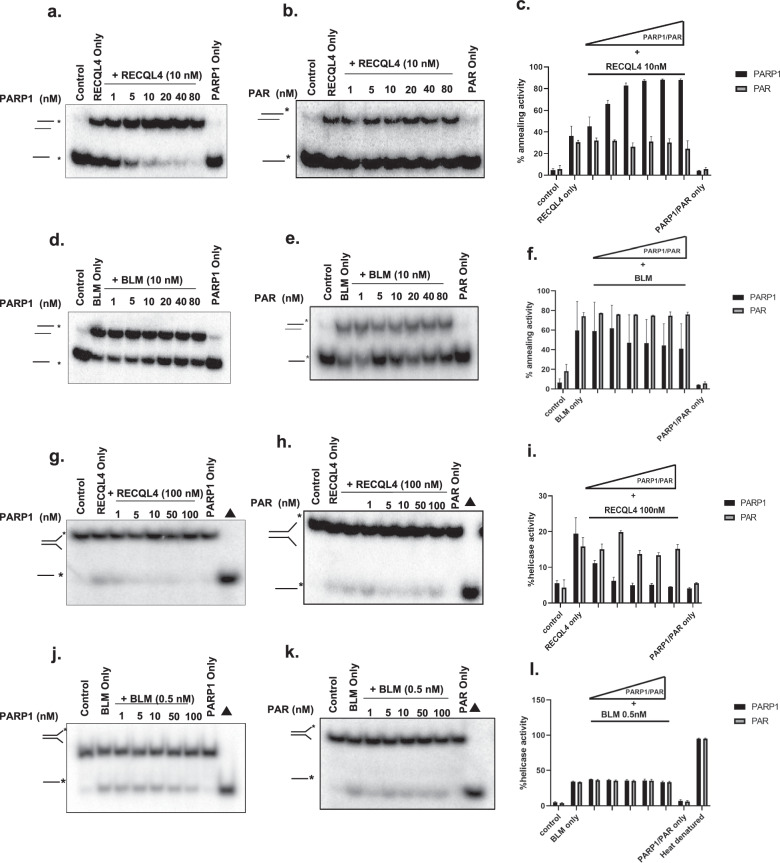
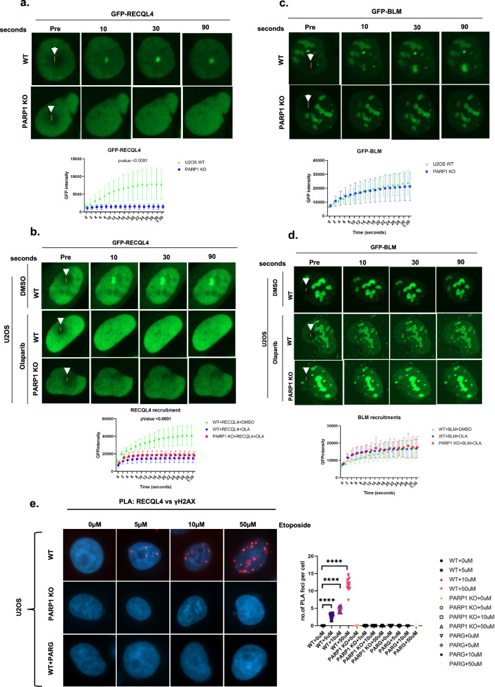
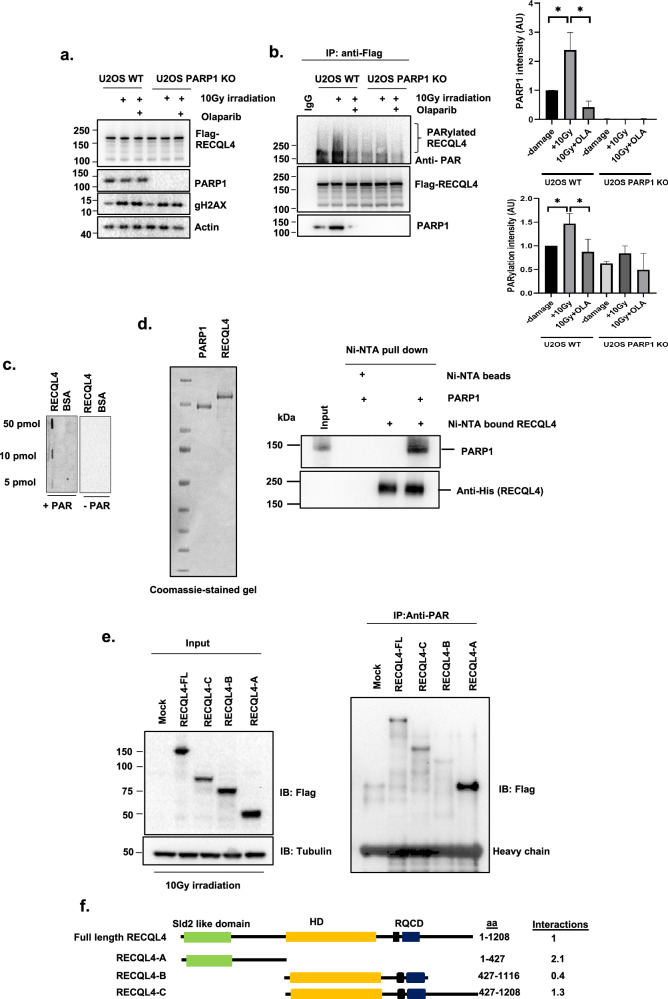
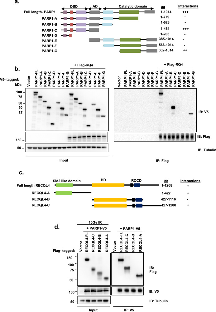
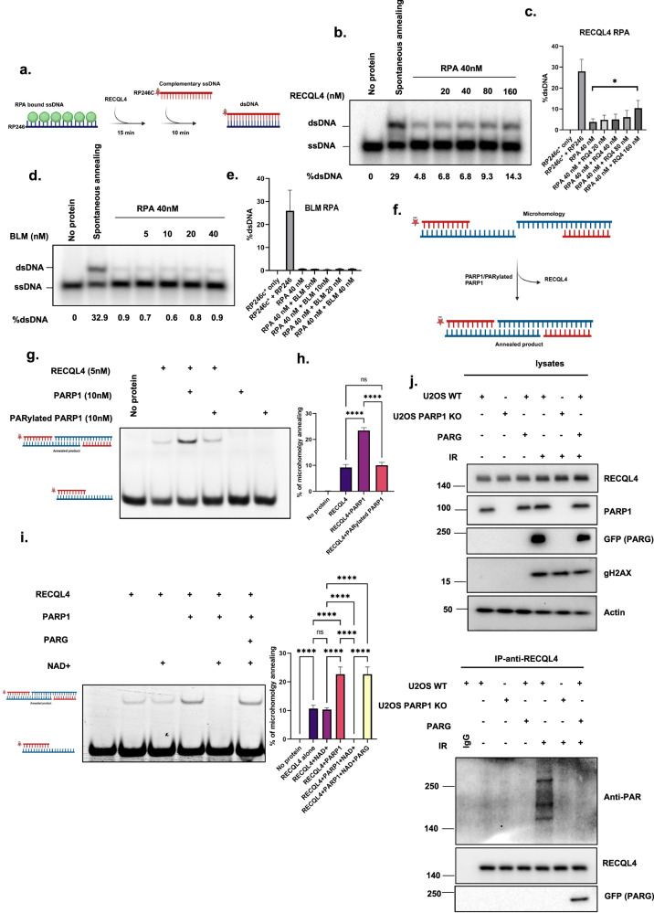
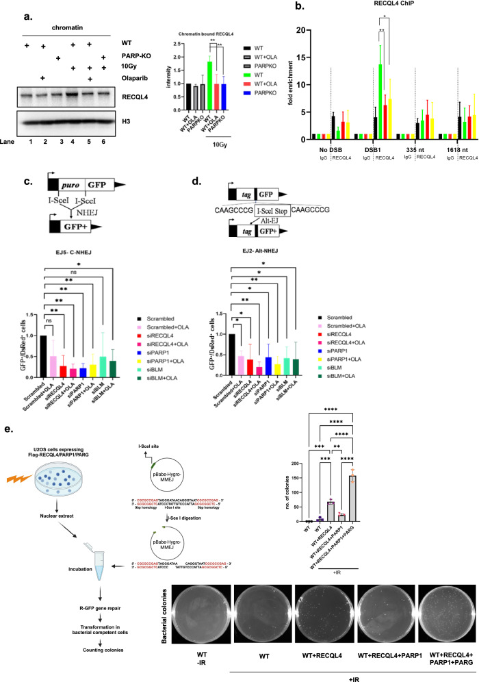
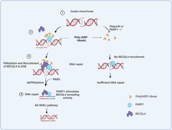
RecQ helicases, highly conserved proteins with pivotal roles in DNA replication, DNA repair and homologous recombination, are crucial for maintaining genomic integrity. Mutations in RECQL4 have been associated with various human diseases, including Rothmund-Thomson syndrome. RECQL4 is involved in regulating major DNA repair pathways, such as homologous recombination and nonhomologous end joining (NHEJ). RECQL4 has more prominent single-strand DNA annealing activity than helicase activity. Its ability to promote DNA damage repair and the precise role of its DNA annealing activity in DNA repair are unclear. Here we demonstrate that PARP1 interacts with RECQL4, increasing its single-stranded DNA strand annealing activity. PARP1 specifically promoted RECQL4 PARylation at both its N- and C-terminal regions, promoting RECQL4 recruitment to DNA double-strand breaks (DSBs). Inhibition or depletion of PARP1 significantly diminished RECQL4 recruitment and occupancy at specific DSB sites on chromosomes. After DNA damage, PARG dePARylated RECQL4 and stimulated its end-joining activity. RECQL4 actively displaced replication protein A from single-stranded DNA, promoting microhomology annealing in vitro. Furthermore, depletion of PARP1 or RECQL4 substantially impacted classical-NHEJ- and alternative-NHEJ-mediated DSB repair. Consequently, the combined activities of PARP1, PARG and RECQL4 modulate DNA repair.


Conflict of interest statement

Competing interests: The authors declare no competing interests.

Figures

Fig. 1
Fig. 1. RECQL4-catalyzed strand annealing activity is stimulated by non-PARylated PARP1.
a, b, The strand annealing activity of RECQL4 (10 nM) examined in the presence of increasing concentrations (0, 1, 5, 10, 20, 40 and 80 nM) of non-PARylated PARP1 (PARP1) (a) or PAR (b) with radiolabeled ssDNA 80 mer DNA and its complimentary single-stranded DNA. c, Graph showing the quantitative results of a and b. d, e, BLM (10 nM) strand annealing activity measured in the presence of increasing concentrations (0, 1, 5, 10, 20, 40 and 80 nM) of non-PARylated PARP1 (PARP1) (d) or PAR (e) with radiolabeled ssDNA 80 mer DNA and its complimentary single-stranded DNA. f, Graph showing the quantification results of d and e. g, h, Helicase activity of RECQL4 (100 nM) measured in the presence of increasing concentrations (1, 5, 10, 50 and 100 nM) of non-PARylated PARP1 (g) or PAR (h) with radiolabeled duplex fork DNA. i, A graph showing the quantitative results of g and h. j, k, BLM helicase activity (0.5 nM) was measured in the presence of increasing concentrations (1, 5, 10, 50 and 100 nM) of non-PARylated PARP1 (j) or PAR (k) with radiolabeled duplex fork DNA. l, Graph showing the quantification results of j and k. PARP1 and PAR alone have no helicase activity and ∆ represents the denatured substrate control. All experiments were repeated at least three times and the error bars represent the s.e.m.
Fig. 2
Fig. 2. PARP1 is required for the early recruitment of RECQL4 to sites of DNA damage.
a, RECQL4 recruitment to laser-induced DNA damage. GFP-tagged RECQL4 was transiently transfected into U2OS cells (WT or PARP1 KO) for 24 h. The cells were targeted with a 21% laser to induce DSBs. The cells were imaged at the indicated time points to observe the recruitment of the proteins to the damaged DNA. The recruitment kinetics of cells from three independent experiments were quantified for GFP–RECQL4. b, RECQL4, showing the same type of cells (U2OS) that were pretreated for 3 h with 5 µM olaparib and then targeted with the 21% laser to induce DSBs. c, GFP-tagged BLM was transiently transfected into U2OS cells (WT or PARP1 KO) for 24 h. The cells were targeted with a 21% laser to induce DSBs. The cells were imaged at the indicated time points to observe the recruitment of the proteins to the damaged DNA. The recruitment kinetics of cells from three independent experiments were quantified for GFP–BLM. d, GFP–BLM image showing the same type of cells (U2OS) that were pretreated for 3 h with 5 µM olaparib and then targeted with the 21% laser to induce DSBs. In ad, the lower graphics represent the relative signal intensities at the laser line calculated using volocity software and plotted versus time. The white arrow indicates the laser striking area. The error bars represent the s.e.m. Two-way analysis of variance (ANOVA) was performed to assess significant differences (*P < 0.05, **P < 0.01 and ***P < 0.001). e, Proportional increase in RECQL4–γH2AX foci in response to etoposide dosage. U2OS cells, including WT, PARP1 KO and PARG-expressing cells, were treated with either dimethylsulfoxide (DMSO) or specified concentrations of etoposide for 2 h. PLA staining was subsequently performed with anti-RECQL4 and anti-γH2AX antibodies. The graph shows the number of PLA foci per cell under various treatment conditions. Statistical analysis was conducted using two-way ANOVA to evaluate significant differences (*P < 0.05, **P < 0.01 and ****P < 0.0001).
Fig. 3
Fig. 3. RECQL4 interacts with PARP1 and PAR.
a, U2OS WT cells were transfected with the indicated plasmids and treated with or without 5 µM olaparib for 3 h before 10 Gy of IR DNA damage. Protein inputs were assessed by western blotting with the indicated antibodies. Actin served as the endogenous loading control. b, Whole-cell extracts from U2OS cells were immunoprecipitated with anti-Flag beads and immunoblotted with anti-PAR, anti-PARP1 and anti-Flag antibodies (left). Densitometric analysis was performed to measure the extent of the RECQL4‒PARP1 interaction and RECQL4 PARylation (right). N = 3, a Student’s t-test (two-sided) was performed to assess statistical significance (*P < 0.05, **P < 0.01 and ***P < 0.001) c, Slot‒blot assays were used to analyze the binding of purified RECQL4 proteins (50, 10 and 5 pmol) to PAR (how much PAR was loaded on a blot) that were dot blotted onto a nitrocellulose membrane. BSA (50, 10 and 5 pmol) was used as a negative control. d, The interaction between RECQL4 and PARP1 was investigated. Purified recombinant RECQL4 bound to Ni-NTA beads and soluble PARP1 were detected via Coomassie-stained gel (left). The interaction between bound RECQL4 and soluble PARP1 was analyzed via western blotting with anti-His (for RECQL4) and anti-PARP1 antibodies (right). e, PAR pulldown assays with RECQL4 fragments. After 10 Gy of IR damage, cell lysates were prepared from cells overexpressing either control Flag-tagged RECQL4 or various Flag-tagged RECQL4 domains. These lysates were incubated with an anti-PAR antibody to pull down PARylated proteins. The immunoprecipitants were then probed with an anti-Flag antibody. f, An illustration of Flag-tagged RECQL4 full-length RECQL4 and its fragments used for the binding assay. The values represent the degree of PARylation, which was determined through densitometry analysis of the pulled-down RECQL4 fragments relative to their initial input. This method quantifies PARylation on RECQL4.
Fig. 4
Fig. 4. Identification of the PARP1 domains required for interaction with RECQL4.
a, An illustration of V5-tagged full-length PARP1 and fragments used for binding assays. The numbers denote amino acid (aa) residues. The results from b are scored in the right column as RECQL4 binding. b, Cell extracts from HEK293T cells expressing V5-tagged full-length or fragments of PARP1 with or without Flag–RECQL4 were immunoprecipitated with an anti-FLAG antibody, followed by immuno blotting (IB) with an anti-V5 antibody. c, An illustration of Flag-tagged full-length RECQL4 and fragments used for binding assays. The numbers denote amino acid residues. Sld2-like domain and HD. The results from d are scored with + in the right column as PARP1 binding. d, After 10 Gy of IR DNA damage, extracts from HEK293T cells expressing Flag-tagged full-length or fragments of RECQL4 with V5-tagged PARP1 were immunoprecipitated with an anti-V5 antibody, followed by western blotting with anti-Flag and anti-V5 antibodies.
Fig. 5
Fig. 5. RECQL4 displaces RPA to promote microhomology-mediated annealing in alt-NHEJ.
a, A schematic diagram of the annealing assay used to study whether RECQL4 stimulates the annealing of RPA-bound ssDNA. b, A representative nondenaturing gel showing ssDNA annealing in the presence of RPA with the indicated amounts of RECQL4. The percentage of dsDNA is indicated. c, A graph showing a quantitative representation of the data in b. d, Representative nondenaturing gel showing ssDNA annealing in the presence of RPA with the indicated amount of BLM. The percentage of dsDNA is indicated. e, Graph showing a quantitative representation of the data in d. f, A schematic diagram of the in vitro end-joining assay. g, A nondenaturing gel showing RECQL4-mediated microhomology annealing in the presence of the mentioned protein concentrations. h, A graph showing a quantitative representation of the data in g. i, A nondenaturing gel showing RECQL4 (5 nM)-mediated microhomology annealing in the presence of PARP1 (10 nM), with or without NAD+ (20 μM), and in the presence or absence of PARG (20 nM). The accompanying graph represents quantitative data derived from three independent experiments. j, GFP–PARG-expressing U2OS WT or PARP1 KO cells subjected to 10 Gy of gamma IR. Western blot analysis was performed for the indicated proteins. IP was conducted with an anti-RECQL4 antibody to assess PARylation with an anti-PAR antibody and to examine PARG interaction with an anti-GFP (PARG) antibody.
Fig. 6
Fig. 6. Loss of RECQL4 diminishes the DSBR pathways NHEJ and alt-NHEJ.
a, The effect of PARP1 and its activity on the chromatin binding of RECQL4 after DNA damage. Chromatin fractions were prepared from U2OS WT/PARP1 KO cells before and after 10 Gy of IR DNA damage with or without 5 µM olaparib treatment. Western blot analysis was performed using an anti-RECQL4 antibody. Anti-H3 served as a chromatin marker and loading control. The graph shows the quantitative representation of chromatin-bound RECQL4. A Student’s t-test (two-sided) was performed to assess statistical significance (*P < 0.05, **P < 0.01 and ***P < 0.001). b, ChIP analysis of RECQL4 occupancy at AsiSi-induced DSBs. ChIP analysis was performed in AsiSi–ER–U2OS cells before (−) and after (+) 4 h of 4-OHT treatment and with or without PARP1 inhibition (Ola) or knockdown (siPARP) using rabbit IgG or anti-RECQL4 antibodies. Enrichment was assessed via real-time qPCR amplification using primers proximal and distal to the AsiSi-induced DSB site on a specific chromosome. The error bars represent the s.e.m. A two-way ANOVA was performed to assess significant differences (*P < 0.05, **P < 0.01 and ***P < 0.001). c, d, Schematic images of the results of the cellular GFP reporter cassette DNA repair assays, with the effects of RECQL4, BLM and PARP1 inhibitors on the efficiency of repairing I-SceI-mediated DSBs through the c-NHEJ (c) and alt-NHEJ (d) pathways. RECQL4 inhibits NHEJ with or without a PARP1 inhibitor (olaparib) 24 h post-siRNA transfection (c). EJ5 cells were cotransfected with plasmids expressing I-SceI and DsRed constructs, and the relative NHEJ efficiency was measured. RECQL4 inhibits alt-NHEJ in PARP1 inhibitor (olaparib)-treated cells (d). Plasmid-transfected cells were treated with olaparib for 4 days, after which NHEJ and alt-NHEJ efficiency were measured. The error bars represent the s.e.m. of three independent experiments. The repair efficiency of each repair pathway is reported relative to the siControl condition, which is set arbitrarily to 1.0. All experiments were repeated at least three times, and the error bars represent the s.e.m. A two-way ANOVA was performed to assess significant differences (*P < 0.05, **P < 0.01 and ***P < 0.001). e, A schematic outline of the MMEJ in vitro assay (left). Mean number ± s.d. of colonies obtained from the in vitro MMEJ assay using nuclear lysates from U2OS cells expressing Flag–RECQL4 alone or with V5-PARP1 in the presence or absence of GFP–PARG after 10 Gy IR from three independent experiments; ***P < 0.001 and ****P < 0.0001 using one-way ANOVA (right top). Representative images of colonies harboring the repaired pBABE-hygro-MMEJ plasmid (right bottom).
Fig. 7
Fig. 7. A model for the mechanism by which PARP1 regulates RECQL4 recruitment to DSB sites.
After DNA damage, RECQL4 interacts with PARP1 and undergoes PARylation, which is essential for its recruitment at DSB sites to promote the alt-NHEJ pathway. PARP1 KO and PARPi treatment results in no PARylation of RECQL4 after DNA damage and hampers its recruitment to DNA damage sites. Furthermore, PARG removes PARylation repressive marks from PARP1 and RECQL4 so that they can perform their role in alt-NHEJ DNA repair. (The model was generated using BioRender).

References

    1. Jeggo, P. A. & Lobrich, M. How cancer cells hijack DNA double-strand break repair pathways to gain genomic instability. Biochem. J.471, 1–11 (2015). - PubMed
    1. Vermeij, W. P., Hoeijmakers, J. H. & Pothof, J. Genome integrity in aging: human syndromes, mouse models, and therapeutic options. Annu. Rev. Pharmacol. Toxicol. 56, 427–445 (2016). - PubMed
    1. Srivastava, M. & Raghavan, S. C. DNA double-strand break repair inhibitors as cancer therapeutics. Chem. Biol.22, 17–29 (2015). - PubMed
    1. Bochman, M. L. Roles of DNA helicases in the maintenance of genome integrity. Mol. Cell. Oncol.1, e963429 (2014). - PMC - PubMed
    1. Bohr, V. A. Rising from the RecQ-age: the role of human RecQ helicases in genome maintenance. Trends Biochem. Sci.33, 609–620 (2008). - PMC - PubMed

LinkOut - more resources