

Comparison of MRI findings for predicting massive hemorrhage during cesarean section in patients with placental malposition: partial placental volume of lower uterine segment is an accurate and objective indicator
- PMID: 39870918
- PMCID: PMC12033094
- DOI: 10.1007/s00404-024-07909-0
Comparison of MRI findings for predicting massive hemorrhage during cesarean section in patients with placental malposition: partial placental volume of lower uterine segment is an accurate and objective indicator
Abstract
Purpose: To comprehensively compare the diagnostic ability and inter-reader agreement of magnetic resonance imaging (MRI) findings for predicting massive hemorrhage after cesarean section in patients with placental malposition, aiming to identify the most reliable and objective indicators.
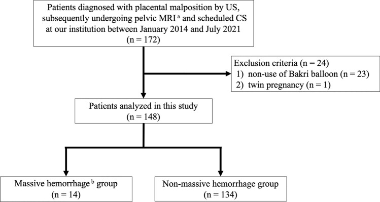
Methods: Totally, 148 consecutive patients with placental malposition underwent MRI and cesarean section at our hospital between January 2014 and July 2021. The patients were divided into massive and non-massive hemorrhage groups. MRI findings of placenta accreta, placental position, and placental volume were evaluated by two radiologists, and inter-reader agreement was calculated. Diagnostic ability for predicting massive hemorrhage was evaluated using receiver operating characteristic analysis.
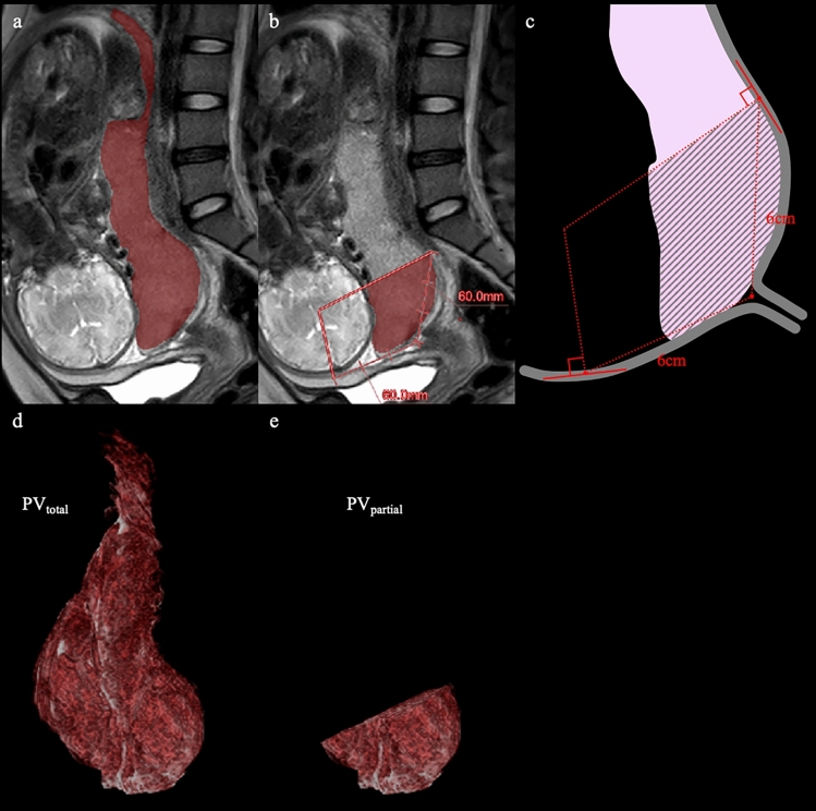
Results: Intraplacental T2 dark bands (100% vs. 58.2%, p = 0.001), placental bulge (50% vs. 3.7%, p < 0.001), loss of retroplacental T2 hypointense line (100% vs. 67.2%, p = 0.01), myometrial thinning (92.9% vs. 57.5%, p = 0.009), total placenta previa (64.3% vs. 23.9%, p = 0.033), and anterior placenta (35.7% vs. 10.5%, p = 0.02) were significantly observed in massive hemorrhage group. Partial placental volume of lower uterine segment (PVpartial) was larger in massive hemorrhage group (166 [108-214] cm3 vs. 70 [43-112] cm3 p < 0.001), had the second highest intraclass correlation coefficient (0.84), and had the highest area under the curve (0.81) for diagnosing massive hemorrhage.
Conclusion: This study revealed PVpartial is an accurate and objective indicator for massive hemorrhage, independent of radiologists' experience. This indicator potentially enables prediction of massive hemorrhage, improving pre-operative planning in high-risk pregnancies.
Keywords: Lower uterine segment; Placenta accreta spectrum; Placenta previa; Placental volume.
© 2025. The Author(s).
Conflict of interest statement
Declarations. Conflict of interest: The authors have no relevant financial or non-financial interests to disclose. Ethical approval: This study was performed in line with the principles of the Declaration of Helsinki. This study was approved by the Ethics Review Committee of National Defense Medical College Hospital (Certification No. 4441) on July 29, 2021. Consent to participate: The records of all patients during the study period were anonymized prior to the analysis. This study was exempt from obtaining informed consent from all participants. Consent to publish: Not applicable.
Figures

Similar articles
-
A novel approach for the early prediction and prevention of placenta accreta spectrum and severe peripartum hemorrhage in patients with complete placenta previa: leveraging three-dimensional placental topography, cervical length, and dilatation parameters on magnetic resonance imaging.BMC Pregnancy Childbirth. 2024 Nov 25;24(1):784. doi: 10.1186/s12884-024-06996-w. BMC Pregnancy Childbirth. 2024. PMID: 39587553 Free PMC article.
-
Nomogram based on clinical characteristics and ultrasound indicators for predicting severe postpartum hemorrhage in patients with anterior placenta previa combined with previous cesarean section: a retrospective case-control study.BMC Pregnancy Childbirth. 2024 Aug 31;24(1):572. doi: 10.1186/s12884-024-06706-6. BMC Pregnancy Childbirth. 2024. PMID: 39217290 Free PMC article.
-
Intraplacental T2-hypointense bands may help predict placental invasion depth and postpartum hemorrhage in placenta accrete spectrum disorders in high-risk gravid patients.Magn Reson Imaging. 2022 Dec;94:73-79. doi: 10.1016/j.mri.2022.09.005. Epub 2022 Sep 16. Magn Reson Imaging. 2022. PMID: 36116713
-
Development and validation of MRI-based scoring models for predicting placental invasiveness in high-risk women for placenta accreta spectrum.Eur Radiol. 2024 Feb;34(2):957-969. doi: 10.1007/s00330-023-10058-8. Epub 2023 Aug 17. Eur Radiol. 2024. PMID: 37589907
-
Placenta accreta, increta, and percreta.Obstet Gynecol Clin North Am. 2013 Mar;40(1):137-54. doi: 10.1016/j.ogc.2012.12.002. Obstet Gynecol Clin North Am. 2013. PMID: 23466142 Review.
References
-
- Bahar A, Abusham A, Eskandar M, Sobande A, Alsunaidi M (2009) Risk factors and pregnancy outcome in different types of placenta previa. J Obstet Gynaecol Can 31:126–131. 10.1016/s1701-2163(16)34096-8 - PubMed
-
- Ishibashi H, Miyamoto M, Soyama H et al (2018) Marginal sinus placenta previa is a different entity in placenta previa: A retrospective study using magnetic resonance imaging. Taiwan J Obstet Gynecol 57:532–535. 10.1016/j.tjog.2018.06.009 - PubMed
-
- Silver RM, Landon MB, Rouse DJ et al (2006) Maternal morbidity associated with multiple repeat cesarean deliveries. Obstet Gynecol 107:1226–1232. 10.1097/01.AOG.0000219750.79480.84 - PubMed
-
- Clark SL, Koonings PP, Phelan JP (1985) Placenta previa/accreta and prior cesarean section. Obstet Gynecol 66:89–92 - PubMed
-
- Tikkanen M, Paavonen J, Loukovaara M, Stefanovic V (2011) Antenatal diagnosis of placenta accreta leads to reduced blood loss. Acta Obstet Gynecol Scand 90:1140–1146. 10.1111/j.1600-0412.2011.01147.x - PubMed
Publication types
MeSH terms
LinkOut - more resources
Full Text Sources
Medical
