. 2025 Jan 27;58(1):8.
doi: 10.1186/s40659-024-00578-y.

hUC-MSC preserves erectile function by restoring mitochondrial mass of penile smooth muscle cells in a rat model of cavernous nerve injury via SIRT1/PGC-1a/TFAM signaling

Affiliations

hUC-MSC preserves erectile function by restoring mitochondrial mass of penile smooth muscle cells in a rat model of cavernous nerve injury via SIRT1/PGC-1a/TFAM signaling

Mengbo Yang et al. Biol Res. .

Abstract

Background: Cavernous nerve injury-induced erectile dysfunction (CNI-ED) is a common complication following radical prostatectomy and severely affects patients' quality of life. The mitochondrial impairment in corpus cavernosum smooth muscle cells (CCSMCs) may be an important pathological mechanism of CNI-ED. Previous studies have shown that transplantation of human adipose derived stem cells (ADSC) can alleviate CNI-ED in a rat model. However, little is known about the effect of human umbilical cord mesenchymal stem cells (hUC-MSC) on CNI-ED. It remains unclear whether hUC-MSC can ameliorate mitochondrial damage in CCSMCs. In this study, we aimed to investigate the impacts of hUC-MSC on the mitochondrial mass and function of CCSMCs, as well as elucidate its underlying molecular mechanism.

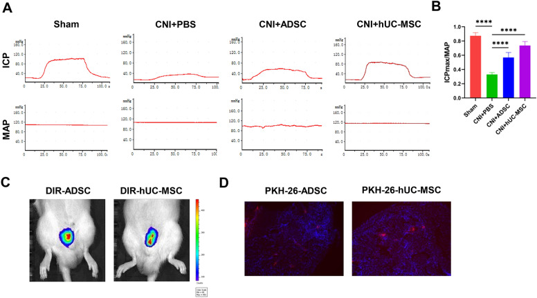
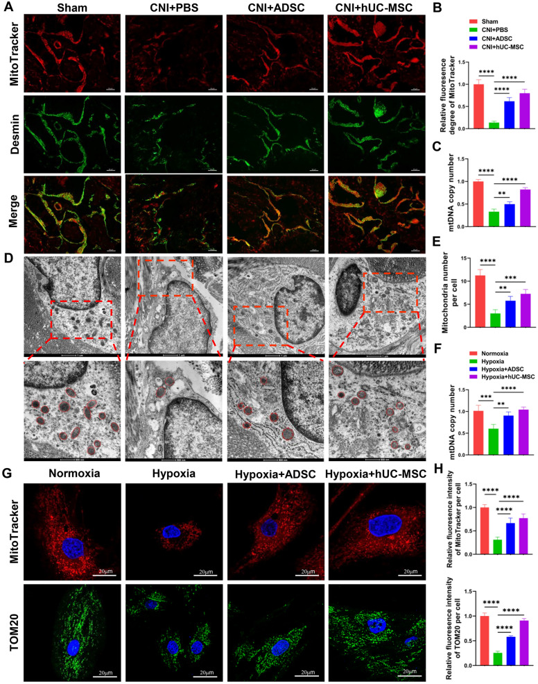
Methods: The CNI-ED rat model was established by bilaterally crushing cavernous nerves. Subsequently, hUC-MSC were transplanted into the cavernosum and ADSC were injected as a positive control group. Erectile function evaluation and histological detection were performed 4 weeks after cell transplantation. In vitro, CCSMCs underwent hypoxia and were then co-cultured with ADSC or hUC-MSC using a transwell system. The mitochondrial mass and function, as well as signaling pathways, were investigated. To explore the role of the SIRT1/PGC-1α/TFAM pathway in regulating mitochondrial biogenesis of CCSMCs, we knocked down SIRT1 by siRNA.

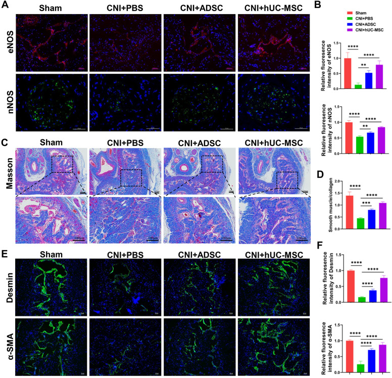
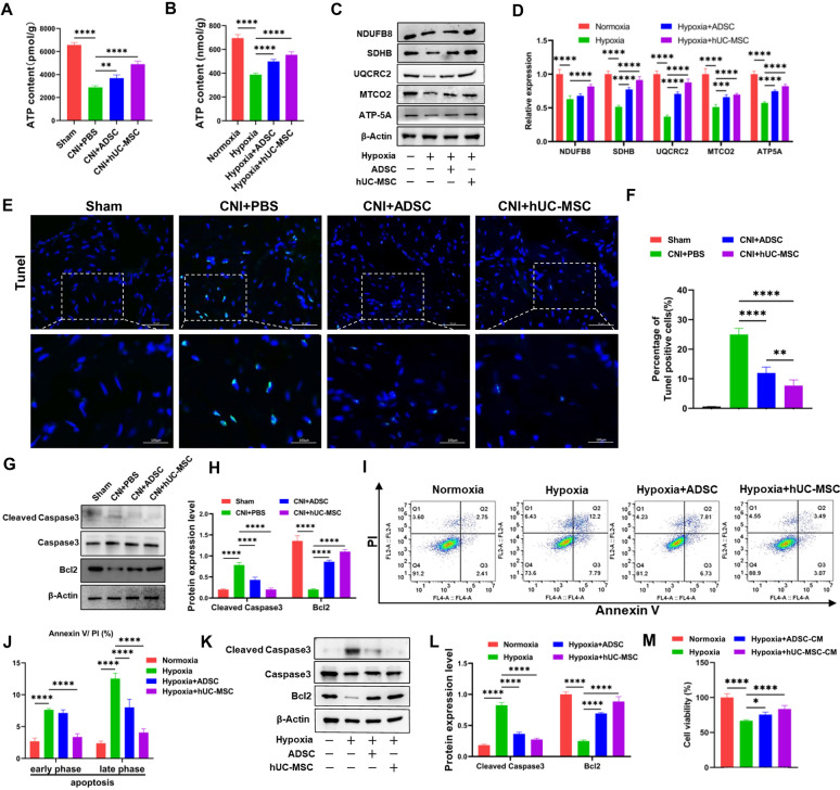
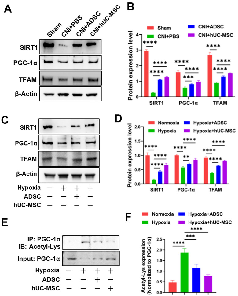
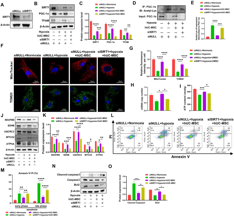
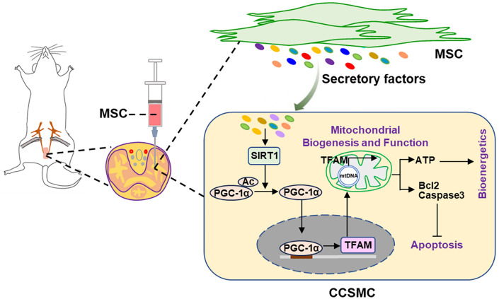
Results: The administration of hUC-MSC significantly improved erectile function of CNI-ED rats and reduced the ratio of collagen to smooth muscle. Specifically, hUC-MSC treatment restored mitochondrial mass and function in CCSMCs injured by CNI or hypoxia, and inhibited the apoptosis of CCSMCs. Mechanistically, the application of hUC-MSC activated SIRT1/PGC-1α/TFAM pathway both in rat penile tissues and CCSMCs. In addition, knockdown of SIRT1 in CCSMCs abolished the protective effects of hUC-MSC on mitochondrial mass and function, while leading to an increase in cellular apoptosis.

Conclusions: hUC-MSC contribute to the recovery of erectile function in CNI-ED rats by restoring mitochondrial mass and function of CCSMCs through the SIRT1/PGC-1α/TFAM pathway. Our present study offers new insights into the role and molecular mechanisms of hUC-MSC in regulating mitochondrial homeostasis, thereby facilitating the restoration of the erectile function in CNI-ED.

Keywords: Cavernous nerve injury-induced erectile dysfunction; Corpus cavernous smooth muscle cell; Mesenchymal stem cell; SIRT1/PGC-1α/TFAM pathway.


Conflict of interest statement

Declarations. Ethics approval and consent to participate: All experiments in this study conform to the Chinese legislation and have been approved by The Human and Animal Research Ethics Committee of Renji Hospital Affiliated to Shanghai Jiao Tong University School of Medicine. Consent for publication: Not applicable. Competing interests: The authors have declared that no conflict of interest exists.

Figures

Fig. 1
Fig. 1
ADSC or hUC-MSC transplantation restores erectile function in CNI-ED rats. A Representative images of the ICP and MAP in each group. B Quantification of maximal ICP to MAP ratio (ICPmax/MAP) of different groups. Data are presented as mean ± SD (n = 6). ****P < 0.0001). C Representative DIR dye-labelled ADSC or hUC-MSC images in corpora cavernosa 24 h after injection. D Representative images of PKH26 dye labeled-ADSC or hUC-MSC in corpora cavernosa 72 h after injection
Fig. 2
Fig. 2
ADSC or hUC-MSC therapy improves the expression of eNOS and nNOS and increased the ingredient of smooth muscle in corpus cavernosum. A Immunostaining of corpora cavernosa with eNOS or nNOS antibody and the representative images are shown. Scale bar: 50 µm. B Quantification of immunofluorescence images of eNOS and nNOS from different groups. C Representative images of Masson trichrome staining of penile midshaft specimen in the four groups. The smooth muscle (red) and collagen (blue) tissues were stained by Masson Trichrome Kit. Scale bar: 100 µm. D Quantification of the ratio of smooth muscle to collagen. E Representative images of immunofluorescence of Desmin and α-SMA in different groups. Scale bar: 50 µm. F Quantification of immunofluorescence images of Desmin and α-SMA. The data of the above quantification of slides images are presented as mean ± SD, at least six representative visual fields in each slide for each group were counted. At least three rats were used in each group. (*p < 0.05, **p < 0.01, ***p < 0.001, ****p < 0.0001)
Fig. 3
Fig. 3
Treatment with ADSC or hUC-MSC improves mitochondrial mass and function in rat corpus cavernosum and in CCSMCs. A Representative images of immunofluorescence of penile cryosections stained using MitoTracker deep red (red) and Desmin (green) in CNI-ED. Scale bar: 50 µm. B Quantification of fluorescence density of the colocalization of MitoTracker deep red and Desmin. At least six representative visual fields were counted of each slide and at least three rats were used in each group. Data are presented as mean ± SD, C Real time analysis of mtDNA copy number in corpus cavernosum in different groups. mtDNA copy number in sham group were set as 1. At least 3 rats in each group were used, and the data are presented as mean ± SD. D Representative images of transmission electron microscopy (TEM) of mitochondria in CCSMCs. E Quantification of mitochondria number in different groups. At least 20 cells in each group were counted. F Quantification of mtDNA copy number in CCSMCs by real-time PCR. mtDNA copy number in normoxia group were set as 1. Data were collected from at least three independent experiments and the data are presented as mean ± SD. G Representative images of penile cryosections stained for MitoTracker deep red (red) and TOM20 (green) in CCSMCs. Scale bar: 20 µm. H Quantification of fluorescence density of MitoTracker deep red and in different groups. At least 20 cells were counted in each group and data are presented as mean ± SD. *p < 0.05, **p < 0.01, ***p < 0.001, ****p < 0.0001
Fig. 4
Fig. 4
ADSC or hUC-MSC therapy alleviates mitochondrial dysfunction in corpus cavernosum and in CCSMCs. A ATP levels in corpus cavernosum of rats after ADSC or hUC-MSC injection. Data are presented as mean ± SD (n = 6). B Intracellular ATP levels in CCSMCs was measured after coculture with ADSC or hUC-MSC. Data were collected from at least three independent experiments, and data are presented as mean ± SD. C Immunoblot analysis of ETC protein levels including NDUFB8, SDHB, UQCRC2, MTCO2 and ATP5A in the lysates of CCSMCs. The level of b-actin was used as internal control of the total protein of cell lysate. Full-length blots of CCSMCs were presented (Additional file 2: Fig. S1). D Quantification of relative protein levels of NDUFB8, SDHB, UQCRC2, MTCO2 and ATP5A levels in CCSMCs. E Representative images of TUNEL staining of corpus cavernosum in different groups. Scale bar: 50 µm. F Quantification of TUNEL positive cells in all groups. At least six represent visual fields were counted for each group and at least 3 rats were used in each group. Data are presented as mean ± SD. G Immunoblot analysis of protein levels of Cleaved Caspase-3, Caspase-3 and Bcl-2 proteins in different groups. The level of b-actin was used as internal control of the total protein of cell lysate. Full-length blots of corpora cavernousa were presented (Additional file 2: Fig. S2). H Quantification of relative protein levels of Cleaved Caspase-3 and Bcl-2 levels in CCSMCs. I Flow cytometry analysis of Annexin V / PI staining of CCSMCs in different groups. J Quantification of apoptotic CCSMCs in different groups. Data were collected from at least three independent experiments and data are presented as mean ± SD. K Immunoblot analysis of protein levels of Cleaved caspase-3, Caspase-3 and Bcl-2 protein in CCSMCs. Full-length blots of CCSMCs were presented (Additional file 2: Fig. S3). L Quantification of relative protein levels of Cleaved caspase-3 and Bcl-2 levels in CCSMCs. M The cell viability of CCSMCs was assayed using Celltiter-Lumi™ Steady Luminescent Cell Viability Assay Kit. Data were collected from at least three independent experiments, and the data are presented as mean ± SD. *p < 0.05, **p < 0.01, ***p < 0.001, ****p < 0.0001
Fig. 5
Fig. 5
SIRT1/PGC-1α/TFAM pathway is upregulated after ADSC or hUC-MSC treatment. A Immunoblot analysis of protein levels of SIRT1, PGC-1α, TFAM in corpora cavernosa of rats. β-Actin expression levels were used as an internal control. Full-length blots of corpora cavernosa were presented (Additional file 2: Fig. S4). B Quantification of relative protein levels of SIRT1, PGC-1α, TFAM levels in corpora cavernosa. C Immunoblot analysis of protein levels of SIRT1, PGC-1α, TFAM expression levels in CCSMCs. β-Actin expression levels were used as an internal control. Full-length blots of CCSMCs were presented (Additional file 2: Fig. S5). D Quantification of relative protein levels of SIRT1, PGC-1α, TFAM levels in CCSMCs. E Immunoprecipitation analysis of acetylation level of PGC-1α in CCSMCs. Full-length blots of CCSMCs were presented (Additional file 2: Fig. S6). F Quantification of acetylation level of PGC-1α in CCSMCs. Acetylated level of PGC-1α was normalized to PGC-1α. *p < 0.05, **p < 0.01, ***p < 0.001, ****p < 0.0001)
Fig. 6
Fig. 6
MSC restore mitochondrial biogenesis and function via SIRT1/PGC-1a/TFAM pathway. A Immunoblot analysis of protein level of SIRT1 expression in CCSMCs after SIRT1 knockdown by siRNA. β-Actin expression levels were used as an internal control. Full-length blots of CCSMCs were presented (Additional file 2: Fig. S7). B Immunoblot analysis of protein levels of SIRT1, PGC-1α, TFAM in CCSMCs of different groups. Full-length blots of CCSMCs were presented (Additional file 2: Fig. S8). C Quantification of relative protein levels of SIRT1, PGC-1α, TFAM levels in different groups. D Immunoprecipitation analysis of acetylation level of PGC-1α in CCSMCs. Full-length blots of CCSMCs were presented (Additional file 2: Fig. S8). E Quantification of acetylation level of PGC-1α in different groups. Acetylation level of PGC-1α were normalized to PGC-1α. F Representative images of mitochondria of CCSMCs stained with MitoTracker deep red (red) or anti-TOM20 monoclonal antibody (green). Scale bar: 20 µm. G Quantification of relative fluorescence intensity of MitoTracker deep red or TOM20 of per cell in CCSMCs in different groups. At least 20 cells were counted for each group and data are presented as mean ± SD. H Quantification of the mtDNA copy number in CCSMCs. Data were collected from at least three independent experiments, and the data are presented as mean ± SD. I Intracellular ATP level in CCSMCs after knockdown of SIRT1 and coculture with hUC-MSCs. Data were collected from at least three independent experiments and data are presented as mean ± SD. J Immunoblot analysis of protein levels of NDUFB8, SDHB, UQCRC2, MTCO2 and ATP5A in CCSMCs in different groups. Full-length blots of CCSMCs were presented (Additional file 2: Fig. S10). K Quantification of relative protein levels of NDUFB8, SDHB, UQCRC2, MTCO2 and ATP5A levels of CCSMCs in different groups. L Flow cytometry analysis of Annexin V / PI staining of CCSMCs in different groups. M Quantification of apoptotic CCSMCs in different groups. Data were collected from at least independent groups, and the data are presented as mean ± SD. N Immunoblot analysis of protein levels of Cleaved caspase-3, Caspase-3 and Bcl-2 protein in CCSMCs. Full-length blots were presented (Additional file 2: Fig. S11). O Quantification of relative protein levels of Cleaved caspase-3 and Bcl-2 levels in CCSMCs. *p < 0.05, **p < 0.01, ***p < 0.001, ****p < 0.0001
Fig. 7
Fig. 7
Working model of MSC in alleviating CNI-ED rats. Rats are subjected to bilateral cavernous nerve crush and followed by intracavernous injection of MSC. The secretions from MSC activate SIRT1 expression, which subsequently deacetylates acetylated PGC-1α and activates PGC-1α. The activated PGC-1α then translocated into the nuclei, where it promotes the transcription of TFAM. TFAM plays a crucial role in mitochondrial biogenesis and maintaining mitochondrial homeostasis, thereby enhancing mitochondrial bioenergetics and reducing cell apoptosis. Ultimately, this process contributes to the restoration of erectile function

Similar articles

Cited by

References

    1. Catalona WJ, Bigg SW. Nerve-sparing radical prostatectomy: evaluation of results after 250 patients. J Urol. 1990;143:538–43. - PubMed
    1. Yafi FA, Jenkins L, Albersen M, Corona G, Isidori AM, Goldfarb S, et al. Erectile dysfunction. Nat Rev Dis Primers. 2016;2:16003. - PMC - PubMed
    1. Haglind E, Carlsson S, Stranne J, Wallerstedt A, Wilderäng U, Thorsteinsdottir T, et al. Urinary incontinence and erectile dysfunction after robotic versus open radical prostatectomy: a prospective, controlled. Nonrandomised Trial Eur Urol. 2015;68:216–25. - PubMed
    1. Hatzimouratidis K, Burnett AL, Hatzichristou D, McCullough AR, Montorsi F, Mulhall JP. Phosphodiesterase type 5 inhibitors in postprostatectomy erectile dysfunction: a critical analysis of the basic science rationale and clinical application. Eur Urol. 2009;55:334–47. - PubMed
    1. Song G, Hu P, Song J, Liu J, Ruan Y. Molecular pathogenesis and treatment of cavernous nerve injury-induced erectile dysfunction: a narrative review. Front Physiol. 2022;13:1029650. - PMC - PubMed

Publication types

MeSH terms

Substances

LinkOut - more resources