

Deltopectoral approach without subscapularis detachment for reverse shoulder arthroplasty. Technique and results of a safe and reproducible subscapularis-sparing approach
- PMID: 39872348
- PMCID: PMC11764111
- DOI: 10.1016/j.xrrt.2024.09.006
Deltopectoral approach without subscapularis detachment for reverse shoulder arthroplasty. Technique and results of a safe and reproducible subscapularis-sparing approach
Abstract
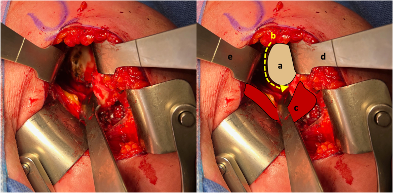
Background: The importance of the subscapularis for reverse total shoulder arthroplasty has been demonstrated, especially for internal rotation and stability. In a deltopectoral approach, a detachment of the subscapularis is performed (tenotomy, tuberosity peeling, or osteotomy), but the tendon is not always repairable at the end. When it is repaired, healing is obtained in only 40%-76% of the cases, with potential consequences for the outcomes. The anterior muscle-sparing (AMS) approach is a deltopectoral approach with preservation of the subscapularis, providing a solution to these problems. We present the surgical technique and results.
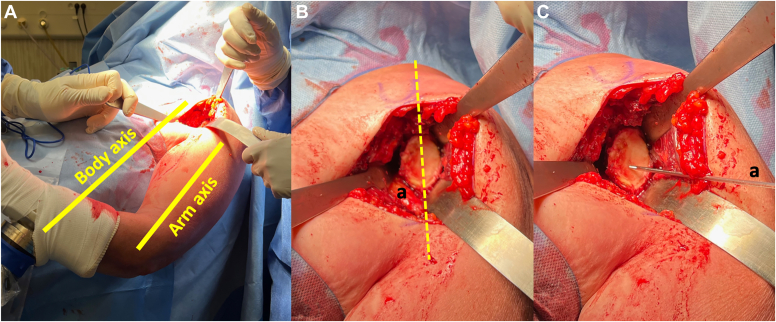
Methods: In a retrospective study, we included our first 45 cases of reverse total shoulder arthroplasty performed with the AMS approach for a degenerative affection of the shoulder (massive rotator cuff tear, cuff tear arthropathy, primary glenohumeral arthritis, or rheumatoid arthritis), excluding traumatic and revision cases. The subscapularis was intact in all the cases. The mean age at inclusion was 74.1 years. No patients were lost at the minimum 24-month follow-up. All the patients underwent a clinical evaluation preoperatively and at the last follow-up, including Constant score, simple shoulder value, pain scale, and range of motion. An X-ray evaluation was conducted postoperatively and at the last follow-up to assess implant positioning and evolution.
Results: There was no intraoperative complication, and the mean operative time was 62 minutes. We observed a significant improvement in Constant score (from 36 to 70, P <.001), simple shoulder value (from 33 to 81, P <.001), pain (from 6.3 to 0.7, P <.001), strength (from 0.5 to 3.5, P <.001), and most of the active mobilities. Regarding internal rotation, 95% of the patients reached level L3 or higher. Glenoid positioning was considered optimal in more than 90% of the cases (inferior tilt and low position) without any occurrence of superior tilt or high position. The osteophytes could be totally removed in 8 out of 9 cases (88.9%). Six postoperative complications (13.3%) were reported: 1 infection (2.2%), 2 cases of traumatic glenoid loosening (4.4%), 2 acromion fractures (4.4%), and 1 hematoma (2.2%). There was no instability. Eighty percent of the patients could return home, with a mean hospital stay of 1.8 days.
Conclusion: The AMS approach is a safe and reproducible technique. The preservation of the subscapularis has potential benefits regarding internal rotation and stability. In the absence of tendon suture, rehabilitation can be started immediately without motion restriction, allowing for a fast recovery and return to autonomy.
Keywords: Deltopectoral approach; Minimally invasive surgery; Recovery; Reverse shoulder arthroplasty; Rotation; Stability; Subscapularis sparing; Surgical technique.
© 2024 The Author(s).
Figures


References
LinkOut - more resources
Full Text Sources
