. 2022 Oct 21;1(6):437-446.
doi: 10.1007/s44164-022-00033-w. eCollection 2022 Dec.

Harvestable tumour spheroids initiated in a gelatin-carboxymethyl cellulose hydrogel for cancer targeting and imaging with fluorescent gold nanoclusters

Affiliations

Harvestable tumour spheroids initiated in a gelatin-carboxymethyl cellulose hydrogel for cancer targeting and imaging with fluorescent gold nanoclusters

Ashkan Kamali Dashtarzheneh et al. In Vitro Model. .

Abstract

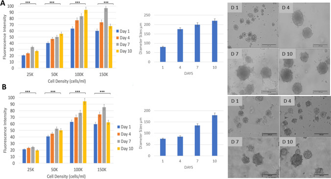
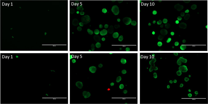
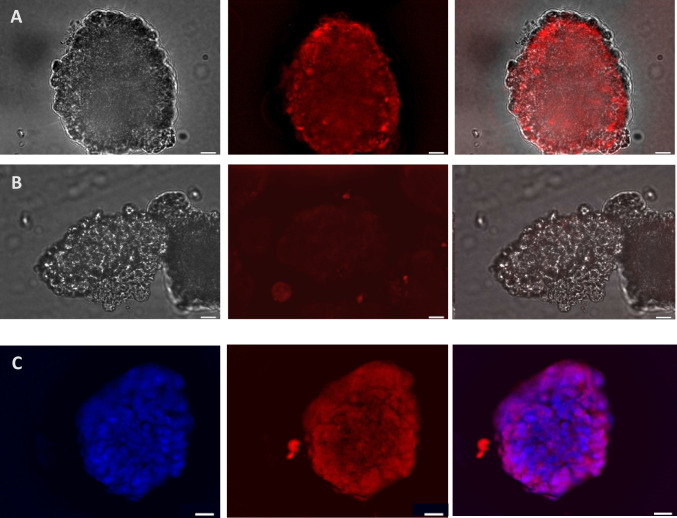
Cancer cell spheroids are the simplest 3D in vitro cancer models and have been extensively used for cancer research. More recently, models have been becoming complex, with the introduction of a matrix and non-cancer cell types to mimic specific tumour aspects. However, applying drugs or agents in matrix-embedded cancer spheroids can be problematic. Most matrices can impede and also bind drugs or visualizing agents non-specifically, in the vicinity of the embedded spheroids. This may interfere with imaging or further analysis without breaking apart the 3D model into its constituents. Here, we developed a combined gelatin-carboxymethyl cellulose (G-CMC) hydrogel for initiating cancer spheroids that enabled intact harvesting pre/post treatment for further investigation, such as targeting and imaging. We combined CMC (1.25%) and gelatin (2.5%) at 25 °C and initiated polymerisation after autoclaving (121 °C) to obtain a mechanical strength (sheer stress) of 38 Pas versus 1.28 Pas for CMC alone. These matrix conditions facilitated separation of the spheroids from the G-CMC, using low centrifugation (100 g). We described growth of colorectal and breast cancer spheroids within the G-CMC matrix (with average diameters of 220 mm and 180 μm for representative cell lines HT29 and MCF7 at 10 days, respectively). As the cancer cells express the surface biomarker calreticulin (CRT), we manufactured anti-calreticulin IgG (anti-CRT) conjugated to fluorescent gold nanoclusters (anti-CRT-AuNC) as a probe. We harvested cancer spheroids and incubated live with the nanoclusters. Imaging demonstrated strong binding of CRT-targeted AuNCs compared to control AuNCs. This novel model preserves cancer spheroid integrity upon isolation and is well suited for targeted imaging and drug delivery of cancer in 3D.

Keywords: 3D in vitro cancer model; Breast cancer spheroids; Calreticulin; Cancer targeting; Carboxymethyl cellulose; Colorectal cancer spheroids; Gelatin; Gold nanoclusters.


Conflict of interest statement

Conflict of interestThe authors declare no competing interests.

Figures

Fig. 1
Fig. 1
a The rheometer comprises a rotating upper cone and a fixed lower plate, with the mixture to be tested placed between the two. To measure viscosity, the upper cone is turned, and resistance recorded. Representative graphical representations of viscosity (blue line) and shear stress (red line) for b G-CMC and c CMC. Repeat experimental measurements were within 5% of these values
Fig. 2
Fig. 2
Growth of cancer spheroids in the 3D G-CMC matrix over 10 days. HT29 colorectal cells (A) and MCF7 breast cells (B) were seeded at a range of concentrations (25–150 × 103cells/ml) for 1,4,7 and 10 days (n = 6). Growth is shown as equivalent to intensity of fluorescence (AlamarBlue™ assay; excitation/emission 530 nm/620 nm). Spheroids, for the 100 × 103/ml cell seeding concentration, were imaged by light microscopy (representative images shown) and diameters calculated, ImageJ. One-way ANOVA, Tukey’s post hoc analysis (***p < 0.05)
Fig. 3
Fig. 3
HT29 3D spheroids (A, C) and MCF7 3D spheroids (B, D) grown in 3D G-CMC matrix for 10 days, harvested by centrifugation (100 g, 5 min) and embedded into fresh G-CMC. Representative images, showing some debris in HT29 cultures. Magnification bars = 250 μm, A, B; 10 μm, C, D
Fig. 4
Fig. 4
Live/dead assay in HT29 3D spheroids (top panel) and MCF7 3D spheroids (bottom panel) grown in 3D G-CMC matrix, over time (representative images). Spheroids were harvested and exposed to fluorescein diacetate (FDA) and propidium iodide (PI), which stain viable cells (green) and dead cells (red). Fluorescent signal is weak in day 1 cultures due to small numbers, for both cell lines. Magnification bar = 200 μm
Fig. 5
Fig. 5
Expression of calreticulin by HT29 spheroids grown in G-CMC matrix for 10 days (representative images). Spheroids were harvested, incubated with anti-CRT-AuNCs for 2 h and imaged live, using fluorescent microscopy. A Uptake of anti-CRT-AuNCs. B Uptake of un-targeted, unconjugated control AuNCs. Both A and B series of images (L-R): light, fluorescence; merged; with NIR red pseudocolour. C Spheroids exposed to anti-CRT-AuNCs (red) were fixed prior to imaging, to allow for staining with nuclear DAPI (blue); L-R: DAPI fluorescence, NC fluorescence; merged. Bar = 10 μm; magnification × 60, immersed objective, Olympus BX63)
Fig. 6
Fig. 6
Expression of calreticulin by MCF7 spheroids grown in G-CMC matrix for 10 days (representative images). Spheroids were harvested, incubated with anti-CRT-AuNCs for 2 h and imaged live, using fluorescent microscopy. A Uptake of anti-CRT-AuNCs. B Uptake of un-targeted, unconjugated control AuNCs. Both A and B series of images (L-R): light, fluorescence; merged; with NIR red pseudocolour. C Spheroids exposed to anti-CRT-AuNCs (red) were fixed prior to imaging, to allow for staining with nuclear DAPI (blue); L-R: DAPI fluorescence, NC fluorescence; merged. Bar = 10 μm; magnification × 60, immersed objective, Olympus BX63)

References

    1. Fischbach C, Chen R, Matsumoto T, Schmelzle T, Brugge JS, Polverini PJ, Mooney DJ. Engineering tumors with 3D methods. Nat Methods. 2007;4:855–66. 10.1038/nmeth1085. - PubMed
    1. Gurski LA, Jha AK, Zhang C, Jia X, Farach-Carson MC. Hyaluronic acid-based hydrogels as 3D matrices for in vitro evaluation of chemotherapeutic drugs using poorly adherent prostate cancer cells. Biomaterials. 2009;30:6076–85. 10.1016/j.biomaterials.2009.07.054. - PMC - PubMed
    1. Mehta G, Hsiao AY, Ingram M, Luker GD, Takayama S. Opportunities and challenges for use of tumor spheroids as models to test drug delivery and efficacy. J Control Release. 2012;164:192–204. 10.1016/j.jconrel.2012.04.045. - PMC - PubMed
    1. Mih JD, Marinkovic A, Liu F, Sharif AS, Tschumperlin DJ. Matrix stiffness reverses the effect of actomyosin tension on cell proliferation. J Cell Sci. 2012;125(24):5974–83. 10.1242/jcs.108886. - PMC - PubMed
    1. Santini MT, Rainaldi G, Romano R, Ferrante A, Clemente S, Motta A, Indovina PL. MG-63 human osteosarcoma cells grown in monolayer and as three-dimensional tumor spheroids present a different metabolic profile: a (1)H NMR study. FEBS Lett. 2004;557:148–54. 10.1016/S0014-5793(03)01466-2. - PubMed

LinkOut - more resources