Editorial
. 2025 Jan 27;17(1):100278.
doi: 10.4240/wjgs.v17.i1.100278.

Rectal ameboma: A new entity in the differential diagnosis of rectal cancer

Affiliations
Editorial

Rectal ameboma: A new entity in the differential diagnosis of rectal cancer

Kemal Bugra Memis et al. World J Gastrointest Surg. .

Abstract

We examined the case report written by Ke et al, describing a rare clinical case. In this editorial, we would like to emphasize the differential diagnosis of rectal masses through a rare case. We describe a case of ameboma, which manifested itself as a mass in the rectum in terms of imaging and rectoscopic features, in an immunocompetent patient who had complaints of constipation and rectal bleeding for weeks. The initial diagnosis suggested malignancy due to imaging and rectoscopic features, but the pathology report reported it as amoebiasis. After ten days of metronidazole and oral amebicide (diloxanide furoate) treatment, the patient's symptoms and radiological findings were successfully regressed.

Keywords: Ameboma; Amoebic colitis; Imaging findings; Immunocompetent patient; Rectal mass.


Conflict of interest statement

Conflict-of-interest statement: All the authors report no relevant conflicts of interest for this article.

Figures

Figure 1
Figure 1
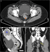
Contrast-enhanced pelvic computed tomography images of a 46-year-old female patient. A: Lesion suggestive of malignancy on the rectal wall, ameboma (orange asterisk), increased density in the pararectal mesenteric tissue (green arrow); B: Only one of the lymph nodes in the pararectal region increased in number (yellow arrow); C: Sagittal image shows increased thickness of the rectal wall and calcified myoma (blue star) in the uterus; D and E: It is the magnified image of coronal plane image (D). This magnified image shows increased thickness of the rectal wall (orange asterisk) and lymph nodes in the coronal section (yellow arrow).
Figure 2
Figure 2
Contrast-enhanced pelvic magnetic resonance images of a 46-year-old female patient. A: Increased wall thickness in sagittal sections on T2-weighted magnetic resonance imaging, almost causing obstruction of the rectal wall (orange circle); B: Heterogeneity in mesocolic adipose tissue (white arrow) and asymmetric significant wall thickening in the rectum (orange asterisk) on T2-weighted axial magnetic resonance imaging; C: Lymph nodes in the perirectal region on diffusion weighted imaging (yellow arrow); D: T1-weighted axial post-contrast magnetic resonance images showing homogeneous contrast enhancement in the rectum with increased wall thickness (orange asterisk) and heterogeneity in mesorectal adipose tissue (white arrow).
Figure 3
Figure 3
Contrast-enhanced pelvic computed tomography images of a 46-year-old female patient after treatment. A: On the axial plane image, the thickness of the rectal wall appears to regress (orange asterisk); B: In the sagittal plane image, calcified myomas in the uterus (blue star) are seen in addition to regression of rectal wall thickening; C: Shrinkage of lymph nodes after treatment (yellow arrow).
Figure 4
Figure 4
The flowchart summarises our diagnostic and treatment process. CT: Computed tomography; MRI: Magnetic resonance imaging.

References

    1. Morán P, Serrano-Vázquez A, Rojas-Velázquez L, González E, Pérez-Juárez H, Hernández EG, Padilla MLA, Zaragoza ME, Portillo-Bobadilla T, Ramiro M, Ximénez C. Amoebiasis: Advances in Diagnosis, Treatment, Immunology Features and the Interaction with the Intestinal Ecosystem. Int J Mol Sci. 2023;24 - PMC - PubMed
    1. Tanaka E, Tashiro Y, Kotake A, Takeyama N, Umemoto T, Nagahama M, Hashimoto T. Spectrum of CT findings in amebic colitis. Jpn J Radiol. 2021;39:558–563. - PubMed
    1. Kinoo SM, Ramkelawon VV, Maharajh J, Singh B. Fulminant amoebic colitis in the era of computed tomography scan: A case report and review of the literature. SA J Radiol. 2018;22:1354. - PMC - PubMed
    1. Omwansa P, Nyatsambo C, Ngwisanyi W, McGrath N, Moeng MS. A case report of colonic Ameboma mimicking colon cancer in an immunocompromised patient. Int J Surg Case Rep. 2023;110:108768. - PMC - PubMed
    1. Nasrallah J, Akhoundi M, Haouchine D, Marteau A, Mantelet S, Wind P, Benamouzig R, Bouchaud O, Dhote R, Izri A. Updates on the worldwide burden of amoebiasis: A case series and literature review. J Infect Public Health. 2022;15:1134–1141. - PubMed

Publication types

LinkOut - more resources