Review
. 2024 Oct 16;5(1):12-22.
doi: 10.1016/j.jointm.2024.07.006. eCollection 2025 Jan.

Nebulized aminoglycosides for ventilator-associated pneumonia: Methodological considerations and lessons from experimental studies

Collaborators, Affiliations
Review

Nebulized aminoglycosides for ventilator-associated pneumonia: Methodological considerations and lessons from experimental studies

Jean-Jacques Rouby et al. J Intensive Med. .

Abstract

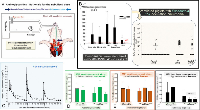
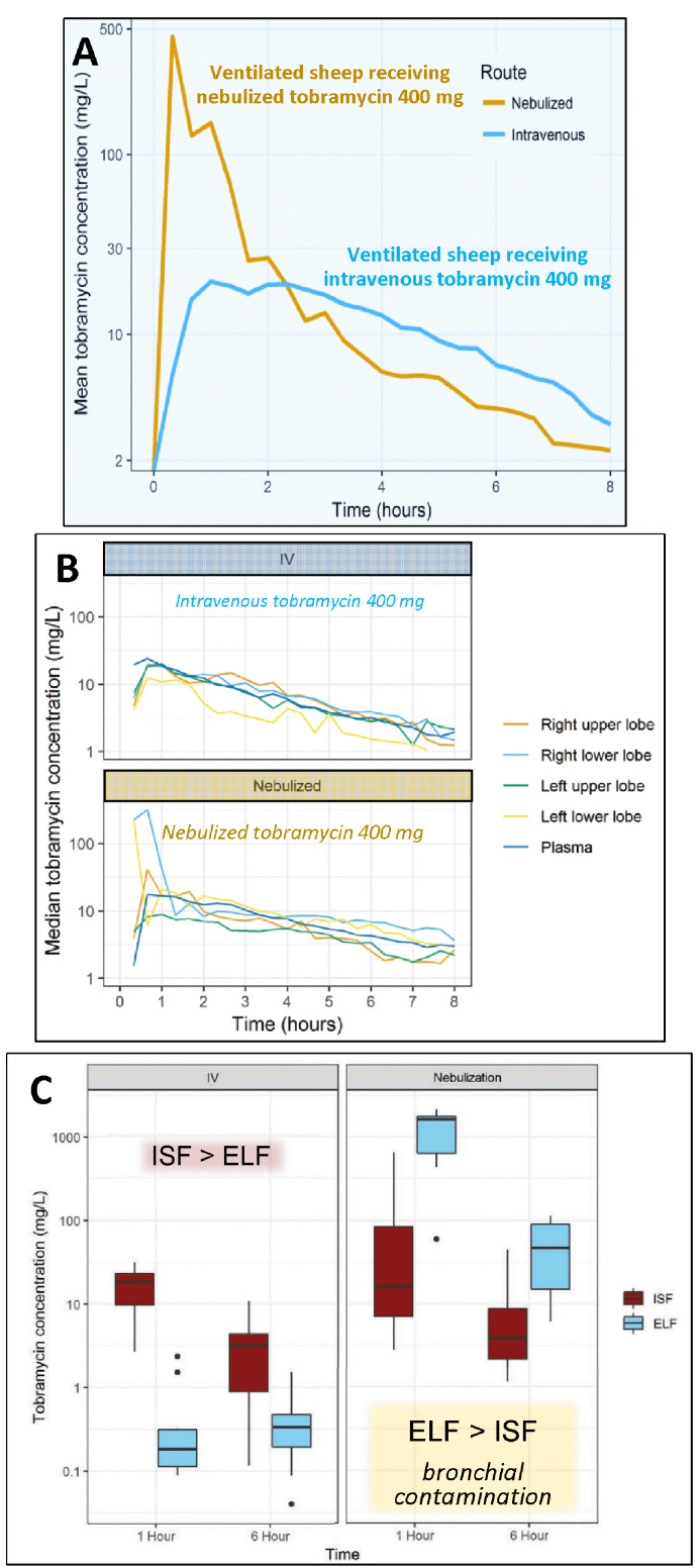
Aminoglycosides are concentration-dependent antibiotics exerting a bactericidal effect when concentrations at the site of infection are equal to or greater than 5 times the minimum inhibitory concentrations (MIC). When administered intravenously, they exhibit poor lung penetration and high systemic renal and ototoxicity, imposing to restrict their administration to 5 days. Experimental studies conducted in anesthetized and mechanically ventilated sheep and pigs provide evidence that high doses of nebulized aminoglycosides induce a rapid and potent bacterial killing in the infected lung parenchyma. They also confirm that the alveolar-capillary membrane, either normal or injured by the infectious process, restricts the penetration of intravenous aminoglycosides in the infected lung parenchyma, precluding a bactericidal effect at the site of infection. However, injury of the alveolar-capillary membrane promotes the systemic diffusion of nebulized aminoglycosides. Based on experimental data obtained in animals with inoculation pneumonia, it challenges the classical belief that nebulization protects against systemic toxicity. Loss of lung aeration decreases the lung penetration of nebulized aminoglycosides. Nevertheless, lung tissue concentrations measured in non-aerated lung regions with severe and extended pneumonia are most often greater than 5 times the MICs, resulting in a bactericidal effect followed by a progressive pulmonary reaeration. It is likely that the penetration into the consolidated lung, results from the bronchial diffusion of nebulized aminoglycosides toward adjacent non-aerated infected alveolar spaces and their penetration into mechanical ventilation-induced intraparenchymal pseudocysts and distended bronchioles. In animals receiving nebulized aminoglycosides, epithelial lining fluid concentrations grossly overestimate lung interstitial fluid concentrations because of the bronchial contamination of the distal tip of the bronchoscope during the bronchoalveolar procedures. Lung microdialysis is the only technique able to accurately assess lung pharmacokinetics in animals with inoculation pneumonia treated by nebulized aminoglycosides. In 2024, the European Investigators Network for Nebulized Antibiotics in Ventilator-associated Pneumonia (ENAVAP) called for the creation of an international research network for Lung Microdialysis applied to Nebulized Antibiotics (LUMINA) to promote multicentered, experimental, randomized, and controlled studies addressing lung pharmacokinetics of intravenous vs. nebulized antibiotics, using different dosing and ventilator settings.

Keywords: Aminoglycosides; Experimental intensive care unit; Lung microdialysis; Nebulized amikacin; Nebulized tobramycin; Ventilator-associated pneumonia.


Figures

Figure 1
Figure 1
Experimental laboratories for studying nebulized aminoglycosides. A and B: The Experimental Intensive Care Unit of the University of Lille, France (Department Hospitalo-Universitaire de Recherche Expérimentale). The equipment includes mechanical ventilators, cardiorespiratory monitoring, strip-chart recorder, electrical infusors, material for endotracheal suctioning and thoracic drainage, fiberscope, material for antibiotic nebulization (mesh nebulizers), and surgical material for postmortem lung biopsies. Physicians and technicians are permanently present throughout experiments on a 24-hour period shift. In the experimental model of inoculation pneumonia, anesthetized piglets are mechanically ventilated in the prone position for periods ranging between 2 days and 3 days (reproduced from Ref. [12] with permission from the publisher). C and D: The Experimental Intensive Care Unit of the University of Barcelona, Spain (Hospital Clinic of Barcelona – Institut d'Investigacions Biomèdiques August Pi i Sunyer). The laboratory has the same facilities as the experimental intensive care unit of Lille. For the aspiration of the oropharyngeal secretions model, pigs are placed in the anti-Trendelenburg position equivalent to the semirecumbent position in humans. The bed is covered by an anti-slip surface and the rear legs are secured to the bed through cohesive bandage (reproduced from Ref. [30] with permission from the authors by A. Soler-Comas). E and F: The Experimental Intensive Care Unit of the University of Queensland, Brisbane, Australia (Centre for Clinical Research). The laboratory is equipped for performing experiments using lung microdialysis: expertise and equipment for performing thoracotomy and inserting microdialysis probes in different pulmonary lobes of anesthetized ewes; microdialysis probes (0.6 mm × 50 mm) with an intercostal catheter connected to the microdialysis probes; microdialysis pumps for saline administration at a flow rate of 0.1–10 µL/min; liquid chromatography-tandem mass spectrometry for analyzing the collected microdialysate containing the antibiotic (reproduced from Ref. [34] with permission from the publisher).
Figure 2
Figure 2
Experimental data that demonstrate the superiority of nebulized over intravenous amikacin for treating Escherichia coli inoculation pneumonia in anesthetized and mechanically ventilated piglets lying in the prone position (physiologic position). A: The rationale for comparing intravenous and inhalation routes was to administer the same dose at the entrance to the respiratory system (distal tip of the endotracheal tube) and at the entrance to the pulmonary circulation (main pulmonary artery). As a consequence 45 mg/kg of nebulized AMK was compared to 15 mg/kg of intravenous AMK (Reproduced from Ref. [42] with permission of the publisher). B: Following AMK nebulization, lung tissue concentrations were much higher than the MIC (4 mg/L) in the upper, middle, and lower lobes. In segment 10, the most dependent and consolidated lung region, AMK lung tissue concentrations were lower than in other lung segments but still equal to 5 times the MIC. Following intravenous AMK, lung tissue concentrations were in the range of MIC. Bactericidal activity (each triangle represents the Escherichia coli lung tissue concentration of a given pulmonary segment) was exclusively observed after nebulization (aerosol group). In animals treated by intravenous AMK, Escherichia coli lung tissue concentrations were not different from those in non-treated animals (Reproduced from Ref. [16] with permission of the publisher). C: AMK serum concentrations following three consecutive 40 mg/kg/day nebulizations in nine anesthetized ventilated piglets with healthy lungs. There is no evidence of any AMK systemic accumulation. D–F: AMK lung tissue concentrations following 1 (n=5), 2 (n=4), and 4 (n=9) 40 mg/kg/day nebulizations in 18 anesthetized ventilated piglets with healthy lungs. There is no evidence of AMK lung tissue accumulation. Reproduced from Ref. [39] with permission of the publisher.
Figure 3
Figure 3
Impact of lung aeration loss and bronchopneumonia severity on amikacin lung tissue concentration and bactericidal efficacy. The effects of lung aeration loss (A) and bronchopneumonia severity (B) were assessed in a series of anesthetized piglets with Escherichia coli inoculation pneumonia treated either by intravenous amikacin 15 mg/kg or by nebulized amikacin 45 mg/kg. C: The method used to quantify lung aeration. An image analyzer computerized system was coupled to a high-resolution color camera, and an optical microscope objective to quantify lung aeration on histologic slices. An interactive software program using a specifically designed computerized program served for the detection of air space structures. Each histologic section was analyzed on a screen of a personal computer connected to the optical microscope and the color camera. Each optical field was analyzed as an automatically delineated rectangular elementary unit with an area of 2.289 mm2. Within the elementary unit, aerated lung structures were automatically identified by a color encoding system (yellow for bronchi, red for alveoli). Air-like structures, such as pulmonary vessels and interlobular septa, were visually detected and manually deselected to include lung aeration alveolar and bronchial air-filled structures only. Lung aeration of the elementary unit expressed as a percentage was computed as the area of alveolar and bronchial air-filled structures divided by the difference between 2.289 mm2 and the area of air-like structures.[40] D: Mild bronchopneumonia characterized by foci of pneumonia centered by an infected bronchiole. Surrounding alveoli are inflammatory but still aerated. E: Severe bronchopneumonia characterized by infection extending to adjacent alveolar spaces without any more aeration. Reproduced from Ref. [17] with permission of the publisher. AMK: Amikacin; BPN: Bronchopneumonia.
Figure 4
Figure 4
Effect of lung aeration loss and bronchopneumonia severity on lung tissue concentration of nebulized antibiotics in anesthetized and ventilated piglets with inoculation pneumonia. The method used to quantify lung aeration is shown in Figure 3. A: Eleven animals with Escherichia coli inoculation pneumonia (MIC=2 mg/L), received two nebulizations of 40 mg/kg amikacin at 24-h intervals. One hour following the second administration, animals were killed, and 55 lung specimens were sampled for assessing amikacin pulmonary concentrations and quantifying lung aeration on histologic sections. In lung segments with lung aeration of 30% or less, mean pulmonary concentrations of amikacin were (18±7) µg/g, ie. 9 times the MIC (MIC, red dashed line). Mean amikacin pulmonary concentrations increased to 20 times the MIC in lung segments whose aeration ranged between 30% and 50% and to 33 times the MIC in lung segments whose aeration was greater than 50%. Reproduced from Ref. [17] with permission of the publisher. B: Six animals with Pseudomonas aeruginosa inoculation pneumonia (MIC=2 mg/L), received three nebulizations of 100,000 IU/kg (8 mg/kg) of colistimethate sodium diluted every 12 h. One hour following the third administration, animals were killed, and 30 lung specimens were sampled for assessing colistin pulmonary concentrations and quantifying pneumonia severity on histologic sections. Severe pneumonia was defined by confluent and purulent pneumonia reducing lung aeration to 30% or less. Mild pneumonia was defined by bronchiolitis, interstitial pneumonia, and foci of bronchopneumonia maintaining lung aeration above 30%. In 7 among the 13 lung segments with severe pneumonia, mean pulmonary concentrations of colistin ranged between 2 and 4 times the MIC, providing a bactericidal effect. In six segments, mean colistin concentrations were equal to or less than MIC, precluding any bactericidal effect. Reproduced from reference Ref. [13] with permission of the publisher. C: Six animals with Peudomonas aeruginosa inoculation pneumonia (MIC=16 mg/L), received ceftazidime nebulizations of 25 mg/kg every 3 h. Three hours following the eighth nebulization, animals were killed, and 30 lung specimens were sampled for assessing ceftazidime pulmonary concentrations and quantifying pneumonia severity on histologic sections. In 3 among the 10 lung segments with severe pneumonia, mean pulmonary concentrations of ceftazidime ranged between 2 and 3 times the MIC, providing a bactericidal effect. In 20 segments, mean ceftazidime concentrations were equal to or less than MIC, precluding any bactericidal effect. Reproduced from Ref. [14] with permission of the publisher. MIC: Minimum inhibitory concentration; IU: International Units.
Figure 5
Figure 5
Computerized tomographic assessment of lung reaeration resulting from 8-day nebulization of ceftazidime and amikacin 25 mg/kg/day in a 53-year-old patient with VAP caused by susceptible Pseudomonas aeruginosa. VAP developed in the postoperative period after the surgical repair of a major thoracoabdominal aneurysm. On the left, six computerized tomographic sections representative of both lungs and acquired before starting nebulized antibiotics are shown with a color-encoding system showing normally aerated lung regions in dark gray, interstitial pneumonia and/or edema in light gray (mild loss of lung aeration), and confluent pneumonia in red (non-aerated lung region). On the right, the same six computerized tomographic sections acquired after 8-day nebulization of ceftazidime and amikacin are shown. Nebulization of antibiotics induced a 1000-mL lung reaeration, predominating in regions of confluent pneumonia associated with a regression of clinical signs of infection and normalization of quantitative bacteriology of protected mini-bronchoalveolar lavage. The patient was successfully extubated. Reproduced from Ref. [43] with permission of the publisher. VAP: Ventilator-associated pneumonia.
Figure 6
Figure 6
Impact of anatomy of airways and alveoli on systemic diffusion of nebulized amikacin. A: Representation of anatomy and histology of airways and alveoli. Airway epithelium and alveolar-capillary membrane offer resistance to lung penetration of nebulized aminoglycosides Reproduced from Ref. [44] with permission of the publisher. B: AMK serum concentrations after 2 AMK nebulizations of 45 mg/kg (yellow circles) and 2 AMK intravenous administration of 15 mg/kg (blue circles) in 18 anesthetized and ventilated piglets with Escherichia coli inoculation pneumonia. Peak concentrations are higher after the intravenous route whereas trough concentrations are higher after nebulization. Reproduced from Ref. [37] with permission of the publisher. C: AMK serum concentrations after 2 AMK nebulizations of 45 mg/kg in 18 anesthetized and ventilated piglets with healthy lungs (green circles) and 10 anesthetized and ventilated piglets with Escherichia coli inoculation pneumonia (black circles). The infectious process injures the airway and alveolar epitheliums and facilitates systemic diffusion of nebulized aminoglycosides. Reproduced from Refs. [[16], [37]] with permission of the publisher.
Figure 7
Figure 7
Lung ISF concentrations assessed with lung microdialysis in sheep, which received either nebulized or intravenous tobramycin. A: Lung ISF concentrations measured in an anesthetized and ventilated sheep, which received nebulized tobramycin 400 mg (yellow curve) and in another sheep, which received intravenous tobramycin 400 mg (blue curve). Reproduced from Ref. [46] with permission of the publisher. B: Individual lung ISF concentrations measured in anesthetized and ventilated ewes after receiving intravenous tobramycin 400 mg (n=5) or nebulized tobramycin 400 mg (n=5). C: Comparison of median lung interstitial and ELF concentrations in 10 anesthetized and ventilated ewes after receiving 400 mg Tobramycin either by the intravenous route (n=5) or by nebulization (n=5). B and C are reproduced from Ref. [34]. with permission of the publisher.

References

    1. Thy M., Timsit J.F., de Montmollin E. Aminoglycosides for the treatment of severe infection due to resistant gram-negative pathogens. Antibiotics (Basel) 2023;12:860. doi: 10.3390/antibiotics12050860. - DOI - PMC - PubMed
    1. Alfieri A., Di Franco S., Donatiello V., Maffei V., Fittipaldi C., Fiore M., et al. Plazomicin against multidrug-resistant bacteria: a scoping review. Life (Basel) 2022;12:1949. doi: 10.3390/life12121949. - DOI - PMC - PubMed
    1. Zivkovic Zaric R., Zaric M., Sekulic M., Zornic N., Nesic J., Rosic V., et al. Antimicrobial treatment of Serratia marcescens invasive infections: systematic review. Antibiotics (Basel) 2023;12:367. doi: 10.3390/antibiotics12020367. - DOI - PMC - PubMed
    1. Tilanus A., Drusano G. Inoculum-based dosing: a novel concept for combining time with concentration-dependent antibiotics to optimize clinical and microbiological outcomes in severe gram negative sepsis. Antibiotics (Basel) 2023;12:1581. doi: 10.3390/antibiotics12111581. - DOI - PMC - PubMed
    1. Karimzadeh I., Abdollahpour-Alitappeh M., Ghaffari S., Mahi-Birjand M., Barkhordari A., Alemzadeh E. Aminoglycosides: single- or multiple-daily dosing? An updated qualitative systematic review of randomized trials on toxicity and efficacy. Curr Mol Med. 2024;24:1358–1373. doi: 10.2174/1566524023666230801160452. - DOI - PubMed

LinkOut - more resources