. 2025 Jan 27;15(1):e70646.
doi: 10.1002/ece3.70646. eCollection 2025 Jan.

Fiddler Crabs (Crustacea: Decapoda: Ocypodidae) From Coastal Ecuador and the Galápagos Islands: Species Descriptions and DNA Barcodes

Affiliations

Fiddler Crabs (Crustacea: Decapoda: Ocypodidae) From Coastal Ecuador and the Galápagos Islands: Species Descriptions and DNA Barcodes

Carl L Thurman et al. Ecol Evol. .

Abstract

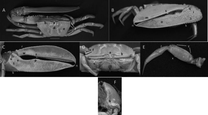
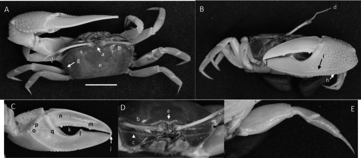
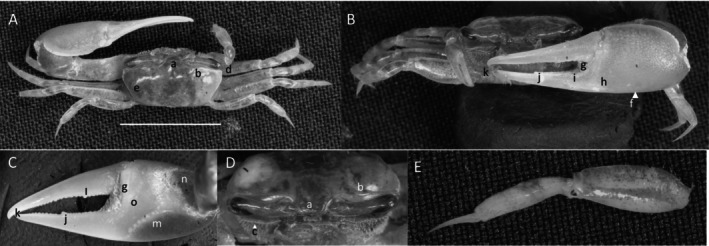
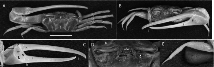
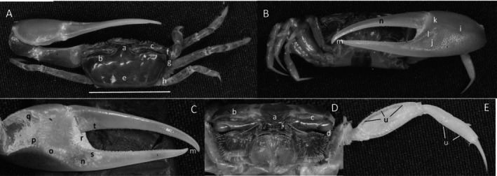
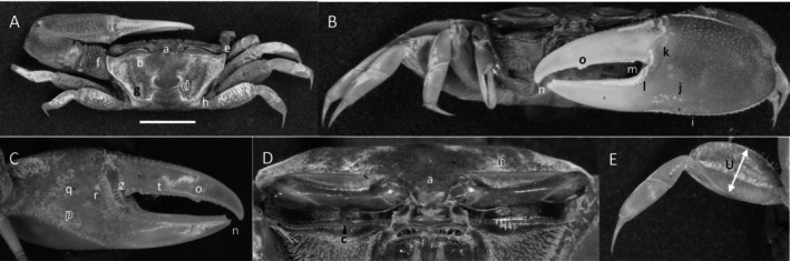
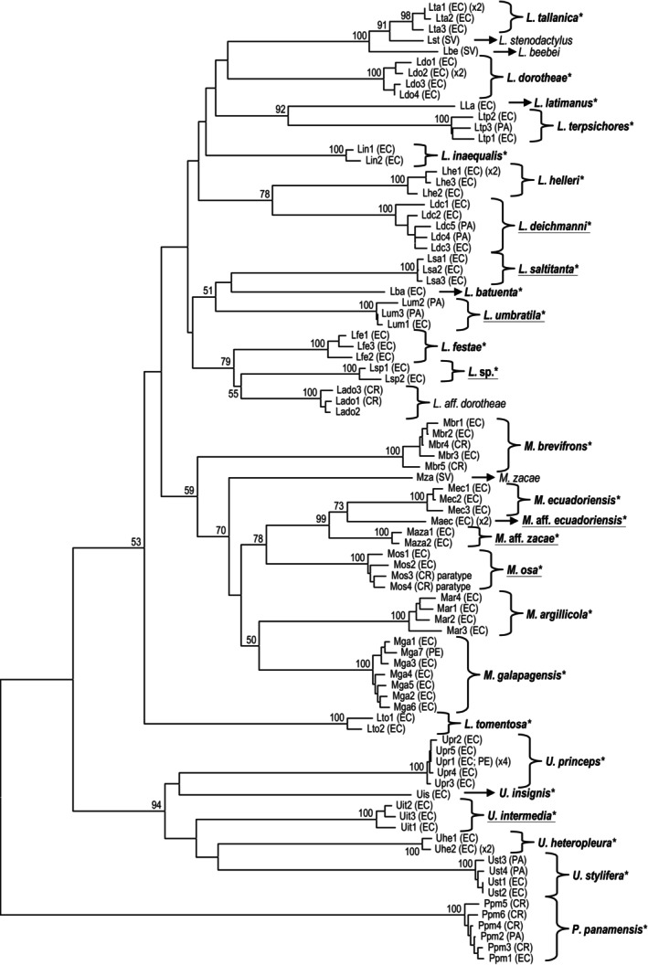
Neotropical regions near the equator are recognized as speciation "hot spots" reflecting their abundant biodiversity. In western South America, the coasts of Panama, Colombia, Ecuador, the Galápagos Archipelago, and northern Peru form the Tropical Eastern Pacific biome. This area has the greatest heterogeneity of sympatric fiddler crab species of any portion of the planet. Since the coastal fauna has not been assessed for almost 50 years, we studied fiddler crab species diversity in Ecuador and on the Galápagos Archipelago. Preserved collecting records for various species were examined at the U.S. National Museum of Natural History, Washington, DC, the American Museum of Natural History, New York, and the Naturalis Biodiversity Center, Leiden, the Netherlands. During a field study, 51 locations were collected resulting in over 870 preserved specimens (120 lots) along the 2237-km (1390 mi) coast of Ecuador and on three Galápagos Islands. A neighbor-joining tree was constructed using the Kimura 2-parameter model with a partial DNA sequence of the cytochrome oxidase-subunit 1 gene (COI) for a barcoding study. Twenty-five taxa were collected during the surveys, while two more were noted from the literature and museum collections. Five published species are new to Ecuador. The species assemblage was divided among four genera: Uca, Leptuca, Minuca, and Petruca. Morphological definitions and photographic images are given for 27 species. COI sequences were obtained for 27 operational taxonomic units from Ecuador, with three morphologically indistinguishable cryptic or pseudocryptic taxa also revealed. Based on species distributions, it appears that the area between Cabo San Lorenzo and Punta Santa Elena serves as a weak barrier separating some "northern" from "southern" taxa. Since coastal Ecuador is undergoing rapid economic development, the construction of maricultural facilities and the deforestation of mangroves promote wholesale habitat destruction. As habitat diversity is reduced, it is expected that there will be, in general, a local decline in fiddler crab species diversity with some taxa becoming rare or extinct.

Keywords: cytochrome c oxidase‐subunit 1; diversity; fiddler crabs; morphology; neighbor‐joining tree.


Conflict of interest statement

The authors declare no conflicts of interest.

Figures

FIGURE 1
FIGURE 1
Collecting locations. Solid dot (•) marks field sites for collection of fiddler crabs in Ecuador and the Galapagos Archipelago during this study. See text. Provinces indicated by large letters: El Oro–ELO, Esmeraldas—ESM, Galapagos Archipelago—GAL, Guayas—GUA, Manabi—MAN, and St Elena—SEL.
FIGURE 2
FIGURE 2
Uca heteropleura (Smith, 1870) (UNI 743) collected Posorja, GUA. (A) Dorsal view of male. Bar = 10 mm scale. (B) Front view. (C) Inner cheliped. (D) Ocular view. (E) Third ambulatory. a—interocular space, b—upper orbital, c—suborbital dentations, d—dorsal lateral angle, e—lateral margin, f—subchelar keel, g—pollex terminal tooth, h—preterminal tooth, i—pollex groove with pubescence, j—dactyl preterminal tooth, k‐ oblique ridge, l—carinal tooth, m—pollex‐gap line of tubercles, n—serrated dorsal edge of pollex, and o—serrations on posteroventral merus.
FIGURE 3
FIGURE 3
Uca insignis (H. Milne Edwards, 1952) (USNM 138577), Puerto Bolivar, ELO. (A) Dorsal view of male. Bar = 10 mm scale. (B) Front view. (C) Inner cheliped. (D) Ocular view. (E) Third ambulatory. (F) Lareal surface. a—frontal region, b—anterior margin, c—lower orbital margin, d—H‐depression, e—venation, f—lateral margin spines, g—venation origin, h—lower lateral carapace margin, i—manus tubracles, j—upper manus spines, k—submanus keel, l—external oblique tubercle row, m—overlap region, n—termianl spine, o—pits, p—ridge of dorsal pollex, q—articulation junction, r—dorsal dactyl ridge, s—dactyl tooth, t—oblique inner manus ridge, u—tubercles on anterior edge of carpal cavity, v—line of tubercles, w—line of larger dactyl tubercles, x—ventral merus spike, y—setae and pubescence, and z—pile on dorsal carpus.
FIGURE 4
FIGURE 4
Uca intermedia von Prahl and Toro,  (UNI 754) collected El Rompido, ESM. (A) Dorsal view of male. Bar = 10 mm scale. (B) Front view of large cheliped. (C) Inner cheliped. (D) Ocular view. (E) Third ambulatory. a—interocular space, b—frontal margin, c—dorsal lateral angle, d—suborbital dentations, e—dorsolateral marigin, f—large bristles, g—submarginal grove, h—terminal pubescence, I—subchela keel, j—row of tubercles, k—proximal gape bristles, l—subterminal pollex tooth, m—terminal tooth, n—proximal dactyl tooth, o—oblique ridge, p—carinal tooth, q—bristles of upper carpal cavity, r—pollex‐gap line of tubercles, and s—“saw‐tooth” dentations of minor chela.
FIGURE 5
FIGURE 5
Uca ornata (Smith, 1870) (USNM 138615), Panama, Gulf of Panama, Panama Bay, Panama Viejo. (A) Dorsal view of male. Bar = 10 mm scale. (B) Front view. (C) Inner cheliped. (D) Ocular view. (E) Third ambulatory. (F) Setose pits on outer pollex. a—frontal region, b—anterior margin, c—anterior‐lateral angle, d—suborbital margin, e—venation, f—lateral margin spines, g‐ knobs on manus, h—setose pits, i—keel, j—row of tubercles, k—tubercles before articulation junction, l‐ terminal spines of dactyl and pollex, m—dorsal ridge of tubercles, n‐ row of teeth on lower dactyl, o—pit, p—large tubercle on edge of carpal cavity, q—line of tubercles on pollex, r—pollex ridge, s—line of dactyl teeth, t—pubescence and setae on merus, u—ventral merus spines, and v—carpal and propodal pubescence.
FIGURE 6
FIGURE 6
Uca princeps (Smith, 1870) (UNI 757) collected Puerto Sitio Nueva, SEL. (A) Dorsal view of male. Bar = 10 mm scale. (B) Front view. (C) Inner cheliped. (D) Ocular view. (E) Third ambulatory. a—interocular space, b—superior orbital margin, c—suborbital dentations, d—antero‐lateral angle, e—depression, f—posterior lateral margin, g—subpollex groove, h—keel of pollex, i—terminal tooth, j—tubercle line of gap, k—dactyl tooth, l—pollex tooth, m—proximal tooth of oblique ridge, n—spine of upper carpal carina, o—inner tubercle line of manus and pollex, and p—small vental spines on merus.
FIGURE 7
FIGURE 7
Uca stilifera (H. Milne Edwards, 1852) (UNI 742) collected Bahia de Caráquez, MAN. (A) Dorsal view of male. Bar = 10 mm scale. (B) Front view. (C) Inner cheliped. (D) Ocular view. (E) Third ambulatory. a—interocular space, b—supraorbital margin, c—lower orbital margin, d—stylet, e—H‐depression, f—anterolateral angle, g—lateral margin, h—keel, i—submarginal groove, j—terminal dactyl and pollex spines, k—proximal gap, l—triangular pollex tooth, m—dactyl tooth, n—inner dactyl sulcus, o—tooth of oblique ridge, p—carina margin, and q—pollex tubracle line.
FIGURE 8
FIGURE 8
Petruca panamensis (Stimpson, 1859) (UNI 752) collected Playa Camerones, ESM. (A) Dorsal view of male. Bar = 10 mm scale. (B) Front view. (C) Inner cheliped. (D) Ocular view. (E) Third ambulatory. a—frontal region, b—upper margin of orbital and eyebrow, c—dentation on suborbital margin, d—anterolateral angle, e—lateral line, f—posterior stria, g—hooked tip of dactyl and pollex, h—dactyl tooth, i—articulation ridge, and j—dense setae.
FIGURE 9
FIGURE 9
Minuca argillicola (Crane, 1941) (UNI 828) collected Rio Agua Clara, Daule, ESM. (A) Dorsal view of male. Bar = 10 mm scale. (B) Front view. (C) Inner cheliped. (D) Ocular view. (E) Third ambulatory. a—frontal region, b—upper orbital margin, c—lower orbital dentations, d—H‐depression, e‐ anterolateral angle, f—long lateral margin, g—inferior stria, h—manus keel, i—proximal dactyl tooth, j—dactyl and pollex terminus, k—triangular sulcus with pubescence, l—pre‐dactyl articulation, m—inner pollex tubercle line, n—width ambulatory merus, and o—pubescence.
FIGURE 10
FIGURE 10
Minuca brevifrons (Stimpson, 1860) (UNI 809) collected Rio Agua Clara, Daule, ESM. (A) Dorsal view of male. Bar = 10 mm scale. (B) Front view. (C) Inner cheliped. (D) Ocular view. (E) Third ambulatory. a—frontal region, b—upper frontal margin, c—suborbital dentations, d—anterolateral angle, e—circular pubescent tufts, f—H‐depression, g—upper manus, h—proximal dactyl tooth, i—dactyl and pollex terminus, j—oblique ridge apex, k—tubracle field, l—predactyl articulation ridge, and m—pubescence of carpus and propodus.
FIGURE 11
FIGURE 11
Minuca ecuadoriensis (Maccagno, 1928) (UNI 811) collected Cayapas‐Mataje, La Tola, ESM. (A) Dorsal view of male. Bar = 10 mm scale. (B) Front view. (C) Inner cheliped. (D) Ocular view. (E) Third ambulatory. a—frontal region, b—upper orbital margin, c—suborbital margin dentations, d—antero‐lateral angle, e—H‐depression, f—keel pubescence, g—pre‐articuation groove, h—tooth on dactyl and pollex, i—terminus of dactyl and pollex, j—apex of oblique ridge, k—tubracle field, l—predactyl ridge, m—tubercles on articulating cuff, and n—setae and pubescence on merus and propodus of ambulatory.
FIGURE 12
FIGURE 12
Minuca galapagenesis (Rathbun, 1902) (UNI 840) collected Bahia de Tortuga, Santa Cruz, GAL. (A) Dorsal view of male. Bar = 10 mm scale. (B) Front view. (C) Inner cheliped. (D) Ocular view. (E) Third ambulatory. a—frontal, b—upper orbital, c—lower orbital dentations, d—anterolatertal angle, e—H‐depression, f—lateral margin, g—posterior stria, h—keel, i—tubercle line on cuff, j—large tooth, k—dactyl and pollex terminus, l—apex of oblique ridge, m—tubercle field, n—predactyl tubercle line, o—tubercle line on inner articulation cuff, and p—carpal setae.
FIGURE 13
FIGURE 13
Minuca osa (Landstorfer and Schubart, 2010) (UNI 820) collected Cayapas Mataje, La Tola, ESM. (A) Dorsal view of male. Bar = 10 mm scale. (B) Front view. (C) Inner cheliped. (D) Ocular view. (E) Third ambulatory. a—frontal region, b—upper orbital margin, c—suborbital margin dentations, d—anterolateral angle, e—circular tufts of setae, f—H‐depression, g—lateral line, h—posterior stria, i—keel of manus, j—pre‐dactyl groove and cuff, k—dactyl teeth, l—teeth on pollex, m—dactyl and pollex terminus, n—oblique ridge apex, o—carina of upper carpal cavity, p—tubercle field, q—pre‐dactyl row of tubercles, r—tubercles on articulation cuff, s—pubescence and setae on merus, carpus and propodus of third ambulatory.
FIGURE 14
FIGURE 14
Leptuca batuenta (Crane, 1941) (UNI 866) collected Portete, Mompeche, ESM. (A) Dorsal view of male. Bar = 10 mm scale. (B) Front view. (C) Inner cheliped. (D) Ocular view. (E) Third ambulatory. a—frontal region, b—upper orbital margin, c—suborbital margin, d—anterolateral angle, e—lateral margin, f—H‐depression, g—submanus keel, h—dactyl articulating cuff, i—tubercle lining of pollex, j—terminal tooth of pollex, k—apex of oblique ridge, l—superior tubercle line of carpal cavity, m–pubescence on upper carpal cavity, n—tubercle line on pollex, o—tubercle line of dactyl articulation cuff.
FIGURE 15
FIGURE 15
Leptuca beebei (Crane, 1941) (UNI 793) collected Estero Salado, Posorja, GUA. (A) Dorsal view of male. Bar = 10 mm scale. (B) Front view. (C) Inner cheliped. (D) Ocular view. (E) Third ambulatory. a—Frontal region, b—upper orbital margin, c—lower orbital dentations, d—antero‐lateral angle, e—posterior‐lateral margin, f—inferior manus keel, g—pubescence of articulation joint, h—manus‐pollex depression, i—line of tubercles following gap, j—dactyl medial tooth, k—dactyl/pollex terminus, l—proximal dactyl tooth, m—oblique ridge, n—pubescence and carina of upper carpal cavity, and o—tubercle line from manus to pollex.
FIGURE 16
FIGURE 16
Leptuca deichmanni (Rathbun, 1935) (UNI 765) collected Playa Achilube, Parque Tematico Marios, ESM. (A) Dorsal view of male. Bar = 10 mm scale. (B) Front view. (C) Inner cheliped. (D) Ocular view. (E) Third ambulatory. a—frontal region, b—upper orbital margin, c—lower orbital dentations, d—antero‐lateral angle, e—H ‐depression, f—tubercle keel, g—outer pollex row, h—dactyl and pollex terminus, i—central tooth, j—tubercle line of dactyl, k—apex of oblique ridge, l—upper carpal bristles, m—first pre‐dactyl line, n—second pre‐dactyl line, and o—third pre‐dactyl tubercle row.
FIGURE 17
FIGURE 17
Leptuca dorotheae (von Hagen, 1968) (UNI 771) collected Rio Mache, San Jose de Chamanga, ESM. (A) Dorsal view of male. Bar = 10 mm scale. (B) Front view. (C) Inner cheliped. (D) Ocular view. (E) Third ambulatory. a—frontal region, b—anterior margin, c—dentations of lower orbital margin, d—antero‐lateral angle, e—H‐depression, f—posterior manus pubescence, g—dactyl junction, h—pollex tubercle, i—dactyl tooth, j—terminus, k—oblique ridge, l—dorsal carina, m—articulation tubercles, n—pre‐dactyl tubercle line, and o—tubercles on edge of pollex.
FIGURE 18
FIGURE 18
Leptuca festae (Nobili, 1901) (UNI 751) collected Isla Santay, Rio Guayas, GUA. (A) Dorsal view of male. Bar = 10 mm scale. (B) Front view. (C) Inner cheliped. (D) Ocular view. (E) Third ambulatory. a—frontal region, b—upper orbital margin, c—dentations of lower orbital margin, d—antero‐lateral angle, e—anterior lateral line, f—H‐depression, g—outer pre‐dactyl tubercle line, h—keel, i—pollex tubercles, j—pollex and dactyl terminus, k—oblique ridge, l—dorsal carina, m—pre‐dactyl tubercles, n—articulation cuff, and o—tubercle line of pollex.
FIGURE 19
FIGURE 19
Leptuca helleri (Rathbun, 1902) (UNI 777) collected Playa los Alemanes, Isla Santa Cruz, GAL. (A) Dorsal view of male. Bar = 10 mm scale. (B) Front view. (C) Inner cheliped. (D) Ocular view. (E) Third ambulatory. a—frontal region, b—upper orbital margin, c—lower orbital margin dentations. d—antero‐lateral angle, e—anterior portion lateral line, f—posterior portion lateral line, g—H‐depression, h—tubercate keel, i—articulating cuff, j—tuberlce line, k—central tooh of pollex, l—dactyl pollex terminus, m—teeth of dactyl, n‐ oblique ridge, o—ridge apex, p—carina of upper carpal cavity, q—pre‐dactyl tubercle line, r—articulation cuff, and s—prominal dactyl pile.
FIGURE 20
FIGURE 20
Leptuca inaequalis (Rathbun, 1935) (UNI 779) collected Malacón, San Lorenzo, ESM. (A) Dorsal view of male. Bar = 10 mm scale. (B) Front view. (C) Inner cheliped. (D) Ocular view. (E) Third ambulatory. a—frontal region, b—upper orbital margin, c—dentation of lower orbital margin, d—patches of pubescence, e—anterolateral angle, f—anterior lateral line, g—posterior lateral line, h—keel, i—pubescence on proximal manus, j—line of tubercles, k—pile at articulation, l—large tubercles, m—oblique ridge, n—dorsal carpal cavity carina, o—grove on inner dactyl, p—sulcus, and q—merus pubescence.
FIGURE 21
FIGURE 21
Leptuca latimanus (Rathbun, 1897) (UNI 784) collected Puerta las Javitas, SEL. (A) Dorsal view of male. Bar = 10 mm scale. (B) Front view. (C) Inner cheliped. (D) Ocular view. (E) Third ambulatory. a‐ frontal region, b—upper orbital margin, c—lower orbital dentations, d—H‐depression, e—antero‐lateral angle, f—anterior lateral line, g—posterior lateral line, h—posterior stria, i—submanus/pollex keel, j—articulation cuff, k—setae in articulation, l—terminus of dactyl and pollex, m—teeth of gap, n—oblique ridge, o—carpal cavity, p—setae, and q—line of tubercles.
FIGURE 22
FIGURE 22
Leptuca saltitanta (Crane, 1941) (UNI 795) collected El Rompido, ESM. (A) Dorsal view of male. Bar = 10 mm scale. (B) Front view. (C) Inner cheliped. (D) Ocular view. (E) Third ambulatory. a—frontal region, b—preorbital sulcus, c—upper orbital margin, d—lower orbital dentations, e—antero‐lateral angle, f—anterior lateral line, g—posterior lateral line, h—H‐depression, i—keel, j—ventral pollex groove, k—manus‐pollex sulcus, l—articulation ridge, m—line of pollex tubercles, n—dactyl groove, o—dactyl and pollex terminus, p—V‐shaped tubercle formation, q—oblique ridge, r—carpal cavity, s—predactyl line of tubercles, and t—line of tubercles and pubescence on merus.
FIGURE 23
FIGURE 23
Leptuca stenodactylus (H. Milne Edwards and Lucas, 1843) (UNI 749) collected Estero Salado, Posorja, GUA. (A) Dorsal view of male. Bar = 10 mm scale. (B) Front view. (C) Inner cheliped. (D) Ocular view. (E) Third ambulatory. a—frontal region, b—preorbital sulcus, c—upper orbital margin, d—lower orbital dentations, e—H‐depression, f—anterior lateral angle, g—antero‐lateral junction, h—posterior stria, i—manus, j—pollex‐manus sulcus, k—articulation ridge, l—line of pollex tubercles, m—dactyl and pollex terminus, n—inner manus‐pollex sulcus, o—oblique ridge, p—apex and carina of carpal cavity, q—upper carina, r—setae at articulation junction, s—line of tubercles on pollex, and t—line of tubercles on dactyl.
FIGURE 24
FIGURE 24
Leptuca tallanica (von Hagen, 1968) (USNM 138838) collected Rio Guayas, Puerto Bolivar, ELO. (A) Dorsal view of male. Bar = 10 mm scale. (B) Front view. (C) Inner cheliped. (D) Ocular view. (E) Third ambulatory. a—frontal region, b—anterior margin, c—lower orbital dentations, d—antero‐lateral angle, e—anterior lateral margin, f—pubescent commas, g—posterior lateral line, h—lateral stria, i—upper manus pubescence‐ filled groove, j—pubescence in triangle, k—keel, l—subterminal tooth, m—line of tubercles, n—pubescence at dactyl junction, o‐ tubercle row on proximal pollex, p—line of large tubercles on dactyl, q—dactyl terminus, r—oblique ridge, s—carina lining upper edge of carpal cavity, t—predactyl line of tubercles, u—articulation tubercles, and v—setae on walking legs.
FIGURE 25
FIGURE 25
Leptuca tenuipedis (Crane, 1941) (USNM 79404) collected Costa Rica, Puntarenas, Nicoya Peninsula, Ballena Bay. (A) Dorsal view of male. Bar = 10 mm scale. (B) Front view. (C) Inner cheliped. (D) Ocular view. (E) Third ambulatory. a—frontal region, b—anterior margin, c—lower orbital dentations, d—antero‐lateral angle, e—anterior lateral margin, f—posterior lateral margin, g—H‐depression, h—tubercles on manus, i—keel, j—submarginal line, k—tubercle line, l—tubercle/tooth, m—obtuse edge, n—dactyl junction, o‐ tubercle line on dorsal dactyl, p—line of large tubercles on ventral dactyl, q—swollen inner manus, r—tubercles, s—row of tubercles on pollex, t—distal tubercle ridge, and u—pubescnce on merus and carpus of walking legs.
FIGURE 26
FIGURE 26
Leptuca terpsichores (Crane, 1941) (UNI 798) collected Malácon, San Lorenzo, ESM. (A) Dorsal view of male. Bar = 10 mm scale. (B) Front view. (C) Inner cheliped. (D) Ocular view. (E) Third ambulatory, (F) Ventral carpal stridulating ridges. a—frontal region, b—upper orbital margin, c—lower orbital dentations, d—pre‐orbital sulcus, e—antero‐lateral angle, f—posterior lateral line, g—H‐depression, h—articulation cuff, i—keel, j—stridulating ridges, k—dactyl tooth, l—terminus of dactyl and pollex, m—proximal row of pollex teeth, n—oblique ridge, o—upper carpal carina, p—tubercle line, q – predactyl cuff tubercles, and r – pollex tubercle row.
FIGURE 27
FIGURE 27
Leptuca tomentosa (Crane, 1941) (UNI 804) collected Simón Bolivar, SEL. (A) Dorsal view of male. Bar = 10 mm scale. (B) Front view. (C) Inner cheliped. (D) Ocular view. (E) Third ambulatory. a—frontal region, b—upper orbital margin, c—lower orbital dentations, d—pubescence on posterior carapace, e—anterolateral angle, f—posterior lateral line, g—postereior stria, h—keel, i—articulating cuff, j—row of pollex teeth, k ‐terminus of dactyl and pollex, l—dactyl teeth, m—sulcus, n—oblique ridge, o—carpal carina, p‐ tubercle patch, q—pre‐dactyl tubercles, r—articulation cuff, s—pile.
FIGURE 28
FIGURE 28
Leptuca umbratila (Crane, 1941) (UNI 805) collected El Rompido, ESM. (A) Dorsal view of male. Bar = 10 mm scale. (B) Front view. (C) Inner cheliped. (D) Ocular view. (E) Third ambulatory. a—frontal region, b—upper orbital margin, c—lower orbital dentations, d—H‐depression with pubescence, e—anterolateral angle, f—anterior lateral margin, g—posterior lateral margin, h—posterior stria, i—keel, j—pollex manus junction, k—articulation cuff, l—line of tubercles, m‐ pile in gap, n—pollex dactyl terminus, o—dactyl tooth, p ‐obliques ridge, q—carpal cavity carina, r‐ first row of tubercles, s—tubercles on articulation cuff, t—row of tubecles on dactyl, and u—width of ambulatory merus.
FIGURE 29
FIGURE 29
A neighbor‐joining COI tree for 30 operational taxonomic units (OTU) of fiddler crabs from Ecuador and other related taxa. Species names marked with “*” indicate specimens from Ecuador, and underlined species names are unidentified species or newly recorded species. EC—Ecuador, SV—El Salvador, CR—Costa Rica, PA—Panama, and PE—Peru. Only bootstrap values > 50% are shown on the nodes. For haplotype names, refer to Table 3.

References

    1. Aerts, K. , Vanagt T., Degraer S., et al. 2004. “Macrofaunal Community Structure and Zonation of an Ecuadorian Sandy Beach (Bay of Valdivia).” Belgium Journal of Zoology 134, no. 1: 17–24. http://core.ac.uk/download/pdf/55703009.pdf.
    1. Ahrens, D. , Ahyong S. T., Ballerio A., et al. 2021. “Is It Time to Describe New Species Without Diagnoses?—A Comment on Sharkey.” Zootaxa 5027, no. 2: 151–159. 10.11646/ZOOTAXA.5027.2.1. - DOI - PubMed
    1. Aoki, M. , and Wada K.. 2013. “Genetic Structure of the Wide‐Ranging Fiddler Crab Uca crassipes in the West Pacific Region.” Journal of the Marine Biological Association of the United Kingdom 93, no. 3: 789–795. 10.1017/S0025315412001178. - DOI
    1. Barnwell, F. H. , and Szelistowski W. A.. 1985. “Twenty‐One Species of Fiddler Crabs (Genus Uca) From a Small Tidal River on the Pacific Coast of Costa Rica.” American Zoologist 25, no. 4: 86A.
    1. Barnwell, F. H. , and Thurman C. L.. 1984. “Taxonomy and Biogeography of the Fiddler Crabs (Ocypodidae: Genus Uca) of the Atlantic and Gulf Coasts of Eastern North America.” Zoological Journal of the Linnaean Society 81, no. 1: 23–87. 10.1111/j.1096-3642.1984.tb02558.x. - DOI

LinkOut - more resources