Review
. 2022 Jan 18;1(1):5-27.
doi: 10.1007/s44164-022-00008-x. eCollection 2022 Feb.

Osteosarcoma tumor microenvironment: the key for the successful development of biologically relevant 3D in vitro models

Affiliations
Review

Osteosarcoma tumor microenvironment: the key for the successful development of biologically relevant 3D in vitro models

João Rodrigues et al. In Vitro Model. .

Abstract

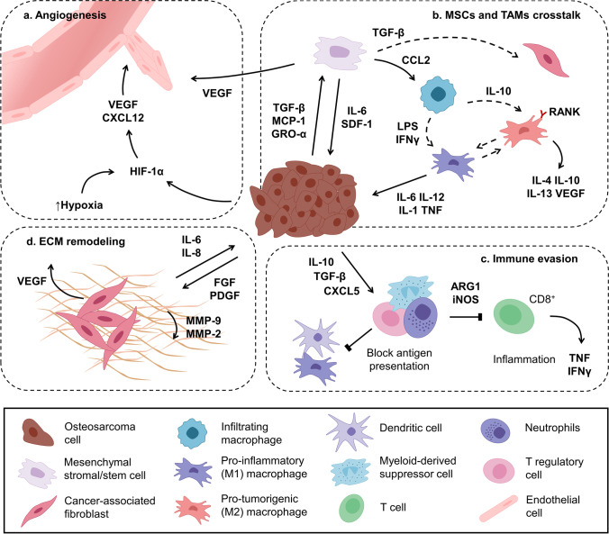
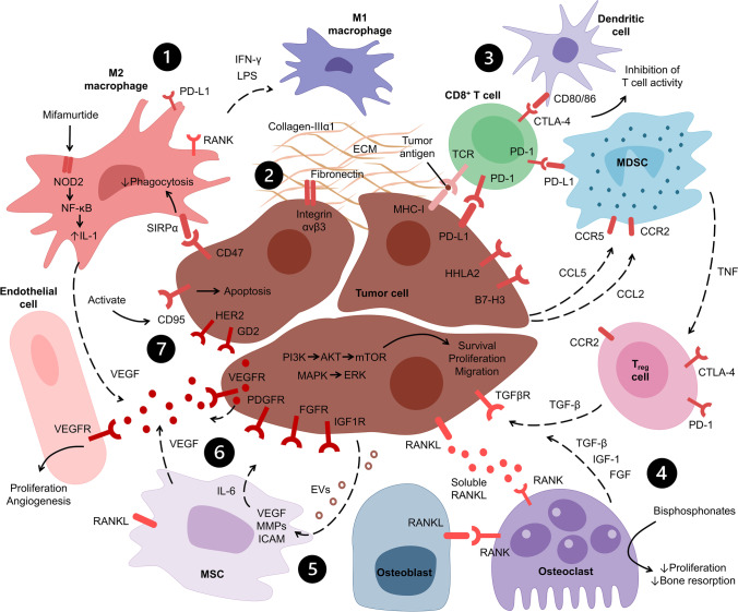
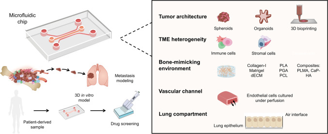
Osteosarcoma (OS) is the most common primary bone cancer in children and young adults. This type of cancer is characterized by a high mortality rate, especially for patients with resistant lung metastases. Given its low incidence, high genetic heterogeneity, the lack of effective targets, and poor availability of relevant in vitro and in vivo models to study the tumor progression and the metastatic cascade, the pathophysiology of OS is still poorly understood and the translation of novel drugs into the market has become stagnant. Due to the importance of the tumor microenvironment (TME) in the development of metastases and the growing interest in targeting TME-specific pathways for novel therapeutics in cancer, models that closely represent these interactions are crucial for a better understanding of cancer-related events. In OS research, most studies rely on oversimplified two-dimensional (2D) assays and complex animal models that do not faithfully recapitulate OS development and progression. In turn, three-dimensional (3D) models are able to mimic not only the physical 3D environment in which cancer cells grow but also involve interactions with the TME, including its extracellular matrix, and thus are promising tools for drug screening studies. In this review, the existing and innovative OS in vitro 3D models are highlighted, focusing on how the TME is crucial to develop effective platforms for OS tumor and metastasis modeling in a physiologically relevant context.

Keywords: 3D in vitro models; Metastasis; Microfluidics; Osteosarcoma; Tumor microenvironment.


Conflict of interest statement

Competing interestsThe authors declare no competing interests.

Figures

Fig. 1
Fig. 1
Bone homeostasis vs. osteosarcoma-related bone remodeling. a Bone homeostasis is a complex process tightly regulated by the RANKL/RANK/OPG signaling pathway. RANKL is secreted by osteoblasts and osteocytes stimulated by PTH, vitamin D3, and prostaglandin E2. RANKL binds to the RANK receptor in the membrane of preosteoclasts differentiated from HSCs due to M-CSF activity, produced by immune cells and osteoblasts. Mature osteoclasts promote bone resorption, which is inhibited by OPG that binds to RANKL, blocking RANK signaling. b In OS, bone remodeling is induced by tumor cells that create a vicious cycle to promote cancer cell survival and proliferation. Tumor cells secrete RANKL, IL-6, IL-11, and TNF-α that stimulate osteoclastogenesis, leading to greater secretion of bone factors that promote tumor cell survival. Moreover, OS cells release factors, such as TGF-β, that promote the secretion of RANKL by osteoblasts and inhibit their activity, decreasing osteoblastic bone formation. Tumor cells also promote the secretion of sclerostin (Scl) from osteocytes that inhibits osteoblast’s activity. This creates a tumor-enhancing environment where the expression of OPG is generally decreased, while the expression of RANKL is normally enhanced. PTHrP, PTH-related protein, SEM4D, semaphorin-4D
Fig. 2
Fig. 2
Osteosarcoma tumor microenvironment interactions. (a) Tumor cells interact with endothelial cells and MSCs within a hypoxic environment, promoting the release of HIF-1α VEGF, which induces angiogenesis. (b) Bone marrow–derived MSCs are attracted to the tumor site by CXCL12 and MCP-1 secreted by OS cells. Within the TME, MSCs are normally “educated” by tumor cells (through TNF signaling) to have pro-tumoral activity and can differentiate into cancer-associated fibroblasts. Circulating macrophages can infiltrate the TME and differentiate into pro-inflammatory M1-like macrophages that have anti-cancer activity, or anti-inflammatory M2-like macrophages that express RANK and promote tumor proliferation and angiogenesis. (c) OS cells promote immunosuppression through the activation of T regulatory cells, myeloid-derived suppressor cells, and neutrophils that suppress the activity of antigen-presenting cells and inhibit cytotoxic CD8+ T cells. (d) Cancer-associated fibroblasts are activated by FGF and PDGF released by cancer cells, and promote ECM remodeling, inducing the formation of a dense matrix structure, which in turn promotes the formation of a necrotic tumor core, leading to VEGF secretion and angiogenesis. ARG1, arginase 1, iNOS, inducible nitric oxide synthase, LPS, lipopolysaccharide, SDF-1, stromal cell–derived factor-1
Fig. 3
Fig. 3
Targeting the osteosarcoma tumor microenvironment. The strategies to target the OS TME to develop novel therapeutics include the following: (1) targeting TAMs by reprogramming M2-like macrophages into an anti-tumorigenic M1 phenotype, and promoting phagocytosis by inhibiting the SIRPα receptor; (2) targeting the ECM by inhibiting the tumorigenic activity of certain molecules (collagen-IIIα1, αvβ3 integrin); (3) using monoclonal antibodies against immune checkpoint inhibitors (PD-1, PD-L1, CTLA-4, HHLA2, B7-H3) to promote the activity of cytotoxic T cells and inhibit the activity of MDSCs and T regulatory cells; (4) targeting osteoclast activity (with bisphosphonates), the RANKL/RANK signaling pathway (with monoclonal antibodies), and the activity of the TGF-β receptor; (5) inhibiting the pro-tumorigenic activity of MSCs; (6) targeting multiple tyrosine kinase receptors (VEGFR, PDGFR, FGFR, IGF1R, HER2) that promote tumor cell survival and proliferation, as well as angiogenesis; (7) targeting other cell surface receptors overexpressed in OS cells (GD2) and promoting tumor cell apoptosis by enhancing the activity of the FAS receptor (CD95). LPS, lipopolysaccharide, MHC-I, major histocompatibility complex I, TCR, T cell receptor
Fig. 4
Fig. 4
Framework for the development of novel osteosarcoma bioengineered models. Schematic representation of how innovative 3D models can be employed to study the metastatic spread of OS cells to the lung on a microfluidic chip adaptable for culture under dynamic conditions. OS cells might be cultured on a separate compartment as spheroids or organoids that can be assembled with other cell types to resemble the TME, namely immune cells (macrophages, lymphocytes) and stromal cells (MSCs, fibroblasts, osteoblasts, osteoclasts), on a bone-mimicking environment composed by natural hydrogels (collagen type I, Matrigel®, decellularized matrices (dECM)), synthetic polymers (PGA, PLA), or composite materials (PLMA, CaP-HA). OS cells growing on this compartment should be able to migrate into the vascular channel with endothelial cells and reach the lung compartment. Importantly, the platform should be adaptable for drug screening studies and to evaluate potential pharmacological targets to inhibit the metastatic spread of OS cells. To further enhance the predictable value, patient-derived tissues could be used, thus harnessing the advantages of personalized medicine and improving the preclinical pipeline before performing animal studies

References

    1. Durfee RA, Mohammed M, Luu HH. Review of osteosarcoma and current management. Rheumatol Ther. 2016;3:221–43. 10.1007/s40744-016-0046-y. - PMC - PubMed
    1. Valery PC, Laversanne M, Bray F. Bone cancer incidence by morphological subtype: a global assessment. Cancer Causes Control. 2015;26:1127–39. 10.1007/s10552-015-0607-3. - PubMed
    1. Kansara M, Teng MW, Smyth MJ, Thomas DM. Translational biology of osteosarcoma. Nat Rev Cancer. 2014;14:722–35. 10.1038/nrc3838. - PubMed
    1. Casali PG, Bielack S, Abecassis N, Aro HT, Bauer S, Biagini R, et al. Bone sarcomas: ESMO–PaedCan–EURACAN clinical practice guidelines for diagnosis, treatment and follow-up. Ann Oncol. 2018;29:iv79–95. 10.1093/annonc/mdy310. - PubMed
    1. Friebele JC, Peck J, Pan X, Abdel-Rasoul M, Mayerson JL. Osteosarcoma: a meta-analysis and review of the literature. Am J Orthop (Belle Mead NJ). 2015;44:547–53. - PubMed

LinkOut - more resources