. 2025 Jan 3;50(4):240-246.
doi: 10.5152/tud.2025.24120.

Population Kinetics and Protein Profiles of Co-Cultured Adult and Fetus Rabbit Bladder Smooth Muscle Cells

Affiliations

Population Kinetics and Protein Profiles of Co-Cultured Adult and Fetus Rabbit Bladder Smooth Muscle Cells

Hayrunisa Kahraman Esen et al. Urol Res Pract. .

Abstract

Objective: Bladder tissue models have been developed using smooth muscle cells (SMCs) on various scaffolds to mimic bladder morphology and physiology. This study investigates the effects of co-culturing fetal and adult SMCs on growth properties and protein profiles to understand cellular interactions and population kinetics.

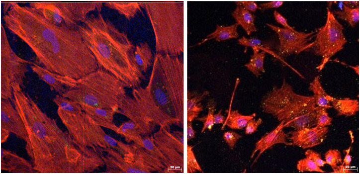
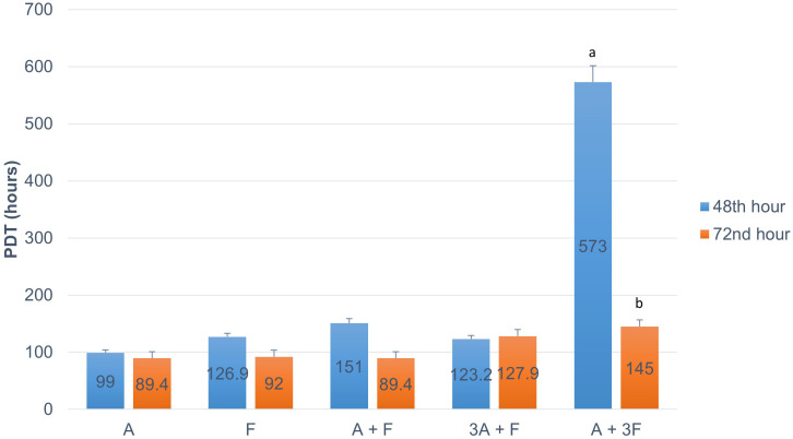
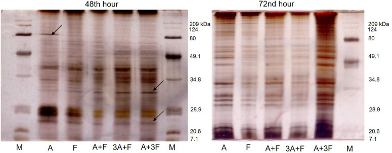
Methods: Bladder tissue samples from 10 adult and 10 fetal New Zealand rabbits were divided into 5 groups: adult SMCs (A), fetal SMCs (F), 50%A+50%F (A+F), 75%A+25%F (3A+F), and 25%A+75%F (A+3F). Population doubling time (PDT) of 3 × 10⁶ cells from each group was measured after 48 and 72 hours. Protein concentrations were estimated by spectrophotometric analysis and analyzed via SDS-PAGE gel electrophoresis. Cells exhibited typical SMC morphology, confirmed by positive staining for α-SMA and MYH11.

Results: Median cell counts of single cultures were significantly higher than co-cultures (P < .05), but cell viability was comparable (P > .05). Population doubling time at 72 hours for A, F, A+F, 3A+F, and A+3F were 89.4, 92.0, 89.4, 127.9, and 145.0 hours, respectively. Protein concentrations were similar between fetal and adult co-cultures (P > .05). Electrophoresis at 48 hours revealed a unique 80kDa band in adult cells and a 32kDa band in co-cultured cells.

Conclusion: Co-culturing resulted in increased PDT, altered protein concentrations, and changes in protein profiles, while each cell population maintained its phenotype. Fetal bladder SMCs maintained their morphology and viability when co-cultured with adult SMCs, resulting in a significant limitation in the cumulative proliferation rate. This may be dependent on alterations of protein profiles of adult and fetal SMCs promoted by rearrangements in co-cultures.

Keywords: Bladder smooth muscle cells; bladder tissue model; population kinetics; protein profiles.


Conflict of interest statement

Declaration of Interests: The authors have no conflict of interest to declare.

Figures

Figure 1.
Figure 1.
Light microscopic images of adult smooth muscle cells (SMCs) (A), fetal SMCs (F), 50% A + 50% F (A+F), 75% A + 25% F (3A+F) and 25% A + 75% F (A+3F).
Figure 2.
Figure 2.
Images of immunostaining show the expression of alpha smooth muscle actin (α-SMA, colored green) and myosin heavy chain 11 (MYH11, colored yellow) in the adult bladder smooth muscle cells (left image) and the fetal bladder smooth muscle cells (right image). Nuclei were counterstained with 4α,6-diamidino-2-phenylin-dole (DAPI, colored blue), and the cytoskeleton was visualized with phalloidin (colored red).
Figure 3.
Figure 3.
Population doubling time of cell populations of adult smooth muscle cells (A), fetal smooth muscle cells (F), 50% A + 50% F (A+F), 75% A + 25% F (3A+F) and 25% A + 75% F (A+3F). (a) P < .001 vs PDTs of other groups incubated for 48 hours. (b) P < .05 vs PDT of cells in the A+3F group incubated for 48 hours. All experiments with cell cultures were repeated 3 times. The numbers on bars represent the mean of PDTs, and error bars represent the standard error.
Figure 4.
Figure 4.
Protein gel electrophoresis of protein extracts from adult smooth muscle cells (A), fetal smooth muscle cells (F), 50% A + 50% F (A+F), 75% A + 25% F (3A+F) and 25% A + 75% F (A+3F) groups after 48 and 72 hours of incubation. Arrows are showing discrete protein bands.

Similar articles

References

    1. Casarin M, Morlacco A, Dal Moro F. Tissue engineering and regenerative medicine in pediatric urology: urethral and urinary bladder reconstruction. Int J Mol Sci. 2022;23(12):6360. (10.3390/ijms23126360) - DOI - PMC - PubMed
    1. Kimuli M, Eardley I, Southgate J. In vitro assessment of decellularized porcine dermis as a matrix for urinary tract reconstruction. BJU Int. 2004;94(6):859 866. (10.1111/j.1464-410X.2004.05047.x) - DOI - PubMed
    1. Horst M, Eberli D, Gobet R, Salemi S. Tissue engineering in pediatric bladder reconstruction-The road to success. Front Pediatr. 2019;7:91. (10.3389/fped.2019.00091) - DOI - PMC - PubMed
    1. Steins A, Dik P, Müller WH, et al. In vitro evaluation of spider silk meshes as a potential biomaterial for bladder reconstruction. PLoS One. 2015;10(12):e0145240. (10.1371/journal.pone.0145240) - DOI - PMC - PubMed
    1. Gasanz C, Raventós C, Morote J. Current status of tissue engineering applied to bladder reconstruction in humans. Actas Urol Esp (Engl Ed). 2018;42(7):435 441. (10.1016/j.acuro.2017.11.005) - DOI - PubMed

LinkOut - more resources