. 2025 Mar 4;13(3):e0216624.
doi: 10.1128/spectrum.02166-24. Epub 2025 Jan 28.

Effective killing of Mycobacterium abscessus biofilm by nanoemulsion delivery of plant phytochemicals

Affiliations

Effective killing of Mycobacterium abscessus biofilm by nanoemulsion delivery of plant phytochemicals

Casey Albano et al. Microbiol Spectr. .

Abstract

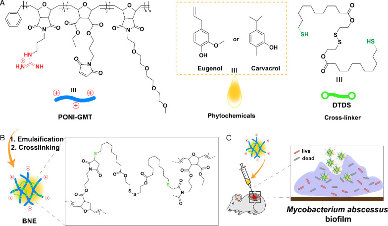
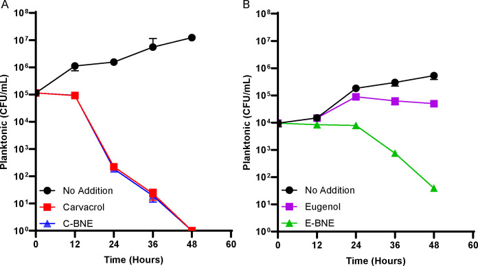
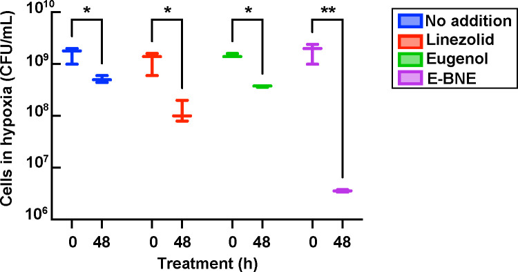
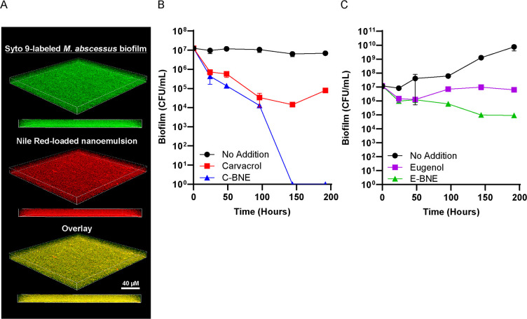
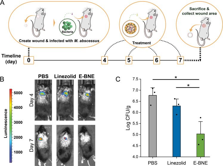
Mycobacterium is an acid-fast, aerobic, non-motile, and biofilm-forming bacterium. The increasing prevalence of mycobacterial infections makes it necessary to find new methods to combat the resistance of bacteria to conventional antibiotics. Mycobacterium abscessus is an emerging pathogen that is intrinsically drug resistant due to several factors, including an impermeable cell envelope, drug efflux pumps, target-modifying enzymes, and the ability to form thick, robust biofilms. Phytochemicals are promising antimicrobials; however, their poor solubility in water and their inability to penetrate biofilms render them inefficient in killing bacterial biofilms. In this study, we demonstrate the efficacy of polymer-stabilized phytochemical nanoemulsions in killing M. abscessus biofilms. These nanoemulsions improve the solubility and stability of the phytochemicals and enable biofilm penetration and eradication. We show that the phytochemical emulsions effectively eliminated M. abscessus in an in vitro biofilm model and killed non-replicating persister cells in the Wayne hypoxia model. These nanoemulsions were also effective in vivo in a wound infection model. These findings demonstrate the potential of polymer-stabilized phytochemical nanoemulsions as a promising alternative to conventional antibiotics for the treatment of mycobacterial infections.

Importance: Mycobacterium abscessus is among the opportunistic bacterial pathogens that cause nontuberculous mycobacterial diseases. The infection caused by M. abscessus is difficult to treat because the bacterium is resistant to many of the currently available antibiotics, limiting chemotherapeutic strategies. Furthermore, it forms biofilms in clinically relevant settings, making the infection difficult to treat. Many phytochemicals have potent antimicrobial activities, but their hydrophobicity limits clinical applications. In this study, we tested a new drug delivery strategy where hydrophobic plant phytochemicals were emulsified with a biodegradable nanosponge. We show that the emulsification makes phytochemicals such as carvacrol and eugenol more effective against M. abscessus biofilms. We further demonstrate that nanoemulsified phytochemicals can kill hypoxia-induced dormant M. abscessus and effectively improve skin wound infection in mice. Our data pave the way to use phytochemical nanosponge as a platform to create synergy by combining other antimycobacterial drugs.

Keywords: Mycobacterium; antimicrobial agents; drug delivery; essential oils; nanoemulsion; phytochemical.


Conflict of interest statement

The authors declare no conflict of interest.

Figures

Fig 1
Fig 1
Fabrication scheme of biodegradable polymeric nanoemulsions incorporating carvacrol and eugenol. (A) Structures of poly(oxanorborneneimide) scaffold bearing guanidine, maleimide, and tetraethyleneglycol monomethyl ether groups (PONI-GMT), phytochemicals (eugenol and carvacrol), and biodegradable dithiol-disulfide (DTDS). (B) Process for emulsification and crosslinking that enhances the stability of the nanoemulsions. (C) Schematic representation of the efficient penetration of BNEs into wound biofilm murine model, leading to the effective killing of residing bacteria.
Fig 2
Fig 2
Characterization of biodegradable nanoemulsion. (A) DLS histogram of C-BNE and E-BNE. (B) Charge characterization of C-BNE and E-BNE through zeta potential, respectively. (C) Transmission electron microscopy images of 12% E-BNE. Due to the evaporation of the oil component during the sample preparation, the nanoemulsion appears irregular in shape under electron microscopy.
Fig 3
Fig 3
Antimycobacterial effects of nanoemulsion-encapsulated phytochemicals on planktonic and biofilm M. smegmatis. M. smegmatis planktonic cultures were grown to the log phase (OD600 = 0.6–0.8) and diluted back to an OD600 of 0.1. Pellicle biofilm was grown for 5 days before initiating the antimycobacterial treatments. (A) Planktonic M. smegmatis treated with 3.1 mM carvacrol or 8% (vol/vol) C-BNE (3.1 mM carvacrol equivalent). (B) Planktonic M. smegmatis treated with 3.1 mM eugenol or 8% (vol/vol) E-BNE (3.1 mM eugenol equivalent). (C) M. smegmatis biofilm treated with 3.1 mM carvacrol or 8% (vol/vol) C-BNE. (D) M. smegmatis biofilm treated with 3.1 mM eugenol or 8% (vol/vol) E-BNE. Time 0 hour indicates when phytochemicals were added. Experiments were done in triplicate, and averages and standard deviations are shown. Note that phytochemicals are essential components of BNE preparations, and thus we cannot test a control BNE that does not contain phytochemicals.
Fig 4
Fig 4
PIM inositol acylation and increased membrane permeability in M. abscessus induced by phytochemicals. (A) The reaction of PIM inositol acylation. A fatty acid is added to the 3-OH of myo-inositol in response to membrane fluidization stress. R1 and R2 are tuberculostearic acid and palmitic acid, respectively, in M. smegmatis, but they have not been determined in M. abscessus. (B) High-performance thin layer chromatography analysis of PIM inositol acylation in response to 1.6 and 3.1 mM of carvacrol or eugenol. (C) Flow cytometry results of M. abscessus treated with DMSO (negative control), 100 mM benzyl alcohol, 3.1 mM carvacrol, and 3.1 mM eugenol. Membrane permeability was determined using TO-PRO-3, a membrane-impermeable fluorescent DNA staining dye.
Fig 5
Fig 5
Nano-emulsified phytochemicals have bactericidal effects on planktonically grown M. abscessus. (A) M. abscessus cultures were treated with no addition, 8% (vol/vol) C-BNE, or 3.1 mM carvacrol. (B) M. abscessus cultures were treated with no addition, 8% (vol/vol) E-BNE, or 3.1 mM eugenol. Time 0 indicates the onset of the treatments. All treatments were performed in triplicate, and standard deviations are shown.
Fig 6
Fig 6
Bactericidal effect of nanoemulsion-encapsulated phytochemicals on M. abscessus cells under hypoxia. Hypoxic M. abscessus cells treated with 28.8 µg/mL linezolid or 3.1 mM eugenol or 8% E-BNE. Time 0 hour indicates when phytochemicals or drugs were added. Experiments were done in triplicate. The bold central line represents the mean, with error bars indicating the standard deviation. Statistical significance was determined by two-way ANOVA, followed by Šídák’s multiple comparisons test. *P < 0.05 and **P < 0.005.
Fig 7
Fig 7
Effect of nanoemulsions on M. abscessus biofilm. (A) M. abscessus biofilm was stained with SYTO 9 green fluorescent DNA stain and incubated with Nile Red-loaded E-BNE for 3 hours. Fluorescence was visualized by confocal fluorescence microscopy. (B) M. abscessus biofilms were grown for 7 days in M63 medium. The biofilm was treated with no addition, 8% (vol/vol) C-BNE, and 3.1 mM carvacrol dissolved in M63. (C) M. abscessus biofilms were grown for 7 days in M63 medium. The biofilm was treated with no addition, 8% (vol/vol) E-BNE, and 3.1 mM eugenol dissolved in M63. All treatments were performed in triplicate, and standard deviations are shown.
Fig 8
Fig 8
M. abscessus skin infection model. (A) Experimental procedure. 108 CFU of luciferase-expressing M. abscessus was inoculated. Treatment was either 85 µM linezolid or E-BNE (containing 3.1 mM eugenol). (B) Luciferase luminescence was measured by an IVIS spectrum-CT imaging system. (C) At day 7, mice were sacrificed and CFU per gram skin lesion was measured in triplicate, with error bars indicating standard deviation. *P < 0.05 determined by one-way ANOVA and Newman-Keuls multiple comparison test.

Similar articles

Cited by

References

    1. Dahl VN, Mølhave M, Fløe A, van Ingen J, Schön T, Lillebaek T, Andersen AB, Wejse C. 2022. Global trends of pulmonary infections with nontuberculous mycobacteria: a systematic review. Int J Infect Dis 125:120–131. doi:10.1016/j.ijid.2022.10.013 - DOI - PubMed
    1. Nessar R, Cambau E, Reyrat JM, Murray A, Gicquel B. 2012. Mycobacterium abscessus: a new antibiotic nightmare. J Antimicrob Chemother 67:810–818. doi:10.1093/jac/dkr578 - DOI - PubMed
    1. Brennan PJ. 2003. Structure, function, and biogenesis of the cell wall of Mycobacterium tuberculosis. Tuberculosis (Edinb) 83:91–97. doi:10.1016/s1472-9792(02)00089-6 - DOI - PubMed
    1. Jankute M, Cox JAG, Harrison J, Besra GS. 2015. Assembly of the mycobacterial cell wall. Annu Rev Microbiol 69:405–423. doi:10.1146/annurev-micro-091014-104121 - DOI - PubMed
    1. Rahlwes KC, Sparks IL, Morita YS. 2019. Cell walls and membranes of actinobacteria. Subcell Biochem 92:417–469. doi:10.1007/978-3-030-18768-2_13 - DOI - PubMed

MeSH terms

LinkOut - more resources