

Immunohistochemical Expression of VEGF and Microvessel Density (CD 34) in Oral Epithelial Dysplasia and Oral Squamous Cell Carcinoma: Original Research
- PMID: 39873996
- PMCID: PMC12082431
- DOI: 10.31557/APJCP.2025.26.1.147
Immunohistochemical Expression of VEGF and Microvessel Density (CD 34) in Oral Epithelial Dysplasia and Oral Squamous Cell Carcinoma: Original Research
Abstract
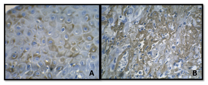
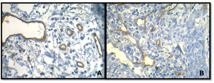
Background: Angiogenesis, the formation of new blood vessels from preexisting ones via capillary sprouting, is a crucial process in tumor growth and metastasis. As a tumor's angiogenic capacity increases, its microvasculature, measured by micro vessel density (MVD), also increases. This study aims to evaluate the expression of Vascular Endothelial Growth Factor (VEGF) and CD34 in oral epithelial dysplasia and oral squamous cell carcinoma through immunohistochemical methods.
Methods: The study analyzed a total of 40 formalin-fixed, paraffin-embedded tissue samples. These included 10 cases of normal buccal mucosa, 15 cases of oral epithelial dysplasia, and 15 cases of oral squamous cell carcinoma. Immunohistochemical staining was performed using monoclonal anti-VEGF and anti-CD34 antibodies. The intensity and area of staining for VEGF were assessed, and the mean MVD was calculated. Statistical analysis was conducted using Pearson's chi-square test and one-way ANOVA.
Results: The expression of VEGF and MVD (indicated by CD34 staining) were significantly higher in oral squamous cell carcinoma compared to oral epithelial dysplasia and normal buccal mucosa.
Conclusion: As tumors grow, angiogenesis increases proportionally with tumor volume and disease progression, contributing to tumorigenesis. VEGF serves as a critical mitogen for tumor vascularization, and MVD can be a useful indicator of disease progression.
Keywords: Angiogenesis; Squamous Cell Carcinoma; carcinogenesis; growth factor; micro vessel density.
Conflict of interest statement
None.
Figures
Similar articles
-
Immunohistochemically Detection of Angiogenesis in Oral Pre-Cancerous Lesions Compared with Oral Invasive Carcinomas.Asian Pac J Cancer Prev. 2018 Jul 27;19(7):1805-1808. doi: 10.22034/APJCP.2018.19.7.1805. Asian Pac J Cancer Prev. 2018. PMID: 30049191 Free PMC article.
-
Evaluation of VEGF, BDNF, TRKB expression in oral epithelial dysplasia, oral verrucous carcinoma and oral squamous cell carcinoma and their role as prognostic indicator.J Cancer Res Ther. 2024 Jul 1;20(5):1376-1383. doi: 10.4103/jcrt.jcrt_2406_22. Epub 2023 Oct 7. J Cancer Res Ther. 2024. PMID: 39412903
-
Angiopoietin-2 expression is correlated with angiogenesis and overall survival in oral squamous cell carcinoma.Med Oncol. 2013;30(2):571. doi: 10.1007/s12032-013-0571-2. Epub 2013 May 7. Med Oncol. 2013. PMID: 23649549
-
Stromelysin 3, Ets-1, and vascular endothelial growth factor expression in oral precancerous and cancerous lesions: correlation with microvessel density, progression, and prognosis.Clin Cancer Res. 2005 Mar 15;11(6):2272-84. doi: 10.1158/1078-0432.CCR-04-0572. Clin Cancer Res. 2005. PMID: 15788677
-
Angiogenesis in benign, pre-malignant and malignant vulvar lesions.Anticancer Res. 2002 Nov-Dec;22(6C):3853-65. Anticancer Res. 2002. PMID: 12553005 Review.
References
-
- Hasina R, Lingen MW. Angiogenesis in oral cancer. J Dent Educ. 2001;65(11):1282–90. - PubMed
-
- Hoeben A, Landuyt B, Highley MS, Wildiers H, Van Oosterom AT, De Bruijn EA. Vascular endothelial growth factor and angiogenesis. Pharmacol Rev. 2004;56(4):549–80. - PubMed
-
- Ribatti D. The crucial role of vascular permeability factor/vascular endothelial growth factor in angiogenesis: a historical review. Br J Haematol. 2005;128(3):303–9. - PubMed
MeSH terms
Substances
LinkOut - more resources
Full Text Sources
Medical
Research Materials