Review
. 2025 May 1;32(3):146-156.
doi: 10.1097/MOH.0000000000000860. Epub 2025 Feb 7.

Histological differences among thrombi in thrombotic diseases

Affiliations
Review

Histological differences among thrombi in thrombotic diseases

Atsushi Yamashita et al. Curr Opin Hematol. .

Abstract

Purpose of review: This review aims to summarize the histological differences among thrombi in acute myocardial infarction, ischemic stroke, venous thromboembolism, and amniotic fluid embolism, a newly identified thrombosis.

Recent findings: Acute coronary thrombi have a small size, are enriched in platelets and fibrin, and show the presence of fibrin and von Willebrand factor, but not collagen, at plaque rupture sites. Symptomatic deep vein thrombi are large and exhibit various phases of time-dependent histological changes. Cancer-associated venous thromboemboli contain invasive cancer cells that penetrate the vascular walls, and small cancer cell aggregates are observed within the thrombi. The thrombus composition in atherosclerotic and cardioembolic ischemic strokes varies from case to case, while the thrombi in cancer-associated ischemic stroke are rich in platelets and fibrin. A pathological study on amniotic fluid embolism identified uterine vein thrombi and massive platelet-rich microthrombi in the lungs.

Summary: Atherothrombus formation is induced by plaque disruption and may occlude a narrow lumen within a short time. Venous thrombi may grow to a large size in a multistage or chronic manner. Cancer cells can directly contribute to venous thrombus formation. The thrombus formation in amniotic fluid embolism may explain the occurrence of consumptive coagulopathy and cardiopulmonary collapse.


Conflict of interest statement

There are no conflicts of interest.

Figures

Box 1
Box 1
no caption available
FIGURE 1
FIGURE 1
Thrombus sizes and compositions in acute myocardial infarction, ischemic stroke, and deep vein thrombosis. (a) Mean thrombus histological cross-sectional areas in acute myocardial infarction (AMI; n = 25) [12], atherosclerotic ischemic stroke (IS; n = 6), cardioembolic ischemic stroke (IS; n = 11), deep vein thrombosis (DVT; n = 89), and pulmonary embolism (PE, n = 14). The thrombus area in AMI was adapted from Okuyama et al.[12] with permission. The thrombus areas in others are original data. (b) Median immunopositive areas for glycoprotein (GP) IIb/IIIa, fibrin, and glycophorin A in aspirated thrombi in acute myocardial infarction (AMI; n = 50), atherosclerotic ischemic stroke (IS; n = 11) [42], cardioembolic ischemic stroke (IS; n = 74) [42]; and deep vein thrombosis (DVT; n = 16) [6]. The thrombus composition in AMI is an original data. The thrombus composition in ischemic stroke was adapted from Shimuzu et al.[42] with permission. The thrombus composition in DVT was adapted from Furukoji et al.[6].
FIGURE 2
FIGURE 2
Representative histological images of the interface between ruptured plaque and coronary thrombi and presence of cancer cells in venous thromboembolism. (a) Immunohistochemical staining for CD68 and tissue factor indicates the presence of macrophages and tissue factor expression in the ruptured plaque. The thrombus on the ruptured plaque is rich in platelets (GPIIb/IIIa), and some fibrin formation is visible within the thrombus (arrows). Fibrin deposition is evident around macrophages and cholesterin clefts, and in the necrotic core. In the high-magnification images of the dashed squares (lower panels), fibrin deposits are evident at the interface between the ruptured plaque and thrombus, and no fibrous matrix is observed in the Azan-stained image. The interface is indicated by the dashed line. GPIIb/IIIa, glycoprotein IIb/IIIa; HE, hematoxylin-eosin; P, ruptured plaque; Th, thrombus. (b) Venous thrombus in the inferior vena cava of a patient with pancreatic adenocarcinoma. The dashed line indicates the interface between the thrombus (Th) and the venous wall (V). Cytokeratin (CK)-positive cancer cells show direct invasion into the thrombus from the venous wall, associated with thrombus formation. The lower panels correspond to the high-magnification images in an area of upper panels. (c) Pulmonary thrombi in a patient with gastric adenocarcinoma. Cancer cell clusters are observed in a variety of sizes. There is no direct cancer invasion from the pulmonary artery (PA). The lower panels correspond to the high-magnification images in an area of upper panels. (d) Representative immunofluorescence image of tissue factor (red)-expressing cancer cells (green) in a venous thrombus from a patient with gastric adenocarcinoma. (e) Representative immunofluorescence image of podoplanin (red)-expressing cancer cells (green) in a pulmonary thrombus from a patient with cutaneous squamous cell carcinoma. Figure (a) was adapted from Yamashita et al.[28]. Figures (b–e) are original data.
FIGURE 2 (Continued)
Representative histological images of the interface between ruptured plaque and coronary thrombi and presence of cancer cells in venous thromboembolism. (a) Immunohistochemical staining for CD68 and tissue factor indicates the presence of macrophages and tissue factor expression in the ruptured plaque. The thrombus on the ruptured plaque is rich in platelets (GPIIb/IIIa), and some fibrin formation is visible within the thrombus (arrows). Fibrin deposition is evident around macrophages and cholesterin clefts, and in the necrotic core. In the high-magnification images of the dashed squares (lower panels), fibrin deposits are evident at the interface between the ruptured plaque and thrombus, and no fibrous matrix is observed in the Azan-stained image. The interface is indicated by the dashed line. GPIIb/IIIa, glycoprotein IIb/IIIa; HE, hematoxylin-eosin; P, ruptured plaque; Th, thrombus. (b) Venous thrombus in the inferior vena cava of a patient with pancreatic adenocarcinoma. The dashed line indicates the interface between the thrombus (Th) and the venous wall (V). Cytokeratin (CK)-positive cancer cells show direct invasion into the thrombus from the venous wall, associated with thrombus formation. The lower panels correspond to the high-magnification images in an area of upper panels. (c) Pulmonary thrombi in a patient with gastric adenocarcinoma. Cancer cell clusters are observed in a variety of sizes. There is no direct cancer invasion from the pulmonary artery (PA). The lower panels correspond to the high-magnification images in an area of upper panels. (d) Representative immunofluorescence image of tissue factor (red)-expressing cancer cells (green) in a venous thrombus from a patient with gastric adenocarcinoma. (e) Representative immunofluorescence image of podoplanin (red)-expressing cancer cells (green) in a pulmonary thrombus from a patient with cutaneous squamous cell carcinoma. Figure (a) was adapted from Yamashita et al.[28]. Figures (b–e) are original data.
FIGURE 3
FIGURE 3
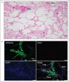
Representative histological images of pulmonary thrombi in amniotic fluid embolism. (a) Representative histological image of amniotic fluid embolism (hematoxylin–eosin staining). Multifocal small thrombi (asterisks) with or without amniotic fluid materials (arrows) are observed in the small pulmonary arteries. (b) Immunofluorescence images of the pulmonary thrombi. The microthrombi in the small pulmonary arteries are composed of platelets (green) and leukocytes (blue; nuclear staining with 4’,6-diamidino-2-phenylindole [DAPI]) without fibrin formation (red). Aggregated platelets are observed in the alveolar capillary vessels (arrows). (c) Schematic illustration of pulmonary microthrombus and uterine vein thrombus formation in amniotic fluid embolism. Although the pathogenesis remains unknown, amniotic fluid flows into the maternal circulation at the uterus, resulting in amniotic fluid embolism. Amniotic fluid entering the uterine veins forms a thrombus with a similar composition to a conventional deep vein thrombus. In the lungs, microthrombi mainly composed platelets and neutrophils show extensive formation alongside amniotic fluid emboli. NETs form in the uterine vein thrombi, but rarely in the pulmonary microthrombi. Mast cells in the perivascular and interstitial space may increase vascular permeability and affect the hemodynamics. The images are original data.
FIGURE 3 (Continued)
Representative histological images of pulmonary thrombi in amniotic fluid embolism. (a) Representative histological image of amniotic fluid embolism (hematoxylin–eosin staining). Multifocal small thrombi (asterisks) with or without amniotic fluid materials (arrows) are observed in the small pulmonary arteries. (b) Immunofluorescence images of the pulmonary thrombi. The microthrombi in the small pulmonary arteries are composed of platelets (green) and leukocytes (blue; nuclear staining with 4’,6-diamidino-2-phenylindole [DAPI]) without fibrin formation (red). Aggregated platelets are observed in the alveolar capillary vessels (arrows). (c) Schematic illustration of pulmonary microthrombus and uterine vein thrombus formation in amniotic fluid embolism. Although the pathogenesis remains unknown, amniotic fluid flows into the maternal circulation at the uterus, resulting in amniotic fluid embolism. Amniotic fluid entering the uterine veins forms a thrombus with a similar composition to a conventional deep vein thrombus. In the lungs, microthrombi mainly composed platelets and neutrophils show extensive formation alongside amniotic fluid emboli. NETs form in the uterine vein thrombi, but rarely in the pulmonary microthrombi. Mast cells in the perivascular and interstitial space may increase vascular permeability and affect the hemodynamics. The images are original data.
FIGURE 4
FIGURE 4
Schematic illustration of coronary atherothrombus and venous thrombus formation. Platelet- and fibrin-rich atherothrombus formation is induced by acute plaque disruption and occludes the narrow lumen within a short time. Intraplaque hemorrhage and fibrin deposition frequently precede plaque rupture. Venous thrombi rich in fibrin and red blood cells form and grow to a large size in a multistage or chronic manner. Cancer-associated venous thrombi contain invasive cancer cells that penetrate the vascular walls, and small cancer cell aggregates are observed within the thrombi. Extracellular vesicles from cancer cells and cancer-associated fibroblasts promote venous thrombus formation. This is an original data.

References

    1. Kelly RV, Cohen MG, Stouffer GA. Mechanical thrombectomy options in complex percutaneous coronary interventions. Catheter Cardiovasc Interv 2006; 68:917–928. - PubMed
    1. Sato Y, Hatakeyama K, Yamashita A, et al. . Proportion of fibrin and platelets differs in thrombi on ruptured and eroded coronary atherosclerotic plaques in humans. Heart 2005; 91:526–530. - PMC - PubMed
    1. Yunoki K, Naruko T, Sugioka K, et al. . Erythrocyte-rich thrombus aspirated from patients with ST-elevation myocardial infarction: association with oxidative stress and its impact on myocardial reperfusion. Eur Heart J 2012; 33:1480–1490. - PubMed
    1. Riegger J, Byrne RA, Joner M, et al. . Histopathological evaluation of thrombus in patients presenting with stent thrombosis. A multicenter European study: a report of the prevention of late stent thrombosis by an interdisciplinary global European effort consortium. Eur Heart J 2016; 37:1538–1549. - PMC - PubMed
    1. Takahashi M, Yamashita A, Moriguchi-Goto S, et al. . Critical role of von Willebrand factor and platelet interaction in venous thromboembolism. Histol Histopathol 2009; 24:1391–1398. - PubMed