. 2025 Feb 4;122(5):e2412029122.
doi: 10.1073/pnas.2412029122. Epub 2025 Jan 28.

Endogenous LRRK2 and PINK1 function in a convergent neuroprotective ciliogenesis pathway in the brain

Affiliations

Endogenous LRRK2 and PINK1 function in a convergent neuroprotective ciliogenesis pathway in the brain

Enrico Bagnoli et al. Proc Natl Acad Sci U S A. .

Abstract

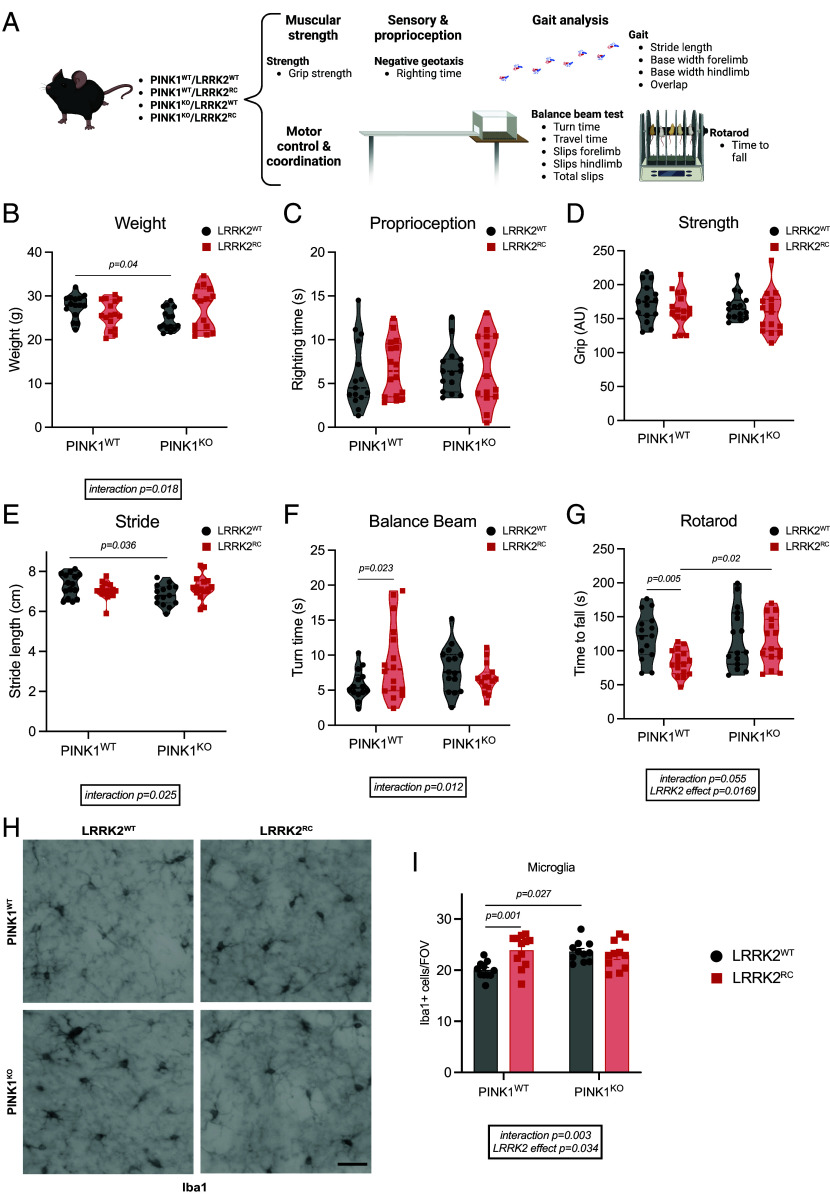
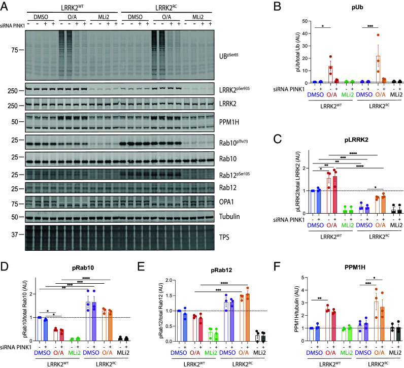
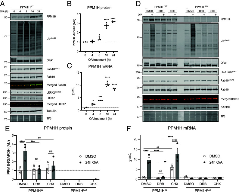
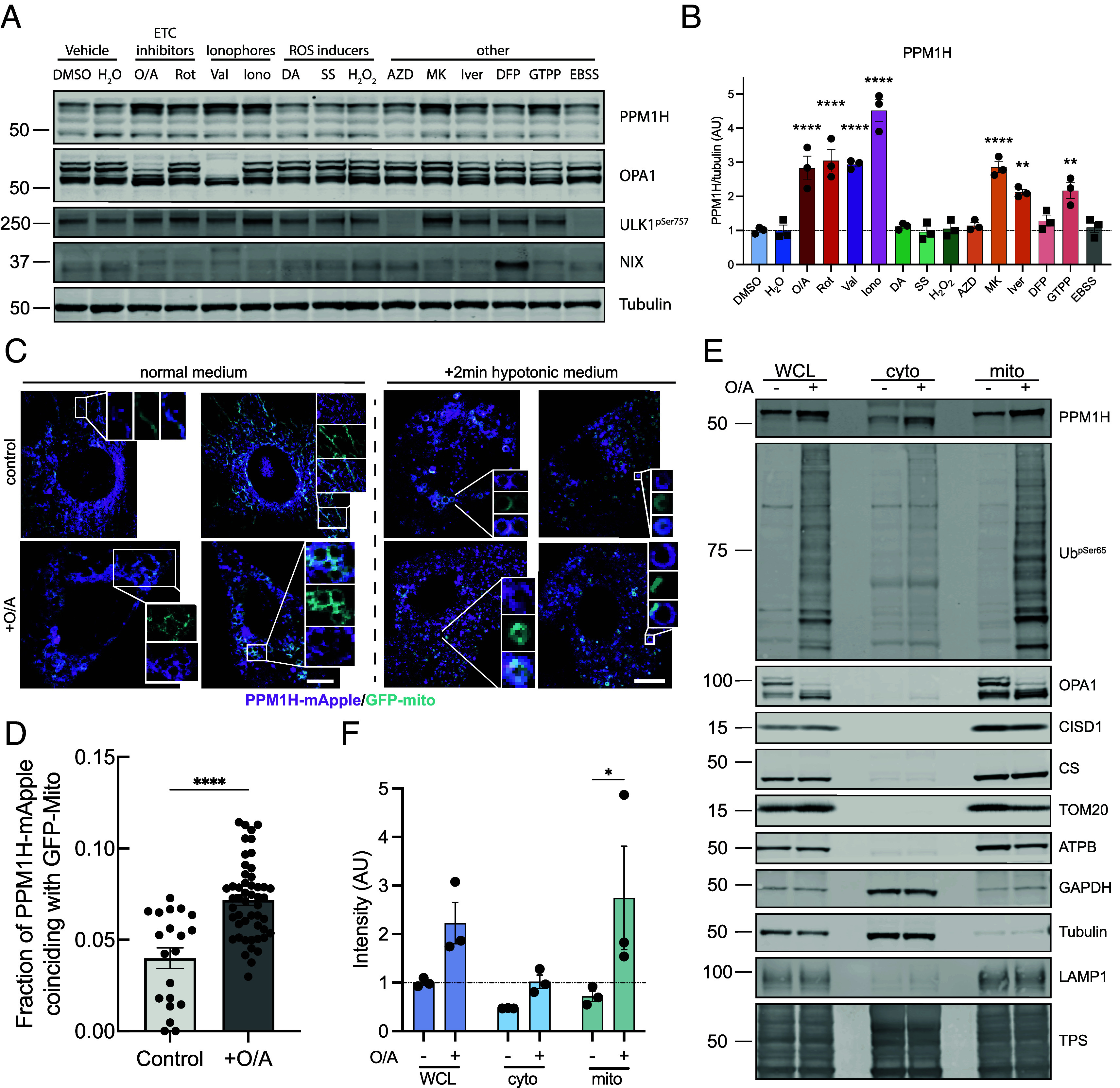
Mutations in Leucine-rich repeat kinase 2 (LRRK2) and PTEN-induced kinase 1 (PINK1) are associated with familial Parkinson's disease (PD). LRRK2 phosphorylates Rab guanosine triphosphatase (GTPases) within the Switch II domain while PINK1 directly phosphorylates Parkin and ubiquitin (Ub) and indirectly induces phosphorylation of a subset of Rab GTPases. Herein we have crossed LRRK2 [R1441C] mutant knock-in mice with PINK1 knock-out (KO) mice and report that loss of PINK1 does not impact endogenous LRRK2-mediated Rab phosphorylation nor do we see significant effect of mutant LRRK2 on PINK1-mediated Rab and Ub phosphorylation. In addition, we observe that a pool of the Rab-specific, protein phosphatase family member 1H phosphatase, is transcriptionally up-regulated and recruited to damaged mitochondria, independent of PINK1 or LRRK2 activity. Parallel signaling of LRRK2 and PINK1 pathways is supported by assessment of motor behavioral studies that show no evidence of genetic interaction in crossed mouse lines. Previously we showed loss of cilia in LRRK2 R1441C mice and herein we show that PINK1 KO mice exhibit a ciliogenesis defect in striatal cholinergic interneurons and astrocytes that interferes with Hedgehog induction of glial derived-neurotrophic factor transcription. This is not exacerbated in double-mutant LRRK2 and PINK1 mice. Overall, our analysis indicates that LRRK2 activation and/or loss of PINK1 function along parallel pathways to impair ciliogenesis, suggesting a convergent mechanism toward PD. Our data suggest that reversal of defects downstream of ciliogenesis offers a common therapeutic strategy for LRRK2 or PINK1 PD patients, whereas LRRK2 inhibitors that are currently in clinical trials are unlikely to benefit PINK1 PD patients.

Keywords: LRRK2; PINK1; brain; ciliogenesis; phosphorylation.


Conflict of interest statement

Competing interests statement:M.M.K.M. is a member of the Scientific Advisory Board of Montara Therapeutics Inc. and scientific consultant to Mission Therapeutics and Merck Sharp and Dohme.

Figures

Fig. 1.
Fig. 1.
LRRK2 signaling pathway in the striatum and cortex is not affected by loss of PINK1 in vivo. (A) Schematic of methodology followed (B) Immunoblot of LRRK2 pathway component in mouse striatum and relative quantification of (C) pSer105/total Rab12, (D) phospho-threonine73/total Rab10, (E) pSer935/total LRRK2, and (F) PPM1H/Vinculin. Similarly in (GK) analysis from the mouse cortex. Each lane was loaded with 40 μg of protein lysate from one mouse. In graphs, the black circle represents PINK1WT while red square PINK1KO animals. Box and whiskers plot, from min to max with the median line. Ordinary two-way ANOVA with Sidak’s multiple comparison test. *P < 0.05, **P < 0.01, ***P < 0.001, ****P < 0.0001.
Fig. 2.
Fig. 2.
Behavioral testing in 10.5-mo-old double-mutant PINK1 knockout/LRRK2 R1441C mutant mice does not suggest genetic interaction in vivo. (A) A battery of different tests was performed to assess motor function in PINK1 KO and LRRK2 [R1441C] knock-in mice (LRRK2RC). (B) Weight at 10.5 mo. (C) Righting time from negative geotaxis test and (D) grip strength. (E) Measure of stride length during gait analysis and (F) time to turn from balance beam. (G) Time to fall from rotarod test. (H) Representative images of the microglial marker Iba1 and (I) quantification of the Iba1 positive cells. In violin plots, the black circle represents LRRK2WT while red squares LRRK2RC mice. Ordinary two-way ANOVA with Sidak’s multiple comparison test. *P < 0.05, **P < 0.01, ***P < 0.001, ****P < 0.0001. N = 15/16 mice per group. (Scale bar, 50 µm.)
Fig. 3.
Fig. 3.
LRRK2 signaling pathway is not affected by mitochondrial damage-induced activation of PINK1. PINK1 was activated with oligomycin/antimycin for 24 h, while LRRK2 was inhibited by 1 h and 30 min MLi2 treatment. (A) Representative immunoblot of PINK1 activation effect on LRRK2 pathway components in LRRK2WT and LRRK2RC MEFs. Quantification from three experimental replicates for pUb, pLRRK2, pRab10, pRab12, and PPM1H is presented in (B–F) respectively. Each lane was loaded with 30 μg of protein lysates from one well of a 6-well plate. In graphs, each dot represents one experimental replicate with two biological replicates. − denotes scramble siRNA while +PINK1 siRNA. Bar graphs show mean ± SEM from three independent experiments. Ordinary two-way ANOVA with Sidak’s multiple comparison test. *P < 0.05, **P < 0.01, ***P < 0.001, ****P < 0.0001.
Fig. 4.
Fig. 4.
Endogenous PPM1H is up-regulated following mitochondrial depolarization by a transcriptional mechanism. (A) Representative immunoblot from PPM1HWT MEFs treated with oligomycin/antimycin A for 0, 4, 8, 16, and 24 h. (B) Quantification of PPM1H protein and (C) PPM1H mRNA from three independent experiments. (D) Representative immunoblot from PPM1HWT and PPM1HKO MEFs upon 24 h treatment with O/A, O/A + DRB, or O/A + CHX. (E) Quantification of PPM1H protein and (F) PPM1H mRNA from three independent experiments. Each lane was loaded with 40 μg of protein lysates. Graphs show mean ± SEM from three independent experiments. For B and C, ordinary one-way ANOVA with Dunnett’s multiple comparison test. For E and F, ordinary two-way ANOVA with Tukey’s multiple comparison test from three independent experiments. Empty circles denote dimethyl sulfoxide (DMSO) control while black circles represent O/A treated samples.
Fig. 5.
Fig. 5.
Endogenous PPM1H is up-regulated by different mitochondrial stressors and recruited to the organelle. (A) Representative immunoblot from PPM1HWT MEFs treated with different mitochondrial and cell stressors for 24 h and (B) quantification of PPM1H/tubulin from N = 3 independent experiment. (C) Live cell imaging of PPM1H-mApple MEF expressing a GFP mitochondrial tag treated with or without oligomycin/antimycin either in normal medium (Left) or hypotonic buffer (Right) and (D) relative quantification of colocalization of PPM1H-mApple and GFP-mito. (E) Immunoblot analysis of whole cell extract, cytoplasmic fraction, and crude mitochondrial fraction in PPM1HWT MEF after mitochondrial depolarization and (F) quantification of PPM1H levels in each of the three cellular compartments. O/A: Oligomycin/Antimycin (1/10 μM) Rot: Rotenone (0. 5 μM); Val: Valinomycin (2 μM); Iono: Ionomycin (5 μM); DA: Dopamine (10 μM); SS: Sodium selenite (7 μM); H2O2: Hydrogen peroxide (50 μM); AZD: AZD8055 (1 μM); MK: MK8722 (10 μM); Iver: Ivermectin (15 μM); DFP: deferiprone (1 mM); GTTP: Gamitrinib-triphenylphosphonium (5 μM); EBSS: Earle’s balanced salt solution. Each lane was loaded with 20 μg (15 μg for mitochondrial fraction) of protein lysates. Graphs show mean ± SEM from three independent experiments. For BD, ordinary one-way ANOVA with Dunnett’s multiple comparison test versus DMSO (all compounds, black circles and *) or H2O (SS, H2O2, DFP, and EBSS, black squares and #). For D, unpaired Mann–Whitney test and for F, ordinary two-way ANOVA with Sidak’s multiple comparison test from three independent experiments (normalized to whole cell DMSO control). (Scale bar, 50 μm.)
Fig. 6.
Fig. 6.
Loss of PINK1 decreases ciliary signaling in mouse striatum in vivo. (A and B) Confocal images of sections of dorsal striatum from 5-mo-old WT, LRRK2RC, PINK1KO, and LRRK2RC/PINK1KO mouse brains. (A) Cholinergic interneurons were labeled using anti-choline acetyltransferase (ChAT) antibody (green), primary cilia were labeled using anti-AC3 antibody (magenta, white arrow), and nuclei were stained with DAPI (blue). (B) Astrocytes were labeled using anti-glial fibrillary acidic protein antibody (green), primary cilia were labeled using anti-ADP ribosylation factor like GTPase 13B antibody (magenta, white arrow) and nuclei were labeled using DAPI (blue). (C) Quantitation of the percentage of ChAT+ neurons containing a cilium and (D) their cilia length. Similar analyses for astrocytes are shown in (E and F). (G) Confocal images to identify ChAT+ neurons and their cilia as in A (Left columns) coupled with RNAscope in situ hybridization to detect GDNF transcripts (Right columns), segregated by ciliation status in WT, LRRK2RC, PINK1KO, and LRRK2RC/PINK1KO mouse brains as indicated. (H) Quantitation of GDNF RNA dots per neuron or (I) segregated as a function of ciliation status. Error bars represent SEM from N = 3, 4 mouse brains, with >30 ChAT+ neurons and 25 astrocytes scored per brain. In bar charts, the black circle represents LRRK2WT while red squares LRRK2RC mice. Ordinary two-way ANOVA with Sidak’s multiple comparison test. *P < 0.05, **P < 0.01, ***P < 0.001, ****P < 0.0001. (Scale bar, 10 µm.)

References

    1. Gilks W. P., et al. , A common LRRK2 mutation in idiopathic Parkinson’s disease. Lancet 365, 415–416 (2005). - PubMed
    1. Paisan-Ruiz C., et al. , Cloning of the gene containing mutations that cause PARK8-linked Parkinson’s disease. Neuron 44, 595–600 (2004). - PubMed
    1. Zimprich A., et al. , Mutations in LRRK2 cause autosomal-dominant parkinsonism with pleomorphic pathology. Neuron 44, 601–607 (2004). - PubMed
    1. Dhekne H. S., et al. , Genome-wide screen reveals Rab12 GTPase as a critical activator of Parkinson’s disease-linked LRRK2 kinase. Elife 12, e87098 (2023). - PMC - PubMed
    1. Vides E. G., et al. , A feed-forward pathway drives LRRK2 kinase membrane recruitment and activation. Elife 11, e79771 (2022). - PMC - PubMed

Substances