. 2025 Mar:80:103511.
doi: 10.1016/j.redox.2025.103511. Epub 2025 Jan 23.

The UCP2/PINK1/LC3b-mediated mitophagy is involved in the protection of NRG1 against myocardial ischemia/reperfusion injury

Affiliations

The UCP2/PINK1/LC3b-mediated mitophagy is involved in the protection of NRG1 against myocardial ischemia/reperfusion injury

Xin-Tao Li et al. Redox Biol. 2025 Mar.

Abstract

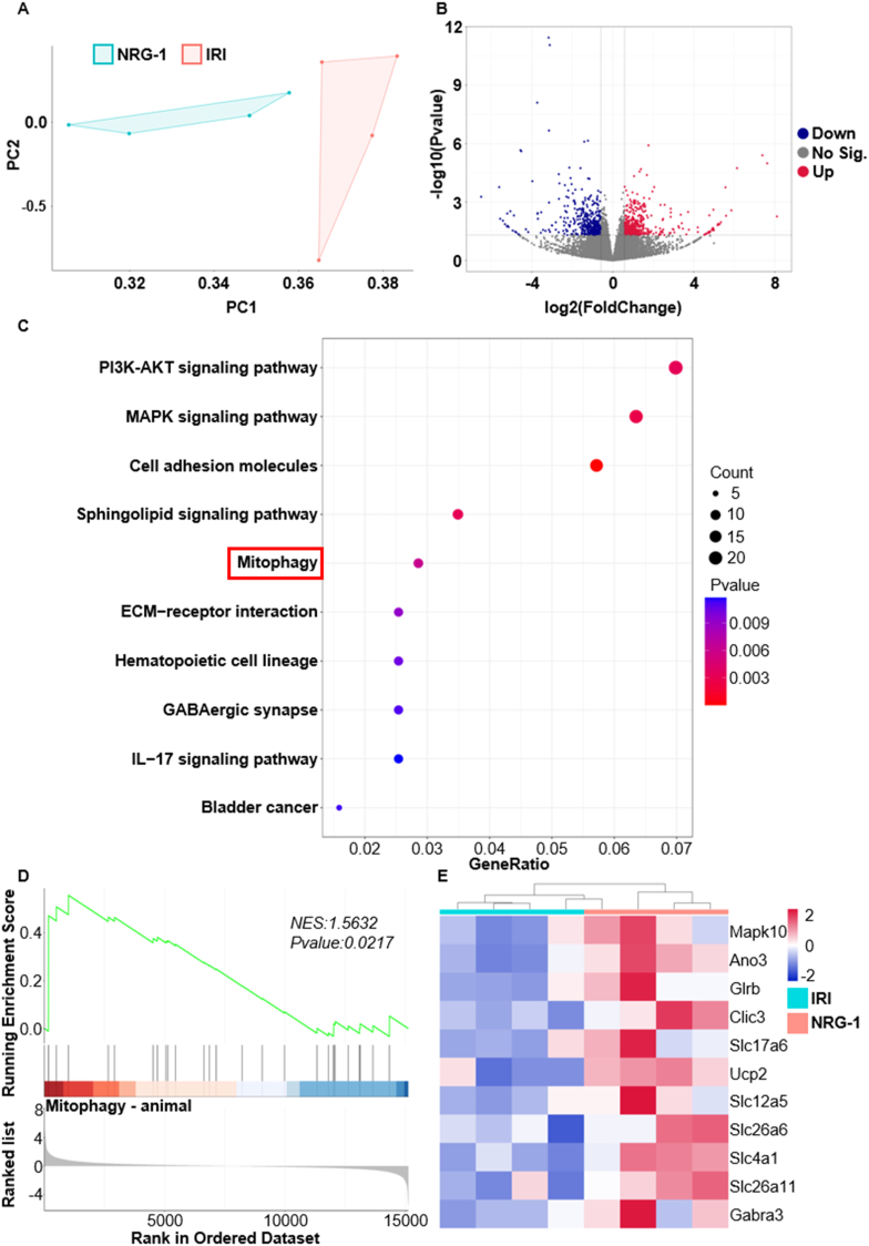
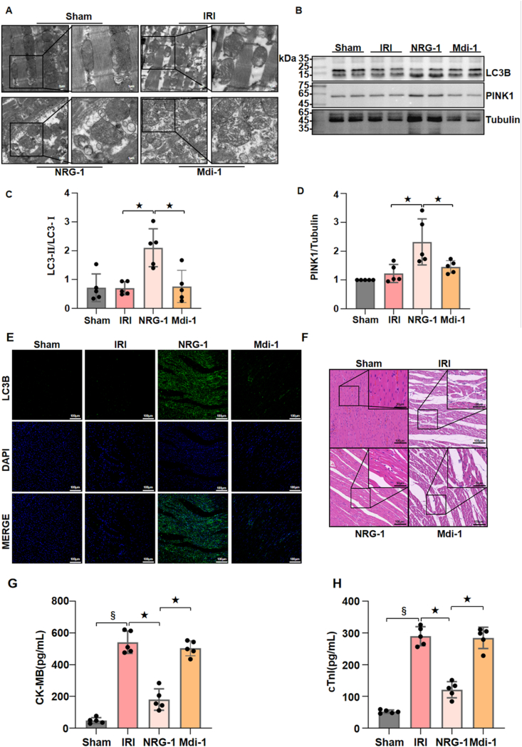
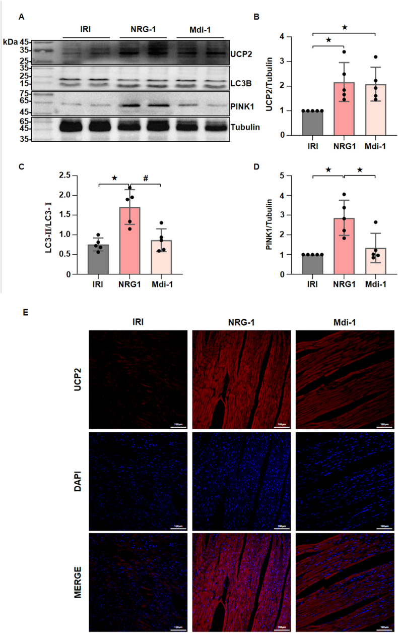
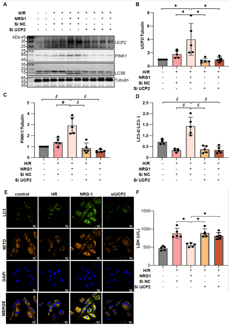
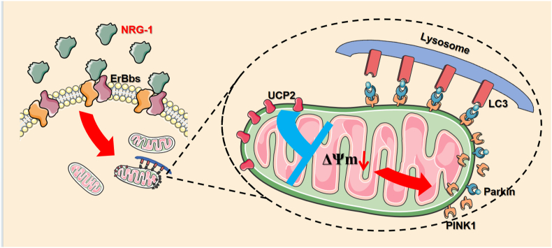
Available evidence indicates that neuregulin-1 (NRG-1) can provide a protection against myocardial ischemia/reperfusion (I/R) injury and is involved in various cardioprotective interventions by potential regulation of mitophagy. However, the molecular mechanisms linking NRG-1 and mitophagy remain to be clarified. In this study, both an in vivo myocardial I/R injury model of rats and an in vitro hypoxia/reoxygenation (H/R) model of H9C2 cardiomyocytes were applied to determine whether NRG-1 postconditioning attenuated myocardial I/R injury through the regulation of mitophagy and to explore the underlying mechanisms. In the in vivo experiment, cardioprotective effects of NRG-1 were determined by infarct size, cardiac enzyme and histopathologic examinations. The potential downstream signaling pathways and molecular targets of NRG-1 were screened by the RNA sequencing and the Protein-Protein Interaction Networks. The expression levels of mitochondrial uncoupling protein 2 (UCP2) and mitophagy-related proteins in both the I/R myocardium and H/R cardiomyocytes were measured by immunofluorescence staining and Western blots. The activation of mitophagy was observed with transmission electron microscopy and JC-1 staining. The KEGG and GSEA analyses showed that the mitophagy-related signaling pathways were enriched in the I/R myocardium treated with NRG-1, and UCP2 exhibited a significant correlation between mitophagy and interaction with PINK1. Meanwhile, the treatment with mitophagy inhibitor Mdivi-1 significant eliminated the cardioprotective effects of NRG-1 postconditioning in vivo, and the challenge with UCP2 inhibitor genipin could also attenuate the activating effect of NRG-1 postconditioning on mitophagy. Consistently, the in vitro experiment using H9C2 cardiomyocytes showd that NRG-1 treatment significantly up-regulated the expression levels of UCP2 and mitophagy-related proteins, and activated the mitophagy, whereas the challenge with small interfering RNA-mediated UCP2 knockdown abolished the effects of NRG-1. Thus, it is conclused that NRG-1 postconditioning can produce a protection against the myocardial I/R injury by activating mitophagy through the UCP2/PINK1/LC3B signaling pathway.

Keywords: Ischemia/reperfusion injury; Mitophagy; Neuregulin-1; Uncoupling protein 2.


Conflict of interest statement

Declaration of competing interest The authors have declared no conflict of interest.

Figures

Image 1
Graphical abstract
Fig. 1
Fig. 1
NRG-1 postconditioning attenuated myocardial I/R injury and inhibited myocardial inflammatory responses. (A) Representative appearances the I/R myocardium with Evans blue and TTC staining. (B) Quantitative analysis of infarct area (n = 5). (C, D) Serum cTnI and CK-MB concentrations (n = 5). (E) Representative pathological appearances of the I/R myocardium with H & E staining (magnification, × 20; inset, × 40; scale bar: 100/50 μm). (F) Representative western blots for interleukin-1 β (IL-1β, 17 kDa) and Tubulin (49 kDa, loading control) in the I/R myocardium. (G) Quantitative analysis of western blotting data for IL-1β in the I/R myocardium (n = 5). The Unpaired Student's t-test for between-group comparisons of data and one-way ANOVA followed with post hoc test (Bonferroni test) for multi-group comparisons. Data represent means ± SD. P < 0.05, #P < 0.001. The unprocessed and extra duplicate images of western blots are provided in supplementry file (Supplementary Fig. 1).
Fig. 2
Fig. 2
The cardioprotection of NRG-1 postconditioning was associated with mitophagy activation. (A, B) The PCA map of genes and volcano map of DEGs in the I/R myocardium from the IRI and NRG-1 groups (n = 4, P < 0.05). (C) The KEGG analysis of DEGs (n = 4, P < 0.05). (D) The GSEA analysis of mitophagy related genes with DEGs (n = 4, p value is indicated on the figure). (E) The heat map of mitophagy related genes in the I/R myocardium from the IRI and NRG-1 groups (n = 4).
Fig. 3
Fig. 3
Inhibiting mitophagy eliminated the cardioprotective effects of NRG-1 postconditioning. (A) Representative TEM images of mitochondria in the I/R myocardium from different groups (magnification, × 5000; inset, × 10000; scale bar: 2/1 μm). (B) Representative western blots for LC3B (15 kDa), PINK1 (63 kDa) and Tubulin (49 kDa, loading control) in the I/R myocardium. (C, D) Quantification of western blotting data for LC3B and PINK1 in the I/R myocardium (n = 5). (E) Representative confocal images of LC3B merged with DAPI in the I/R myocardium (magnification, × 20; cale bar: 100 μm). (F) Representative pathological appearances of the I/R myocardium with H&E staining (magnification, × 20; inset, × 40; scale bar: 100/50 μm). (G, H) Serum concentrations of cTnI and CK-MB (n = 5). The Unpaired Student's t-test for between-group comparisons of data and one-way ANOVA followed with post hoc test (Bonferroni test) for multi-group comparisons. Data represent means ± SD.P < 0.05, #P < 0.001, §P < 0.0001. The unprocessed and extra duplicate images of western blots are provided in supplementry file (Supplement Fig. 2).
Fig. 4
Fig. 4
The regulatory effect of NRG-1 on mitophagy was associated with UCP2. (A) The Venn Diagram of the shared genes from the DEGs and PCGs associated with mitophagy (n = 4). (BE) The correlation analysis between shared genes and mitophagy (n = 4, p value is indicated on the figure). (F)The PPI analysis between UCP2 and mitophagy-related proteins (the connection between the two indicates evidence of interaction, the thickness of the connection correlates with the strength of the evidence).
Fig. 5
Fig. 5
The UCP2 was located at the upstream of myocardial mitophagy to mediate the regulatory role of NRG-1 postconditioning. (A) Representative western blots for UCP2 (33 kDa), LC3B (15 kDa), PINK1 (63 kDa) and Tubulin (49 kDa, loading control) in the I/R myocardium. (BD) The quantitative analyses of western blotting data for UCP2, LC3B and PINK1 in the I/R myocardium (n = 5). (E) Representative confocal images of UCP2 merged with DAPI in the I/R myocardium (magnification, × 20; cale bar: 100 μm). The Unpaired Student's t-test for between-group comparisons of data and one-way ANOVA followed with post hoc test (Bonferroni test) for multi-group comparisons. Data represent means ± SD.P < 0.05, #P < 0.001. The unprocessed and extra duplicate images of western blots are provided in supplementry file (Supplementary Fig. 3).
Fig. 6
Fig. 6
NRG-1 postconditioning induced mitophagy in the cardiomyocytes subjected to H/R procedures. (A) Representative western blots for UCP2 (33 kDa), PINK1 (63 kDa) and Tubulin (49 kDa, loading control) in the H/R cardiomyocytes. (B, C) The quantitative analyses of western blotting data for UCP2 and PINK1 in the H/R cardiomyocytes (n = 5). (D) Representative confocal images of JC-1 staining merged with DAPI in the H/R cardiomyocytes (magnification, × 60; cale bar: 20 μm). The Unpaired Student's t-test for between-group comparisons of data and one-way ANOVA followed with post hoc test (Bonferroni test) for multi-group comparisons. Data represent means ± SD. P < 0.05, #P < 0.001. The unprocessed and extra duplicate images of western blots are provided in supplementry file (Supplement Fig. 4).
Fig. 7
Fig. 7
NRG-1 promoted mitophagy in the H/R cardiomyocytes by upregulating the UCP2-PINK1-LC3B signaling pathway. (A) Representative western blots for UCP2 (33 kDa), PINK1 (63 kDa), LC3B (15 kDa) and Tubulin (49 kDa, loading control) in the H/R cardiomyocytes with or without UCP2 siRNA challenge. (BD) The quantitative analyses of western blotting data for UCP2, PINK1 and LC3B in the H/R cardiomyocytes (n = 5). (E) Representative confocal images of colocalization of LC3B and mitochondria and merged with DAPI in the H/R cardiomyocytes (yellow fluorescence indicates the occurrence of colocalization; magnification, × 60; cale bar: 20 μm). (F) The LDH activity in the supernatants of the cultured cardiomyocytes (n = 5). The Unpaired Student's t-test for between-group comparisons of data and one-way ANOVA followed with post hoc test (Bonferroni test) for multi-group comparisons. Data represent means ± SD. P < 0.05, #P < 0.001, §P < 0.0001. The unprocessed and extra duplicate images of western blots are provided in supplementry file (Supplement Fig. 5).
Fig. 8
Fig. 8
NRG-1 postconditioning attenuated myocardial I/R injury via the UCP2-PINK1-LC3B signaling pathway. (A) Representative western blots for UCP2 (33 kDa), LC3B (15 kDa), PINK1 (63 kDa) and Tubulin (49 kDa, loading control) in the I/R myocardium. (BD) The quantitative analyses of western blotting data for UCP2, LC3B and PINK1 in the I/R myocardium (n = 5). (E) Representative confocal images of UCP2 merged with DAPI in the I/R myocardium (magnification, × 20; cale bar: 100 μm). (F) Representative pathological appearances of the I/R myocardium with H & E staining (magnification, × 20; inset, × 40; scale bar: 100/50 μm). (G, H) Serum cTnI and CK-MB concentrations (n = 5). The Unpaired Student's t-test for between-group comparisons of data and one-way ANOVA followed with post hoc test (Bonferroni test) for multi-group comparisons. Data represent means ± SD. P < 0.05; #P < 0.001; §P < 0.0001. The unprocessed and extra duplicate images of western blots are provided in supplementry file (Supplement Fig. 6).
Fig. 9
Fig. 9
Schematic illustration exhibiting the molecular mechanism that NRG-1 postconditioning activates mitophagy to provide a protection against myocardial I/R injury. The NRG-1 treatment upregulates the UCP2 expression in the mitochondria of cardiomyocytes. The UCP2 is able to reduce the mitochondrial membrane potential (MMP), which subsequently leads to the accumulation of the PINK1 complex on the outer membrane of mitochondria. This process induces the lysosomal surface recognizer LC3B to bind to the complex, ultimately resulting in the activation of mitophagy.

References

    1. Galli M., Niccoli G., De Maria G., Brugaletta S., Montone R.A., Vergallo R., Benenati S., Magnani G., D'Amario D., Porto I., et al. Coronary microvascular obstruction and dysfunction in patients with acute myocardial infarction. Nat. Rev. Cardiol. 2024;21:283–298. doi: 10.1038/s41569-023-00953-4. - DOI - PubMed
    1. Yellon D.M., Hausenloy D.J. Myocardial reperfusion injury. N. Engl. J. Med. 2007;357:1121–1135. doi: 10.1056/NEJMra071667. - DOI - PubMed
    1. de Miranda D.C., de Oliveira Faria G., Hermidorff M.M., Dos Santos Silva F.C., de Assis L.V.M., Isoldi M.C. Pre- and post-conditioning of the heart: an overview of cardioprotective signaling pathways. Curr. Vasc. Pharmacol. 2021;19:499–524. doi: 10.2174/1570161119666201120160619. - DOI - PubMed
    1. Guo Y.F., Zhang X.X., Liu Y., Duan H.Y., Jie B.Z., Wu X.S. Neuregulin-1 attenuates mitochondrial dysfunction in a rat model of heart failure. Chin. Med. J. 2012;125:807–814. doi: 10.3760/cma.j.issn.0366-6999.2012.05.015. - DOI - PubMed
    1. Bersell K., Arab S., Haring B., Kuhn B. Neuregulin1/ErbB4 signaling induces cardiomyocyte proliferation and repair of heart injury. Cell. 2009;138:257–270. doi: 10.1016/j.cell.2009.04.060. - DOI - PubMed

MeSH terms

LinkOut - more resources