. 2025 Jan 28;9(1):25.
doi: 10.1038/s41698-024-00769-6.

A morphometric signature to identify ductal carcinoma in situ with a low risk of progression

Collaborators, Affiliations

A morphometric signature to identify ductal carcinoma in situ with a low risk of progression

Marcelo Sobral-Leite et al. NPJ Precis Oncol. .

Abstract

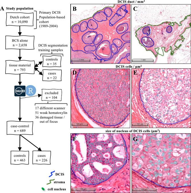
Ductal carcinoma in situ (DCIS) may progress to ipsilateral invasive breast cancer (iIBC), but often never will. Because DCIS is treated as early breast cancer, many women with harmless DCIS face overtreatment. To identify features associated with progression, we developed an artificial intelligence-based DCIS morphometric analysis pipeline (AIDmap) on hematoxylin-eosin-stained (H&E) tissue sections. We analyzed 689 digitized H&Es of pure primary DCIS of which 226 were diagnosed with subsequent iIBC and 463 were not. The distribution of 15 duct morphological measurements was summarized in 55 morphometric variables. A ridge regression classifier with cross validation predicted 5-years-free of iIBC with an area-under the curve of 0.67 (95% CI 0.57-0.77). A combined clinical-morphometric signature, characterized by small-sized ducts, a low number of cells and a low DCIS/stroma ratio, was associated with outcome (HR = 0.56; 95% CI 0.28-0.78). AIDmap has potential to identify harmless DCIS that may not need treatment.


Conflict of interest statement

Competing interests: The authors declare no competing interests.

Figures

Fig. 1
Fig. 1. AIDmap workflow.
HALO deep learning neural network was trained to recognize morphological structures in H&E whole-slides images (WSIs) (details in methods section). 1: The first classifier was trained to annotate the fibroglandular tissue (stroma), leaving adipocytes outside (green line). 2: DCIS classification was applied within the annotated stroma, by detecting pixels that reached more than 90% of confidence of composing a DCIS duct (red areas in the image heatmap). 3: Next, a nuclear segmentation sensing hematoxylin staining was applied within the DCIS regions to detect all nuclear structures. After these three steps, HALO provided tables containing the area, perimeter and spatial coordinates of stroma, DCIS and nuclear objects that were imported to R studio. 4: A True/False computational filtering was applied according to the nuclear perimeter, area and circular shape factor in order to eliminate false nuclear objects. And a True/False filtering was applied on DCIS objects, according the density of cells and average minimal nuclear distance (min. nucl. dist.) within the duct, to eliminate false DCIS ducts detected by HALO. Finally, morphological measurements for each DCIS duct were obtained.
Fig. 2
Fig. 2. Sample and classification details.
A Flow chart of the study population of patients diagnosed with primary DCIS in the Netherlands between 1989 and 2004. For training in the HALO AI module, 57 H&E sections from DCIS treated with BCS alone were used for automated stroma and DCIS segmentation. In total, 689 H&E WSI were successfully analyzed and their images revealed the variability on the density of DCIS ducts: large (B) and small (C) duct size (mm2); density of DCIS cells within the ducts: high (D) and low (E) DCIS cells/μm2; and average size of DCIS nucleus of the cells within the DCIS ducts: large (F) and small (G) DCIS nucleus (μm2); among other morphometric variables that varied among the samples.
Fig. 3
Fig. 3. Analysis of the morphometric variables.
A Heatmap with the Spearman’s rank correlation coefficients between the 55 variables obtained from the AIDmap in each H&E slide. Row side colors represent the parameters used to calculate each morphometric variable. Abbreviations are listed in Supplementary table 2. Receiver operating characteristic (ROC) curves and area under the curve (AUC) calculations from the generalized linear models to predict absence of iIBC event during follow up after 5, 10 or 15 years (BD, respectively).
Fig. 4
Fig. 4. Morphometric signature of DCIS.
A Volcano plot showing the odds ratios (OR) and the p values (p) of the 55 morphometric variables, obtained from linear regression analysis according the iIBC status during follow-up. B Heatmap of the hierarchical cluster analysis of the 30 morphometric variables statistically associated in the volcano plot. Row side colors in blue degrees represent the categories of iIBC events during follow-up. The dendrogram colors highlights the 4 groups sharing morphometric similarities.
Fig. 5
Fig. 5. Comparative morphology between the AIDmap signatures.
Morphometric signature of DCIS. Representative examples of H&E images with segmentation marks. A larger image and a magnification of the boxed area is shown for, respectively, the blue profile (A, B), the red profile (C, D), the green profile (E, F) and the orange profile (G, H).
Fig. 6
Fig. 6. Characteristics of the 4 morphometric signatures.
Differences are illustrated in the violin plots of the distribution of 4 morphometric variables among the morphometric signatures: DCIS/stroma area ratio (A) and the total number of cells inside DCIS ducts (B). The iIBC risk curve for the patients classified with one of the morphometric signatures (C), and the forest plots from the Cox multivariate regression models estimating the risk of iIBC progression during follow-up (D).

Update of

References

    1. Ringberg, A., Palmer, B., Linell, F., Rychterova, V. & Ljungberg, O. Bilateral and multifocal breast carcinoma. A clinical and autopsy study with special emphasis on carcinoma in situ. Eur. J. Surg. Oncol17, 20–29 (1991). - PubMed
    1. Maxwell, A. J. et al. Risk factors for the development of invasive cancer in unresected ductal carcinoma in situ. Eur. J. Surg. Oncol.44, 429–435 (2018). - PubMed
    1. Ryser, M. D. et al. Cancer outcomes in DCIS patients without locoregional treatment. J. Natl Cancer Inst.111, 952–960 (2019). - PMC - PubMed
    1. Myers, E. R. et al. Benefits and harms of breast cancer screening: a systematic review. JAMA314, 1615–1634 (2015). - PubMed
    1. Falk, R. S., Hofvind, S., Skaane, P. & Haldorsen, T. Second events following ductal carcinoma in situ of the breast: a register-based cohort study. Breast cancer Res. Treat.129, 929–938 (2011). - PubMed

LinkOut - more resources